Stimulatory effects of TGFα in granulosa cells of bovine small antral follicles
- PMID: 35772748
- PMCID: PMC9246657
- DOI: 10.1093/jas/skac105
Stimulatory effects of TGFα in granulosa cells of bovine small antral follicles
Abstract
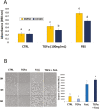
Intraovarian growth factors play a vital role in influencing the fate of ovarian follicles. They affect proliferation and apoptosis of granulosa cells (GC) and can influence whether small antral follicles continue their growth or undergo atresia. Transforming growth factor-alpha (TGFα), an oocyte-derived growth factor, is thought to regulate granulosa cell function; yet its investigation has been largely overshadowed by emerging interest in TGF-beta superfamily members, such as bone morphogenetic proteins (BMP) and anti-Mullerian hormone (AMH). Here, effects of TGFα on bovine GC proliferation, intracellular signaling, and cytokine-induced apoptosis were evaluated. Briefly, all small antral follicles (3-5 mm) from slaughterhouse specimens of bovine ovary pairs were aspirated and the cells were plated in T25 flasks containing DMEM/F12 medium, 10% FBS, and antibiotic-antimycotic, and incubated at 37 °C in 5% CO2 for 3 to 4 d. Once confluent, the cells were sub-cultured for experiments (in 96-, 12-, or 6-well plates) in serum-free conditions (DMEM/F12 medium with ITS). Exposure of the bGC to TGFα (10 or 100 ng/mL) for 24 h stimulated cell proliferation compared to control (P < 0.05; n = 7 ovary pairs). Proliferation was accompanied by a concomitant increase in mitogen-activated protein kinase (MAPK) signaling within 2 h of treatment, as evidenced by phosphorylated ERK1/2 expression (P < 0.05, n = 3 ovary pairs). These effects were entirely negated, however, by the MAPK inhibitor, U0126 (10uM, P < 0.05). Additionally, prior exposure of the bGC to TGFα (100 ng/mL) failed to prevent Fas Ligand (100 ng/mL)-induced apoptosis, as measured by caspase 3/7 activity (P < 0.05, n = 7 ovary pairs). Collectively, the results indicate TGFα stimulates proliferation of bGC from small antral follicles via a MAPK/ERK-mediated mechanism, but this action alone fails to prevent apoptosis, suggesting that TGFα may be incapable of promoting their persistence in follicles during the process of follicular selection/dominance.
Keywords: apoptosis; bovine; follicle; granulosa cell; mitogen-activated protein kinase (MAPK) signaling; transforming growth factor-alpha.
Plain language summary
A variety of hormones regulate ovarian function in the cow, thus influencing fertility. One such hormone, transforming growth factor-alpha, TGFα, is expressed by the oocyte (egg) of the bovine ovary; yet little other information about the actions of this molecule on ovarian cells is available. In this study, we determined that although TGFα directly stimulates growth and proliferation of cells of the bovine ovary, and does so via specific signaling mechanisms, it fails to prevent immune-mediated programmed cell death. The latter observation diminishes the importance of TGFα relative to other oocyte-derived hormones in terms of ovarian function and overall animal fertility.
© The Author(s) 2022. Published by Oxford University Press on behalf of the American Society of Animal Science. All rights reserved. For permissions, please e-mail: journals.permissions@oup.com.
Figures


References
MeSH terms
Substances
Grants and funding
LinkOut - more resources
Full Text Sources
Research Materials
Miscellaneous

