Review
. 2022 Jul 1;100(7):skac143.
doi: 10.1093/jas/skac143.

History, insights, and future perspectives on studies into luteal function in cattle

Affiliations
Review

History, insights, and future perspectives on studies into luteal function in cattle

Cecily V Bishop et al. J Anim Sci. .

Abstract

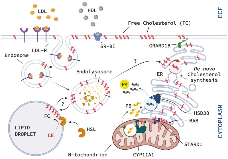
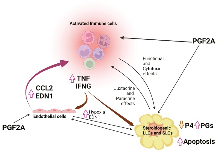
The corpus luteum (CL) forms following ovulation from the remnant of the Graafian follicle. This transient tissue produces critical hormones to maintain pregnancy, including the steroid progesterone. In cattle and other ruminants, the presence of an embryo determines if the lifespan of the CL will be prolonged to ensure successful implantation and gestation, or if the tissue will undergo destruction in the process known as luteolysis. Infertility and subfertility in dairy and beef cattle results in substantial economic loss to producers each year. In addition, this has the potential to exacerbate climate change because more animals are needed to produce high-quality protein to feed the growing world population. Successful pregnancies require coordinated regulation of uterine and ovarian function by the developing embryo. These processes are often collectively termed "maternal recognition of pregnancy." Research into the formation, function, and destruction of the bovine CL by the Northeast Multistate Project, one of the oldest continuously funded Hatch projects by the USDA, has produced a large body of evidence increasing our knowledge of the contribution of ovarian processes to fertility in ruminants. This review presents some of the seminal research into the regulation of the ruminant CL, as well as identifying mechanisms that remain to be completely validated in the bovine CL. This review also contains a broad discussion of the roles of prostaglandins, immune cells, as well as mechanisms contributing to steroidogenesis in the ruminant CL. A triadic model of luteolysis is discussed wherein the interactions among immune cells, endothelial cells, and luteal cells dictate the ability of the ruminant CL to respond to a luteolytic stimulus, along with other novel hypotheses for future research.

Keywords: bovine; corpus luteum; luteolysis; steroidogenesis.

Plain language summary

The corpus luteum (CL) forms on the ovary from the cellular remnants of the follicle following ovulation. The function of the CL is to produce progesterone that is required for successful pregnancy. In the absence of an embryo or sufficient embryonic signaling, the uterus will release a prostaglandin that kills the CL in a process called luteolysis. Therefore, the CL and the embryo share a symbiotic relationship, each requiring the other to be healthy and functional for survival. The Northeast Multistate Project, one of the oldest in the nation, has produced a large body of evidence that has enhanced our understanding of how the CL functions, its regulation, and the impact of ovarian activity on fertility of cattle. This review highlights some of the important advances made in the understanding of the ruminant CL.


Figures

Figure 1.
Figure 1.
Cholesterol sources and steps in luteal cell progesterone (P4) synthesis. Exogenous cholesterol packaged in either high-density lipoproteins (HDLs) delivered directly to the plasma membrane as free cholesterol (FC) via the scavenger receptor class B type I (SR-BI/SCARB1), or alternatively low-density lipoproteins (LDLs) undergo endocytosis after binding to the LDL-receptor (LDL-R). Endosomal vesicles containing LDL fuse with lysosomes to form endolysosomes that facilitate proteolysis and release of FC that enriches the endosomal membranes. From the endosome, cholesterol can take paths to lipid droplets for storage, or the endoplasmic reticulum (ER) and mitochondria; these cellular transport mechanisms for FC remain to be defined. Plasma membrane cholesterol can directly be transferred to the ER by GRAM domain-containing 1B (GRAMD1B). The ER is also the endogenous site de novo cholesterol synthesis. Activation of hormone-sensitive lipase can mobilize endogenous cholesterol stores by converting stored cholesterol esters (CEs) to FC, that enters the endosomal traffic. FC in the ER membranes can be transferred to the outer mitochondrial membrane (OMM) via the mitochondria associate membranes (MAMs). Transfer of FC from the OMM to the inner mitochondrial membrane (IMM) is performed by the steroidogenic acute regulatory protein (STARD1). This mitochondrial import of FC is considered the rate-limiting step in steroidogenesis. FC in the IMM gains access to CYP11A1 enzyme that converts cholesterol to pregnenolone (P5). Pregnenolone that exits the mitochondria is converted to P4 by 3β-hydroxysteroid dehydrogenase (HSD3B; HSD3B2) that resides in the ER. The luteal cells release or secrete the P4 for a variety of local and systemic actions crucial for pregnancy. (Created in Biorender.com).
Figure 2.
Figure 2.
Proposed triadic model of luteolysis highlighting putative interactions among steroidogenic cells, endothelial cells, and immune cells within the corpus luteum. PGF2A initiates a series of luteolytic events that are sustained by interactions between activated immune cells, endothelial cells, and steroidogenic cells. For instance, PGF2A in vivo provokes a decrease in P4 secretion by the CL while simultaneously augmenting CCL2 and ET1 expression by luteal endothelial cells and the production of PGF2A by steroidogenic cells. CCL2 promotes recruitment of immune cells from the general circulation, while ET1 invokes vasoconstriction and hypoxia of the tissue. As a consequence, immune cells (i.e., largely monocytes or macrophages and T-lymphocytes) migrate and accumulate within the CL, and then become activated in response to MHC class II expression, co-stimulation, and the elaboration of proinflammatory molecules (e.g., prostaglandins) by the steroidogenic cells. The activated immune cells drastically reduce CL size by secreting cytokines that induce apoptosis of all cell types (especially steroidogenic cells), engulfing dying cells and cell debris by phagocytosis, and likely promoting the process of tissue repair. Lines in the figure indicate the hypothesized actions of PGF2A in this process: effects of PGF2A on immune cells and endothelial cells directly; and reciprocal interactions between steroidogenic cells and endothelial cells that culminate in luteolysis. (Created in Biorender.com).

Similar articles

Cited by

References

    1. Alila, H. W., and Hansel W.. . 1984a. Origin of different cell types in the bovine corpus luteum as characterized by specific monoclonal antibodies. Biol. Reprod. 31:1015–1025. - PubMed
    1. Alila, H. W., and Hansel W.. . 1984b. Induction of lymphopenia causes luteal dysfunction in cattle. Biol. Reprod. 31:671–678. doi:10.1095/biolreprod31.4.671 - DOI - PubMed
    1. Anderson, L. L., Hard D. L., Carpenter L. S., Awotwi E. K., Diekman M. A., Trenkle A. H., and Cho J.. . 1999. Pregnancy, parturition, and lactation in hypophyseal stalk-transected beef heifers. J. Endocrinol. 163:463–475. doi:10.1677/joe.0.1630463 - DOI - PubMed
    1. Arakane, F., Kallen C. B., Watari H., Foster J. A., Sepuri N. B. V., Pain D., Stayrook S. E., Lewis M., Gerton G. L., and Strauss J. F.. . 1998. The mechanism of action of steroidogenic acute regulatory protein (StAR): StAR acts on the outside of mitochondria to stimulate steroidogenesis. J. Biol. Chem. 273:16339–16345. doi:10.1074/jbc.273.26.16339 - DOI - PubMed
    1. Arakane, F., King S. R., Du Y., Kallen C. B., Walsh L. P., Watari H., Stocco D. M., and Strauss J. F.. . 1997. Phosphorylation of steroidogenic acute regulatory protein (StAR) modulates its steroidogenic activity. J. Biol. Chem. 272:32656–32662. doi:10.1074/JBC.272.51.32656 - DOI - PubMed