. 2022 Jul;43(7):1060-1067.
doi: 10.3174/ajnr.A7551. Epub 2022 Jun 30.

Arterioectatic Spinal Angiopathy of Childhood: Clinical, Imaging, Laboratory, Histologic, and Genetic Description of a Novel CNS Vascular Pathology

Affiliations

Arterioectatic Spinal Angiopathy of Childhood: Clinical, Imaging, Laboratory, Histologic, and Genetic Description of a Novel CNS Vascular Pathology

T Abruzzo et al. AJNR Am J Neuroradiol. 2022 Jul.

Abstract

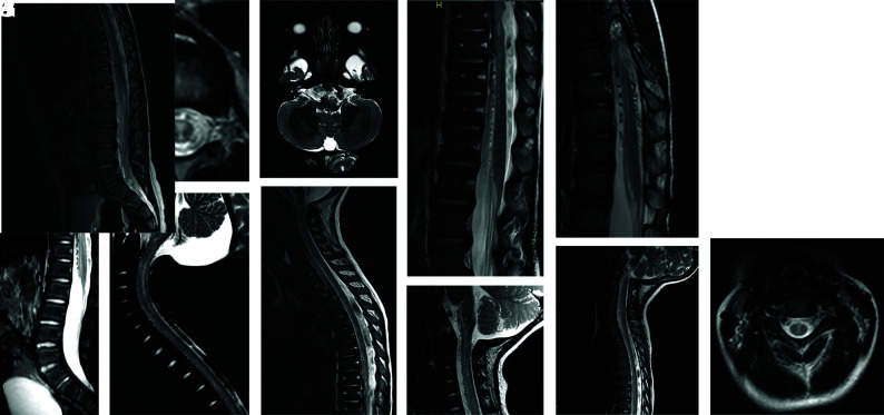
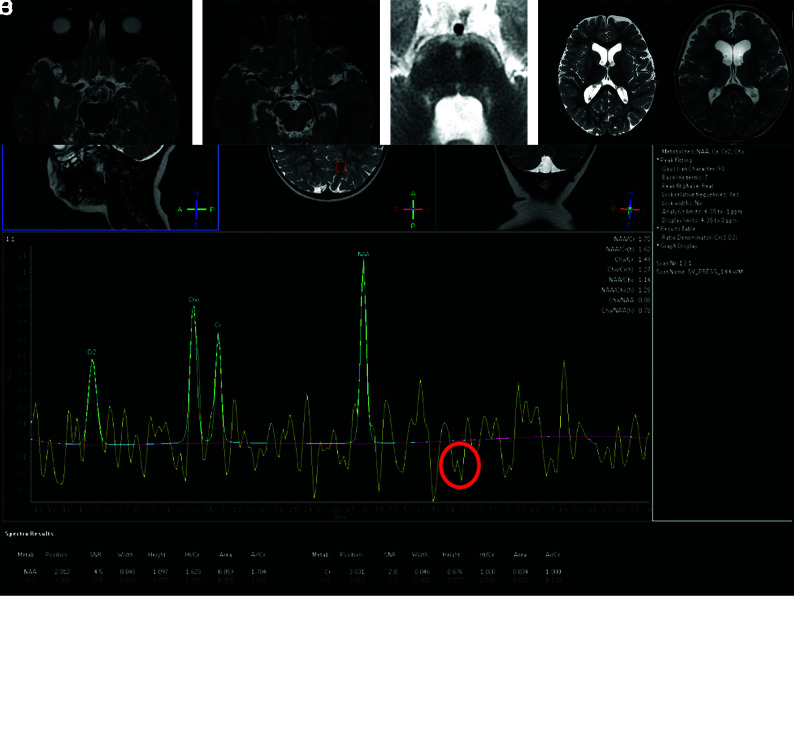
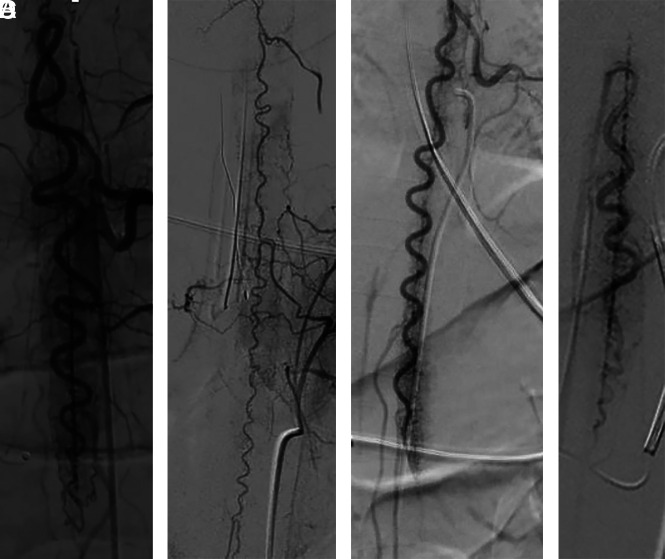
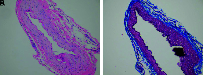
Pediatric patients with myelopathy expressing intradural spinal vascular ectasia without arteriovenous shunting were studied at four tertiary referral neuropediatric centers. Patients were identified by retrospective review of institutional records and excluded if spinal vascular pathology could be classified into a previously described category of spinal vascular malformation. Four patients meeting the study criteria were enrolled in the study. Clinical, magnetic resonance imaging, catheter-directed angiography, laboratory, histological and genetic data were analyzed to characterize the disease process and elucidate underlying pathomechanisms. Our study revealed a highly lethal, progressive multi-segmental myelopathy associated with a unique form of non-inflammatory spinal angiopathy featuring diffuse enlargement and tortuosity of spinal cord arteries, spinal cord hyperemia, and spinal cord edema (Arterioectatic Spinal Angiopathy of Childhood). The condition was shown to mimic venous congestive myelopathy associated with pediatric spinal cord arteriovenous shunts on MRI but to have distinct pathognomonic findings on catheter-directed angiography. Clinicopathological, genetic, and neuroimaging features, which are described in detail, closely overlap with those of mitochondrial disease.


Figures

FIG 1.
FIG 1.
Spine MR imaging findings. A, Midline T2-weighted sagittal MR image of the spine in patient 1 shows abnormal T2-weighted hyperintense signal in the thoracic spinal cord and conus medullaris. Spinal cord volume expansion is also present. There is conspicuous abnormal perimedullary vascular ectasia anterior to the conus medullaris and thoracic spinal cord. B, Axial T2-weighted image of the midthoracic spine shows the distribution of abnormal T2-weighted signal hyperintensity within the central and anterior spinal cord. C, Midline T2-weighted sagittal MR image of the cervical spine reveals abnormal T2-weighted hyperintense signal in the central cervical spinal cord and ventral medulla. There is diffuse spinal cord swelling affecting the region of the cervical enlargement. D, Axial T2-weighted image of the brain at the level of the medulla shows T2-signal hyperintensity in the pyramids, more conspicuous on the left. E, Midline T2-weighted sagittal MR image of the cervical and thoracic spine in patient 4 shows abnormal T2-weighted hyperintense signal in the ventral cervical and thoracic spinal cord and medulla. There is spinal cord volume expansion, prominent in the region of the cervical enlargement, and abnormal perimedullary vascular ectasia anterior to the thoracic spinal cord. F, Midline T2-weighted sagittal MR image of the lower thoracic and lumbar spine reveals abnormal T2-weighted hyperintense signal in the anterior spinal cord. There is diffuse spinal cord swelling, severely affecting the region of the lumbar enlargement. Note marked ectasia of the perimedullary vessels anterior to the spinal cord. G, Midline T2-weighted sagittal MR image of the cervical spine in patient 3 shows abnormal T2-weighted hyperintense signal in the central and anterior cervical spinal cord as well as in the ventral medulla. Mild expansion of cervical spinal cord volume is also present. Conspicuous abnormal perimedullary vascular ectasia anterior to the cervical spinal cord is demonstrated. H, Midline T2-weighted sagittal MR image of the lower thoracic spine reveals abnormal T2-weighted hyperintense signal in the central and anterior thoracic spinal cord and conus medullaris, with mild associated volume expansion. Note marked perimedullary vascular ectasia surrounding the spinal cord. I, Midline T2-weighted sagittal MR image of the cervical and thoracic spine in patient 2 shows abnormal T2-weighted hyperintense signal in the cervical and thoracic spinal cord. The region of the cervical enlargement is severely affected and shows mild volume expansion. J, Midline T2-weighted sagittal image of the lower thoracic and lumbar spine shows abnormal T2-weighted signal hyperintensity within the anterior spinal cord. There is conspicuous perimedullary vascular ectasia anterior to the conus medullaris. K, Axial T2-weighted MR image of the cervical spine reveals abnormal T2-weighted hyperintense signal in the central cervical spinal cord.
FIG 2.
FIG 2.
Brain imaging findings. A, Reference T2-weighted sagittal, axial, and coronal MR images of the brain from patient 1 and MR spectroscopy. There is mild diffuse cerebral parenchymal volume loss with commensurate ex vacuo ventriculomegaly. Note placement of a sampling voxel for MR spectroscopy in the subcortical white matter of the mesial left parietal lobe. MR spectroscopy shows an abnormal inverted doublet peak between 1.3 and 1.4 parts per million, corresponding to lactate (red circle). B, T2-weighted axial image of the brain shows ectasia of the basilar artery and bilateral intradural internal carotid arteries. C, T2-weighted axial image of the brain shows ectasia of the MCAs and posterior cerebral arteries bilaterally. D, Symmetric T2-weighted signal hyperintensity in patient 4 is demonstrated in the anterior edge of the pons, pontine raphe, and middle cerebellar peduncles. E, Axial T2-weighted images at level of foramen of Monro in patients 1 and 4 show mild cerebral parenchymal volume loss with ex vacuo dilation of the cerebral ventricles and cortical sulci.
FIG 3.
FIG 3.
Catheter-directed angiography findings. A, Frontal projection of a left T11 posterior intercostal artery angiogram in the early arterial phase from patient 1 shows marked ectasia of the dominant lower thoracic RMA and descending ramus of the anterior spinal artery (ASA). The ascending ramus of the ASA is mildly enlarged. The left posterolateral spinal artery is mildly enlarged. There is a uniform, unusually intense parenchymal capillary blush and early appearance of parenchymal draining veins. Note striking ectasia of the central sulcal penetrators. B, Frontal projection of a left supreme intercostal artery angiogram in the early arterial phase from patient 4 shows moderate diffuse ectasia of the ASA and central sulcal penetrators. Two RMAs are shown to be moderately ectatic. Note patchy intensification of the parenchymal capillary blush. C, Frontal projection of left posterior intercostal artery angiogram in the early arterial phase from patient 3 shows marked ectasia of the dominant lower thoracic RMA and descending ramus of the ASA. There is striking ectasia of the central sulcal penetrators. There is a uniform, unusually intense parenchymal capillary blush most prominent in the region of the conus medullaris and early appearance of parenchymal draining veins. D, Frontal projection of the right posterior intercostal artery angiogram in the early arterial phase from patient 2 shows ectasia of the dominant lower thoracic RMA and descending ramus of the ASA. Note striking ectasia of central sulcal penetrators.
FIG 4.
FIG 4.
RMA histopathology. Patient 1. A, Hematoxylin and eosin–stained axial cross-section perpendicular to the long axis of the vessel at medium power (original magnification ×50) shows a normal trilaminar arterial wall architecture with no evidence of inflammation. B, Trichrome-stained axial cross-section perpendicular to long axis of the vessel at medium power (original magnification ×50) delineates normal elastic tissue architecture and integrity of the artery.

Similar articles

Cited by

References

    1. Rodesch G, Hurth M, Alvarez H, et al. . Classification of spinal cord arteriovenous shunts: proposal for a reappraisal–the Bicêtre experience with 155 consecutive patients treated between 1981 and 1999. Neurosurgery 2002;51:374–79; discussion 79–80 10.1227/00006123-200208000-00013 - DOI - PubMed
    1. Consoli A, Smajda S, Trenkler J, et al. . Intradural spinal cord arteriovenous shunts in the pediatric population: natural history, endovascular management, and follow-up. Childs Nerv Syst 2019;35:945–55 10.1007/s00381-019-04108-0 - DOI - PubMed
    1. Cullen S, Alvarez H, Rodesch G, et al. . Spinal arteriovenous shunts presenting before 2 years of age: analysis of 13 cases. Childs Nerv Syst 2006;22:1103–10 10.1007/s00381-006-0075-x - DOI - PubMed
    1. Rodesch G, Hurth M, Alvarez H, et al. . Spinal cord intradural arteriovenous fistulae: anatomic, clinical, and therapeutic considerations in a series of 32 consecutive patients seen between 1981 and 2000 with emphasis on endovascular therapy. Neurosurgery 2005;57:973–83; discussion 73–83 10.1227/01.neu.0000181314.94000.cd - DOI - PubMed
    1. Parikh S, Saneto R, Falk MJ, et al. . A modern approach to the treatment of mitochondrial disease. Curr Treat Options Neurol 2009;11:414–30 10.1007/s11940-009-0046-0 - DOI - PMC - PubMed

Publication types