. 2022 Jul;43(7):978-983.
doi: 10.3174/ajnr.A7557. Epub 2022 Jun 30.

Diffuse Calvarial Hyperostosis and Spontaneous Intracranial Hypotension: A Case-Control Study

Affiliations

Diffuse Calvarial Hyperostosis and Spontaneous Intracranial Hypotension: A Case-Control Study

J C Babcock et al. AJNR Am J Neuroradiol. 2022 Jul.

Abstract

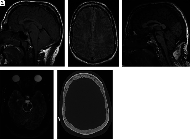
Background and purpose: Diagnosing spontaneous intracranial hypotension and associated CSF leaks can be challenging, and additional supportive imaging findings would be useful to direct further evaluation. This retrospective study evaluated whether there was a difference in the prevalence of calvarial hyperostosis in a cohort of patients with spontaneous intracranial hypotension compared with an age- and sex-matched control population.

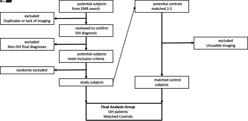
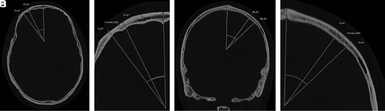
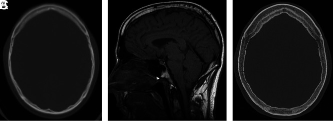
Materials and methods: Cross-sectional imaging (CT of the head or brain MR imaging examinations) for 166 patients with spontaneous intracranial hypotension and 321 matched controls was assessed by neuroradiologists blinded to the patient's clinical status. The readers qualitatively evaluated the presence of diffuse or layered calvarial hyperostosis and measured calvarial thickness in the axial and coronal planes.

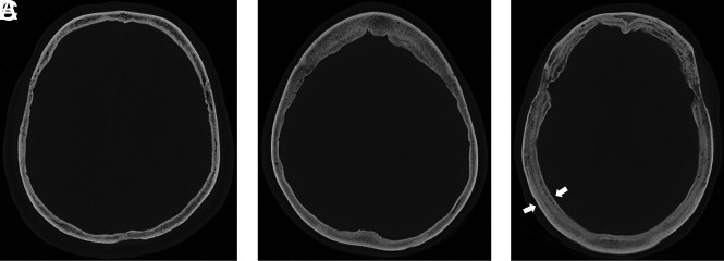
Results: A significant difference in the frequency of layered hyperostosis (31.9%, 53/166 subjects versus 5.0%, 16/321 controls, P < .001, OR = 11.58) as well as the frequency of overall (layered and diffuse) hyperostosis (38.6%, 64/166 subjects versus 13.2%, 42/321 controls, P < .001, OR = 4.66) was observed between groups. There was no significant difference in the frequency of diffuse hyperostosis between groups (6.6%, 11/166 subjects versus 8.2%, 26/321 controls, P = .465). A significant difference was also found between groups for calvarial thickness measured in the axial (P < .001) and coronal (P < .001) planes.

Conclusions: Layered calvarial hyperostosis is more prevalent in spontaneous intracranial hypotension compared with the general population and can be used as an additional noninvasive brain imaging marker of spontaneous intracranial hypotension and an underlying spinal CSF leak.


Figures

FIG 1.
FIG 1.
Flow chart defining the process of selecting the subject inclusion cohort. EMR indicates electronic medical record.
FIG 2.
FIG 2.
Hyperostosis examples. Normal calvarial thickness (A). Axial bone kernel and bone window CT image with a representative example of normal calvarial thickness. Diffuse calvarial hyperostosis (B). Axial bone kernel and bone window CT image demonstrates diffuse thickening of the calvaria. Layered calvarial hyperostosis (C). Axial bone kernel and bone window CT image demonstrates calvarial thickening with discrete enlargement of the inner and outer tables (white arrows), producing a layered appearance.
FIG 3.
FIG 3.
Example of calvarial thickness measurements obtained in the same patient in the axial and coronal planes. Full-field (A) and zoomed (B) axial bone kernel and bone window CT images demonstrate a sample axial thickness measurement obtained 25°–35° off midline. Full-field (C) and zoomed (D) coronal bone kernel and bone window CT images demonstrate a sample coronal thickness measurement obtained 35°–45° off midline.
FIG 4.
FIG 4.
Development of hyperostosis in a 67-year-old man with 2 decades of waxing and waning SIH symptoms with a history of remote CSF leak at C2–C4. A, Axial CT head image at 45 years of age shows qualitatively normal calvarial thickness. B, Sagittal T1-weighted MR image at 48 years of age demonstrates severe brain sag and a suggestion of developing layered hyperostosis. Diffuse pachymeningeal thickening and enhancement are also present (not shown). C, The most recent axial head CT at 67 years of age demonstrates new layered calvarial hyperostosis.
FIG 5.
FIG 5.
A 53-year-old man who developed refractory papilledema and rebound intracranial hypertension following repair of a CSF-venous fistula at T8–T9 at 51 years of age. Rebound intracranial hypertension symptoms began 3 weeks following treatment. Sagittal T1-weighted (A) and axial FLAIR (B) MR images at 29 years of age demonstrate brain sag with normal baseline qualitative calvarial thickness, respectively. Sagittal T1-weighted (C) and axial T2-weighted (D) MR images at 51 years of age demonstrate improvement in brain sag with new posterior globe flattening indicative of papilledema, respectively. E, Preoperative stereotactic CT image before ventriculoperitoneal shunt placement demonstrates new layered calvarial hyperostosis.

References

    1. Schievink WI. Spontaneous intracranial hypotension. N Engl J Med 2021;385:2173–78 10.1056/NEJMra2101561 - DOI - PubMed
    1. Schievink WI, Maya M, Prasad RS, et al. Spontaneous spinal cerebrospinal fluid‐venous fistulas in patients with orthostatic headaches and normal conventional brain and spine imaging. J Headache Pain 2021;61:387–91 10.1111/head.14048 - DOI - PubMed
    1. Brinjikji W, Savastano L, Atkinson J, et al. A novel endovascular therapy for CSF hypotension secondary to CSF-venous fistulas. AJNR Am J Neuroradiol 2021;42:882–87 10.3174/ajnr.A7014 - DOI - PMC - PubMed
    1. Schievink WI. Spontaneous spinal cerebrospinal fluid leaks and intracranial hypotension. JAMA 2006;295:2286–96 10.1001/jama.295.19.2286 - DOI - PubMed
    1. Johnson DR, Carr CM, Luetmer PH, et al. Diffuse calvarial hyperostosis in patients with spontaneous intracranial hypotension. World Neurosurg 2021;146:e848–53 10.1016/j.wneu.2020.11.066 - DOI - PubMed

MeSH terms

Supplementary concepts

LinkOut - more resources