Review
. 2022 Jun 30;7(1):206.
doi: 10.1038/s41392-022-01070-3.

Targeting fibrosis, mechanisms and cilinical trials

Affiliations
Review

Targeting fibrosis, mechanisms and cilinical trials

Manyu Zhao et al. Signal Transduct Target Ther. .

Abstract

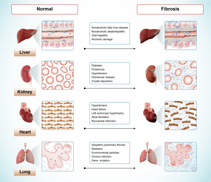
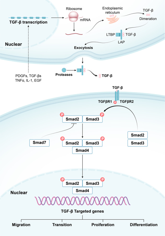
Fibrosis is characterized by the excessive extracellular matrix deposition due to dysregulated wound and connective tissue repair response. Multiple organs can develop fibrosis, including the liver, kidney, heart, and lung. Fibrosis such as liver cirrhosis, idiopathic pulmonary fibrosis, and cystic fibrosis caused substantial disease burden. Persistent abnormal activation of myofibroblasts mediated by various signals, such as transforming growth factor, platelet-derived growth factor, and fibroblast growh factor, has been recongized as a major event in the occurrence and progression of fibrosis. Although the mechanisms driving organ-specific fibrosis have not been fully elucidated, drugs targeting these identified aberrant signals have achieved potent anti-fibrotic efficacy in clinical trials. In this review, we briefly introduce the aetiology and epidemiology of several fibrosis diseases, including liver fibrosis, kidney fibrosis, cardiac fibrosis, and pulmonary fibrosis. Then, we summarise the abnormal cells (epithelial cells, endothelial cells, immune cells, and fibroblasts) and their interactions in fibrosis. In addition, we also focus on the aberrant signaling pathways and therapeutic targets that regulate myofibroblast activation, extracellular matrix cross-linking, metabolism, and inflammation in fibrosis. Finally, we discuss the anti-fibrotic drugs based on their targets and clinical trials. This review provides reference for further research on fibrosis mechanism, drug development, and clinical trials.


Conflict of interest statement

The authors declare no competing interests.

Figures

Fig. 1
Fig. 1
The aetiology of fibrosis in different tissues or organs
Fig. 2
Fig. 2
The activation of HSCs regulated by other cells in liver fibrosis. Extracellular components from injured hepatocytes, Kupffer cells, macrophages, NK cells, T and B lymphocytes modulate HSCs activation via various cytokines. LSECs inhibit or promote the activation of HSCs in different conditions. NK cells kill activated HSCs in IFNγ and TRAIL-dependent ways. TRAIL, TNF-related apoptosis-inducing ligand
Fig. 3
Fig. 3
The interactions among cells involved in lung fibrosis. Injured alveolar epithelial cells activate macrophages, neutrophils, and eosinophils, resulting in the secretion of cytokines, such as TGF-β, IL-1β, and TNF-α. These cytokines mediate the differentiation of fibroblasts into myofibroblasts and the epithelial-mesenchymal transition, which result in the ECM deposition at the injury site
Fig. 4
Fig. 4
Interactions between growth factors-associated signaling pathways and a summary of related target drugs. PDGFs binding to PDGFRs activates the JAK/STAT, PI3K/AKT, and RAS/ERK signals. FGFs binding to FGFRs activates PI3K/AKT and RAS/ERK signals. CTGF binding to FGFR2 (promoting FGF2 and FGF4 binding to FGFR2) activates RAS/ERK signaling, and CTGF binding to LRP6 activates WNT/β-catenin signaling. Drugs targeting these signaling pathways are listed. EMT: epithelial-mesenchymal transition
Fig. 5
Fig. 5
Overview of canonical TGF-β/Smad signaling pathway. Various cytokines stimulate the transcipiton of TGF-β, such as PDGFs, TGF-βs, TNF-α, IL-1β, and EGF. Pro-TGF-β is synthesized in the ribosome and endoplasmic reticulum. After dimeration, LAP binds to mature TGF-β and attaches to LTBP, entering the intercellular space through exocytosis. Actived TGF-β is released by proteases, and binds to TGFβR2 and TGFβR1. Phosphorylated TGFβR2 phosphorylates TGFβR1. TGFβR1 subsequently triggers the phosphorylation of Smad2/3, which interact with Smad4 and enter the nucleus to activate the expression of target genes. Smad7 is a negative regulator of TGF-β/Smad signaling. LAP, latency-associated peptide; LTBP, latent TGF-β binding protein
Fig. 6
Fig. 6
Molecular signaling pathways of NASH and a summary of related target drugs. FFA, free fatty acid; TG, triglycerides

References

    1. Zhao X, et al. Targeting metabolic dysregulation for fibrosis therapy. Nat. Rev. Drug Disco. 2020;19:57–75. doi: 10.1038/s41573-019-0040-5. - DOI - PubMed
    1. Global burden of 369 diseases and injuries in 204 countries and territories, 1990-2019: a systematic analysis for the Global Burden of Disease Study 2019. Lancet. 396, 1204–1222 (2020). - PMC - PubMed
    1. Shu DY, Lovicu FJ. Myofibroblast transdifferentiation: The dark force in ocular wound healing and fibrosis. Prog. Retin Eye Res. 2017;60:44–65. doi: 10.1016/j.preteyeres.2017.08.001. - DOI - PMC - PubMed
    1. Rockey DC, Bell PD, Hill JA. Fibrosis-a common pathway to organ injury and failure. N. Engl. J. Med. 2015;372:1138–1149. doi: 10.1056/NEJMra1300575. - DOI - PubMed
    1. Tomasek JJ, et al. Myofibroblasts and mechano-regulation of connective tissue remodelling. Nat. Rev. Mol. Cell Biol. 2002;3:349–363. doi: 10.1038/nrm809. - DOI - PubMed

Publication types

MeSH terms