

Human placental extract activates a wide array of gene expressions related to skin functions
- PMID: 35773304
- PMCID: PMC9246867
- DOI: 10.1038/s41598-022-15270-y
Human placental extract activates a wide array of gene expressions related to skin functions
Abstract
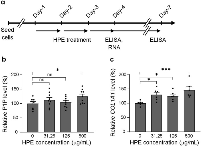
As skin aging is one of the most common dermatological concerns in recent years, scientific research has promoted treatment strategies aimed at preventing or reversing skin aging. Breakdown of the extracellular matrix (ECM), such as collagen and elastin fibers, in the skin results in decreased skin elasticity and tension. Cutaneous cells, especially fibroblasts in the dermis layer of the skin, mainly produce ECM proteins. Although clinical studies have demonstrated that placental extract (PE) has positive effects on skin health, the molecular mechanisms by which PE acts against skin aging are still largely unknown. In this study, we performed RNA-sequence analysis to investigate whether human PE (HPE) alters ECM-related gene expression in normal human dermal fibroblast (NHDF) cells. Gene ontology analysis showed that genes related to extracellular matrix/structure organization, such as COL1A1, COL5A3, ELN, and HAS2 were highly enriched, and most of these genes were upregulated. We further confirmed that the HPE increased the type I collagen, proteoglycan versican, elastin, and hyaluronan levels in NHDF cells. Our results demonstrate that HPE activates global ECM-related gene expression in NHDF cells, which accounts for the clinical evidence that the HPE affects skin aging.
© 2022. The Author(s).
Conflict of interest statement
P-Y.C., L-C.C., and K.K. were employed by Melsmon Pharmaceutical Co. Ltd.. Y.N. declares no competing interests.
Figures


Similar articles
-
Use of versican variant V3 and versican antisense expression to engineer cultured human skin containing increased content of insoluble elastin.J Tissue Eng Regen Med. 2017 Jan;11(1):295-305. doi: 10.1002/term.1913. Epub 2014 Jun 19. J Tissue Eng Regen Med. 2017. PMID: 24945362
-
Anti-aging effects of Piper cambodianum P. Fourn. extract on normal human dermal fibroblast cells and a wound-healing model in mice.Clin Interv Aging. 2016 Jul 29;11:1017-26. doi: 10.2147/CIA.S107734. eCollection 2016. Clin Interv Aging. 2016. PMID: 27536082 Free PMC article.
-
Polyphenol treatments increase elastin and collagen deposition by human dermal fibroblasts; Implications to improve skin health.J Dermatol Sci. 2021 May;102(2):94-100. doi: 10.1016/j.jdermsci.2021.03.002. Epub 2021 Mar 16. J Dermatol Sci. 2021. PMID: 33766446
-
[Role of the extracellular matrix in extrinsic skin aging].Hautarzt. 2011 Aug;62(8):591-7. doi: 10.1007/s00105-011-2133-x. Hautarzt. 2011. PMID: 21681543 Review. German.
-
Molecular Mechanisms of Dermal Aging and Antiaging Approaches.Int J Mol Sci. 2019 Apr 29;20(9):2126. doi: 10.3390/ijms20092126. Int J Mol Sci. 2019. PMID: 31036793 Free PMC article. Review.
Cited by
-
The revolutionary role of placental derivatives in biomedical research.Bioact Mater. 2025 Mar 19;49:456-485. doi: 10.1016/j.bioactmat.2025.03.011. eCollection 2025 Jul. Bioact Mater. 2025. PMID: 40177109 Free PMC article. Review.
-
Human Placenta Extract (HPH) Suppresses Inflammatory Responses in TNF-α/IFN-γ-Stimulated HaCaT Cells and a DNCB Atopic Dermatitis (AD)-Like Mouse Model.J Microbiol Biotechnol. 2024 Oct 28;34(10):1969-1980. doi: 10.4014/jmb.2406.06045. Epub 2024 Sep 11. J Microbiol Biotechnol. 2024. PMID: 39252632 Free PMC article.
-
Antioxidant Capacity and Protective Effect of Cow Placenta Extract on D-Galactose-Induced Skin Aging in Mice.Nutrients. 2022 Nov 3;14(21):4659. doi: 10.3390/nu14214659. Nutrients. 2022. PMID: 36364921 Free PMC article.
-
Human Placental Extract Delays In Vitro Cellular Senescence through the Activation of NRF2-Mediated Antioxidant Pathway.Antioxidants (Basel). 2022 Aug 10;11(8):1545. doi: 10.3390/antiox11081545. Antioxidants (Basel). 2022. PMID: 36009264 Free PMC article.
References
-
- Krämer U, Schikowski T. Recent Demographic Changes and Consequences for Dermatology. In: Gilchrest BA, Krutmann J, editors. Skin Aging. Springer-Verlag; 2006.
-
- Mesa-Arango AC, Flórez-Muñoz SV, Sanclemente G. Mechanisms of skin aging. Iatreia. 2017;30:160–170. doi: 10.17533/udea.iatreia.v30n2a05. - DOI
-
- Yagi M, Yonei Y. Glycative stress and skin aging. Glycative Stress Res. 2018;5:50–54.
MeSH terms
Substances
LinkOut - more resources
Full Text Sources
Medical
Research Materials
Miscellaneous
