Meta-Analysis
. 2022 Oct;76(10):512-524.
doi: 10.1111/pcn.13442. Epub 2022 Aug 28.

Neural correlates of N-back task performance and proposal for corresponding neuromodulation targets in psychiatric and neurodevelopmental disorders

Affiliations
Meta-Analysis

Neural correlates of N-back task performance and proposal for corresponding neuromodulation targets in psychiatric and neurodevelopmental disorders

Lucia Mencarelli et al. Psychiatry Clin Neurosci. 2022 Oct.

Abstract

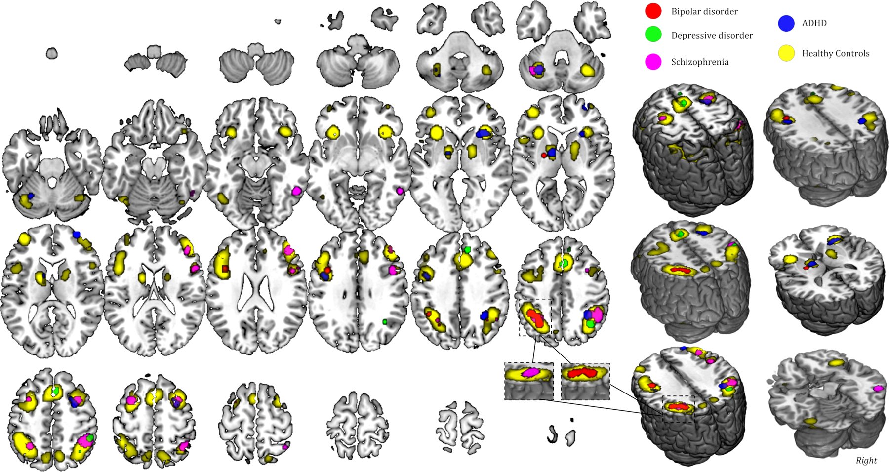
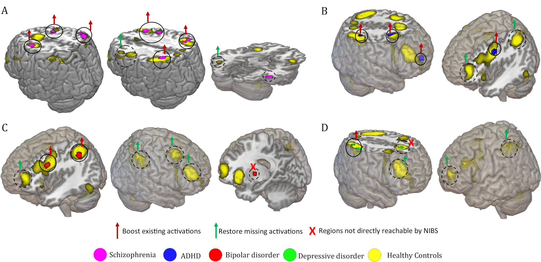
Aim: Working memory (WM) deficit represents the most common cognitive impairment in psychiatric and neurodevelopmental disorders, making the identification of its neural substrates a crucial step towards the conceptualization of restorative interventions. We present a meta-analysis focusing on neural activations associated with the most commonly used task to measure WM, the N-back task, in patients with schizophrenia, depressive disorder, bipolar disorder, and attention-deficit/hyperactivity disorder. Showing qualitative similarities and differences in WM processing between patients and healthy controls, we propose possible targets for cognitive enhancement approaches.

Methods: Selected studies, following the Preferred Reporting Items for Systematic Reviews and Meta-Analyses guidelines, were analyzed through the activation likelihood estimate statistical framework, with subsequent generation of disorder-specific N-back activation maps.

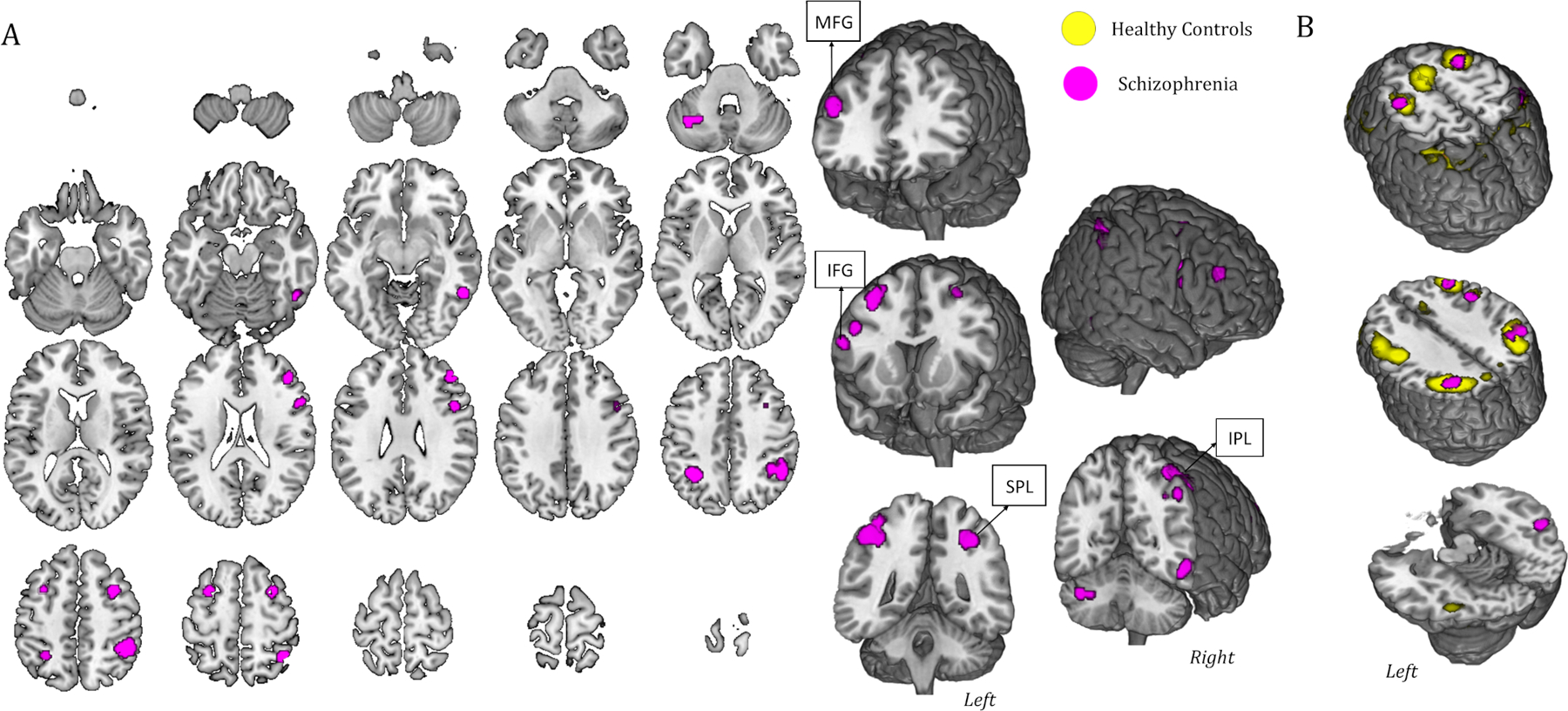
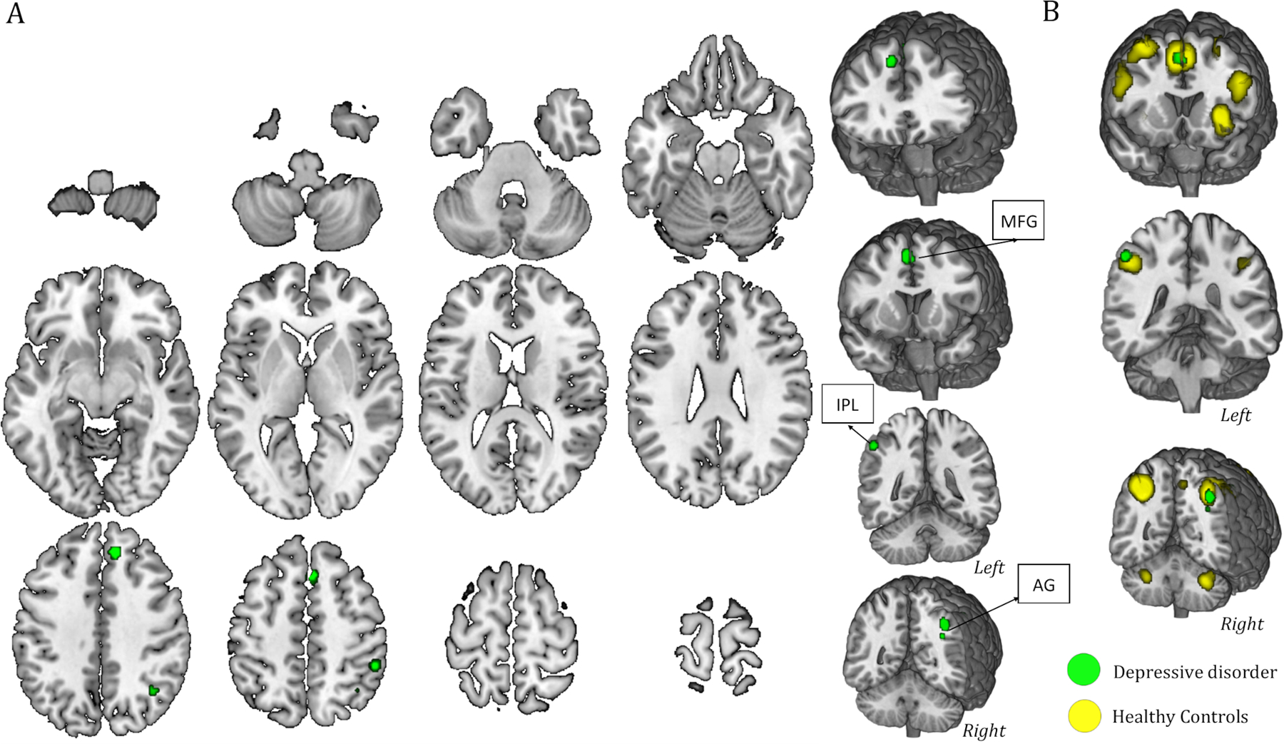
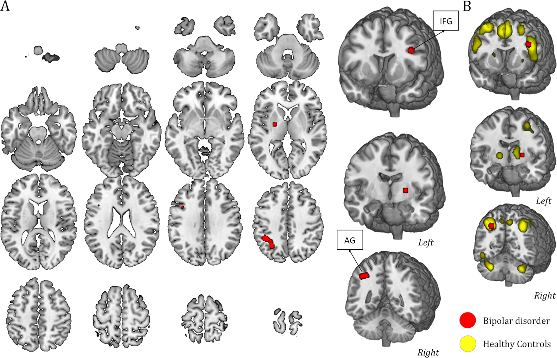
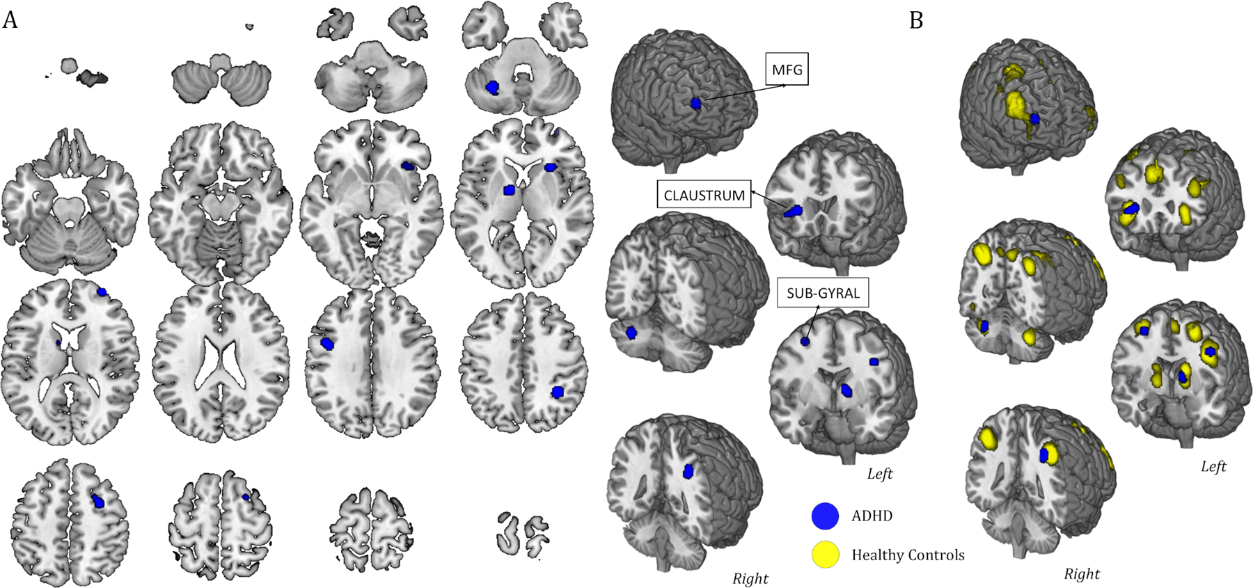
Results: Despite similar WM deficits shared across all disorders, results highlighted different brain activation patterns for each disorder compared with healthy controls. In general, results showed brain activity in frontal, parietal, subcortical, and cerebellar regions; however, reduced engagement of specific nodes of the fronto-parietal network emerged in patients compared with healthy controls. In particular, neither bipolar nor depressive disorders showed detectable activations in the dorsolateral prefrontal cortices, while their parietal activation patterns were lateralized to the left and right hemispheres, respectively. On the other hand, patients with attention-deficit/hyperactivity disorder showed a lack of activation in the left parietal lobe, whereas patients with schizophrenia showed lower activity over the left prefrontal cortex.

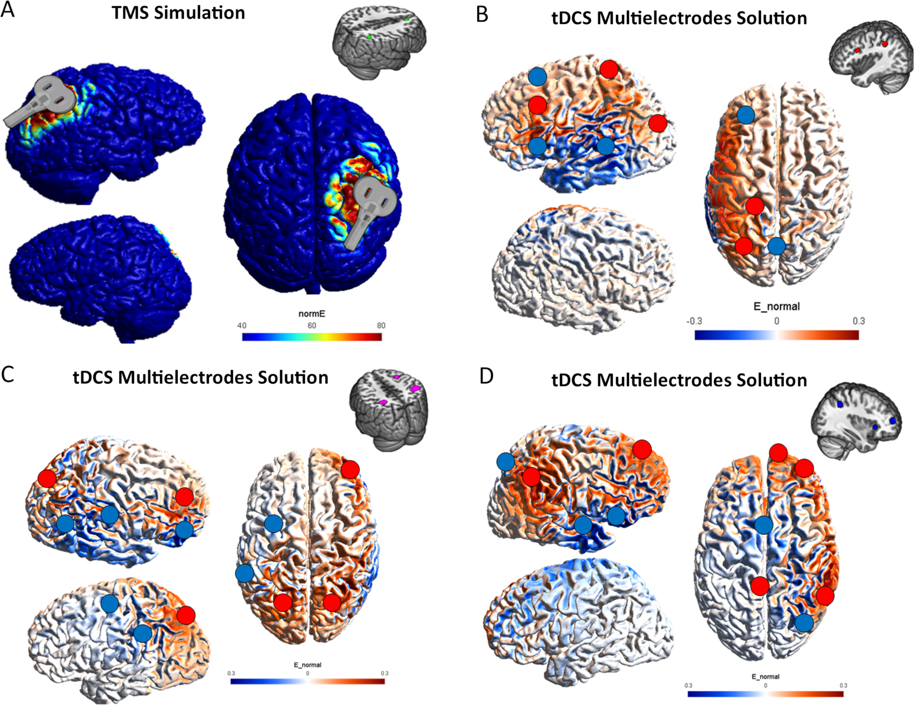
Conclusion: These results, together with biophysical modeling, were then used to discuss the design of future disorder-specific cognitive enhancement interventions based on noninvasive brain stimulation.

Keywords: ADHD; ALE metanalysis; bipolar disorder; depressive disorder; schizophrenia.


Conflict of interest statement

DISCLOSURE STATEMENT

Simone Rossi is a consultant for Neurocare Italy and EBNeuro Italy. All the other authors report no conflict of interest.

Figures

Figure 1.
Figure 1.. Activation maps of the N-back task in Schizophrenia.
(A) The map refers to 13 studies in which the N-back task was performed by patients with schizophrenia (SZ). A complete set of coordinates for each cluster is available in Table 1. (B) Some significant slides of the brain activation overlap between healthy subjects (yellow) and SZ patients (pink) are shown. Note: MFG: Middle Frontal Gyrus; IFG: Inferior Frontal Gyrus; SPL: Superior Parietal Lobule; IPL: Inferior Parietal Lobule.
Figure 2.
Figure 2.. Activation maps of the N-back task in Depressive Disorder.
(A) The map refers to 10 studies that analyzed neural substrates during the N-back task in patients with depressive disorder. A complete set of coordinates for each cluster is available in Table 2. (B) Qualitative overlap with the healthy subjects’ map (in yellow) is presented. Note: MFG: Middle Frontal Gyrus; IPL: Inferior Parietal Lobule; AG: Angular Gyrus.
Figure 3.
Figure 3.. Activation maps of the N-back task in Bipolar Disorder.
(A) The map refers to 8 studies in which the N-back task was performed by patients with bipolar disorder. A complete set of coordinates for each cluster is available in Table 3. (B) Significant slides of the qualitative overlap with the healthy subjects’ map (in yellow) are reported. Note: IFG: Inferior Frontal Gyrus; AG: Angular Gyrus.
Figure 4.
Figure 4.. Activation maps of the N-back task in ADHD.
(A) The map refers to 5 studies in which the N-back task was performed by patients with Attention Deficit Hyperactivity Disorder (ADHD). A complete set of coordinates for each cluster is available in Table 4. (B) Qualitative overlap with the healthy subjects’ map (yellow) is shown. Note: MFG: Middle Frontal Gyrus.
Figure 5.
Figure 5.. Overlap between the considered disorders and healthy subjects.
The map shows a qualitative overlap of activation during the N-back task between the four pathologies considered in this study and the general healthy map corresponding to the data shown in Figure 1 and Table 1 in (18). The map is shown on a template brain in MNI space. MNI= Montreal Neurologic Institute.
Figure 6.
Figure 6.. Possible targets for NIBS.
Cortical regions that could be used as targets for Non-Invasive Brain Stimulation (NIBS) are shown. Areas of overlap between healthy subjects and patients are highlighted with continuous line circles and red arrows, whereas areas that show activation only in the healthy subjects’ map are underlined by dashed line circles and green arrows. Deep cortical regions and subcortical regions are depicted with an X since they are not accessible directly through NIBS. Areas of non-overlap with healthy subjects’ map could be an expression of compensatory mechanisms or dysfunctional activity, future investigation should be conducted to detect the stimulation polarity (inhibitory or excitatory). (A) SZ; (B) BD; (C) ADHD; (D) DD.
Figure 7.
Figure 7.. Optimization montages for each clinical cohort.
Stimulation montages aimed at boosting the nodes still active during the N-back task are proposed with different Non-Invasive Brain Stimulation (NIBS) techniques. In panel A, we suggest stimulating patients with depression disorder on the right angular gyrus through Transcranial Magnetic Stimulation (TMS), based on the ALE map. In panels B, C, and D, we propose multielectrode transcranial Electrical Stimulation (tES) solutions aimed at improving the activity on two or three nodes of the network triggered by the N-back task in patients with Schizophrenia, Bipolar Disorder, and Attention Deficit Hyperactivity Disorder. The weighted map and the E-field (V/m) for each montage are presented (NormE for TMS simulation and NormalE to show polarity for tES solutions—information about the specific montages and fields resulted are reported in the Supplementary Materials).

Similar articles

Cited by

References

    1. Baddeley A Working memory. Science 1992. January-31;255(5044):556–9. - PubMed
    1. Baddeley A, Della Sala S, Robbins TW, Baddeley A. Working Memory and Executive Control [and Discussion]. Philos Trans Biol Sci 1996;351(1346):1397–404. - PubMed
    1. Forbes NF, Carrick LA, Mcintosh A, Lawrie S. Working memory in schizophrenia: A meta-analysis. Psychol Med 2008. Nov 1;39:889–905. - PubMed
    1. Lee J, Park S. Working memory impairments in schizophrenia: a meta-analysis. J Abnorm Psychol 2005. November;114(4):599–611. - PubMed
    1. Van Snellenberg JX, Girgis RR, Horga G, van de Giessen E, Slifstein M, Ojeil N, et al. Mechanisms of Working Memory Impairment in Schizophrenia. Biol Psychiatry 2016. Oct 15;80(8):617–26. - PMC - PubMed

Publication types