. 2022 Apr 8;13(5):2695-2706.
doi: 10.1364/BOE.456198. eCollection 2022 May 1.

Photoacoustic microscopy of vascular adaptation and tissue oxygen metabolism during cutaneous wound healing

Affiliations

Photoacoustic microscopy of vascular adaptation and tissue oxygen metabolism during cutaneous wound healing

Naidi Sun et al. Biomed Opt Express. .

Abstract

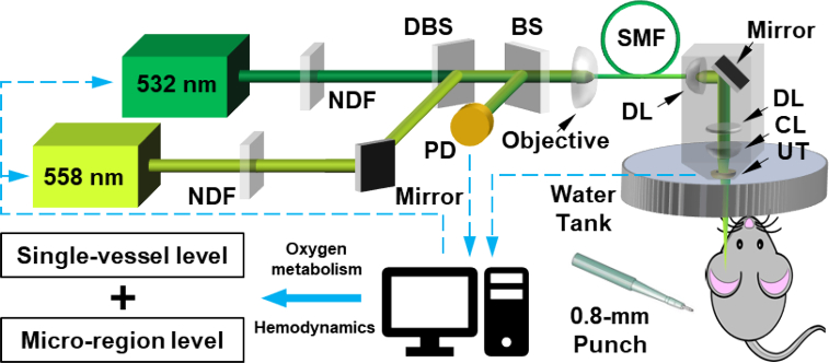
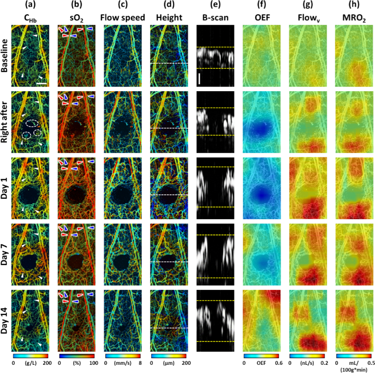
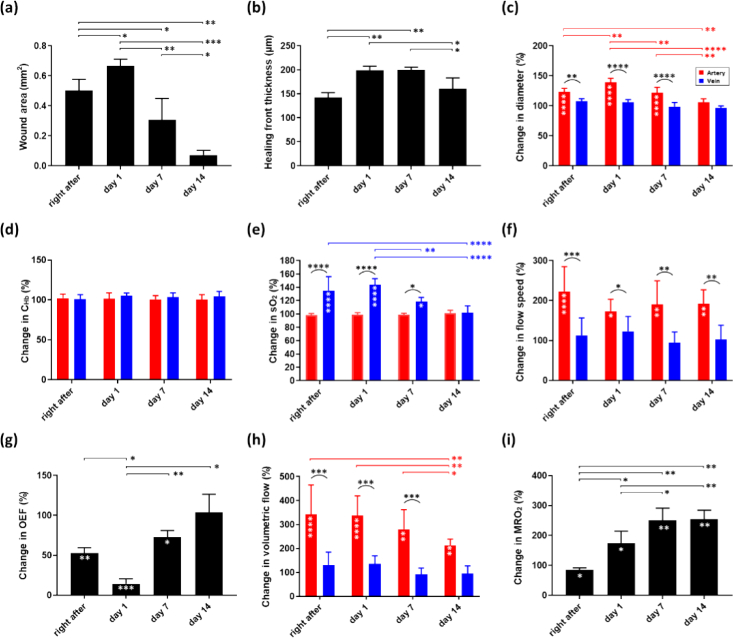
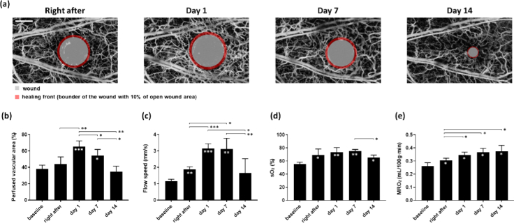
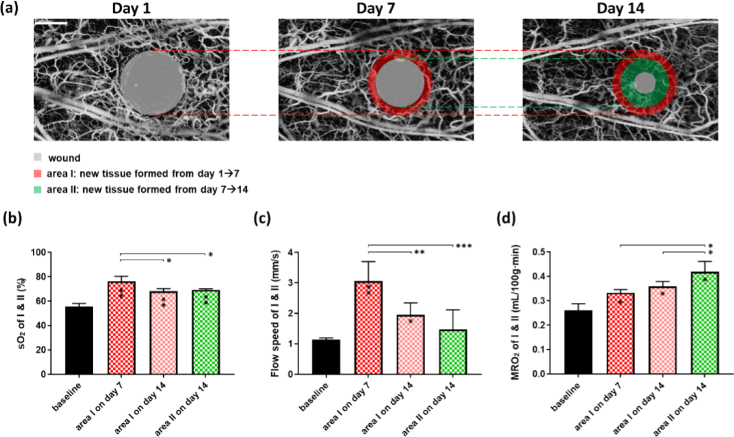
Cutaneous wounds affect millions of people every year. Vascularization and blood oxygen delivery are critical bottlenecks in wound healing, and understanding the spatiotemporal dynamics of these processes may lead to more effective therapeutic strategies to accelerate wound healing. In this work, we applied multi-parametric photoacoustic microscopy (PAM) to study vascular adaptation and the associated changes in blood oxygen delivery and tissue oxygen metabolism throughout the hemostasis, inflammatory, proliferation, and early remodeling phases of wound healing in mice with skin puncture wounds. Multifaceted changes in the vascular structure, function, and tissue oxygen metabolism were observed during the 14-day monitoring of wound healing. On the entire wound area, significant elevations of the arterial blood flow and tissue oxygen metabolism were observed right after wounding and remained well above the baseline over the 14-day period. On the healing front, biphasic changes in the vascular density and blood flow were observed, both of which peaked on day 1, remained elevated in the first week, and returned to the baselines by day 14. Along with the wound closure and thickening, tissue oxygen metabolism in the healing front remained elevated even after structural and functional changes in the vasculature were stabilized. On the newly formed tissue, significantly higher blood oxygenation, flow, and tissue metabolism were observed compared to those before wounding. Blood oxygenation and flow in the new tissue appeared to be independent of when it was formed, but instead showed noticeable dependence on the phase of wound healing. This PAM study provides new insights into the structural, functional, and metabolic changes associated with vascular adaptation during wound healing and suggests that the timing and target of vascular treatments for wound healing may affect the outcomes.


Conflict of interest statement

The authors declare no conflicts of interest.

Figures

Fig. 1.
Fig. 1.
Schematic of the multi-parametric PAM. NDF, neutral-density filter; DBS, dichroic beamsplitter; PD, photodiode; BS, beam sampler; SMF, regular single-mode fiber; DL, doublet; CL, correction lens; UT, ring-shaped ultrasonic transducer.
Fig. 2.
Fig. 2.
Longitudinal comprehensive PAM assessments of structural, functional, and oxygen-metabolic changes in the vascular tissue during wound healing, including (a) the hemoglobin concentration (CHb), (b) the oxygen saturation of hemoglobin (sO2), (c) blood flow speed, (d–e) tissue height and thickness, (f) oxygen extraction fraction (OEF), (g) volumetric blood flow (Flowv), and (h) the metabolic rate of oxygen (MRO2). White arrows in (a): vessels remodeled after inducing the puncture wound in the mouse ear. Dashed white circles in (a): hemorrhage. Red and blue arrows in (b): feeding arteries and draining veins analyzed in Fig. 3, respectively. The “0” position in (d) denotes the bottom of the ear vasculature. The locations of the B-scans in (e) are indicated by the white dashed lines in (d). Horizontal scale bar in (a): 500 µm. Vertical scale bar in (e): 50 µm.
Fig. 3.
Fig. 3.
(a–b) Healing of the wound tissue and associated changes in (c–f) the structure and function of the feeding arteries and draining veins of the wound area and (g–i) tissue oxygen metabolism over the 14-day monitoring. White asterisks within the bars indicate the p values between the baseline (data not shown) and the specific time point. Paired t-test was used in (a), (b), (g) and (i), while two-way ANOVA test was used in (c-f) and (h). *, **, ***, and **** represent p < 0.05, p < 0.01, p < 0.001, and p < 0.0001, respectively. Data are presented as mean ± SD. n = 4.
Fig. 4.
Fig. 4.
(a) Illustration of wound healing process and the definition of the healing front. (b-e) Changes in perfused vascular area, flow speed, sO2, and MRO2 in the wound healing front. White asterisks within the bars indicate the p values between the baseline and the specific time point. Paired t-test was used for all comparisons. *, **, ***, and **** represent p < 0.05, p < 0.01, p < 0.001, and p < 0.0001, respectively. Data are presented as mean ± SD. Scale bar: 500 µm. n = 4.
Fig. 5.
Fig. 5.
(a) Illustration of the newly formed tissues. (b-d) Changes in sO2, flow speed, and MRO2 of the newly formed tissues during the wound healing process. Paired t-test was used for all comparisons. *, **, ***, and **** represent p < 0.05, p < 0.01, p < 0.001, and p < 0.0001, respectively. Data are presented as mean ± SD. Scale bar: 500 µm. n = 4.

References

    1. Nussbaum S. R., Carter M. J., Fife C. E., DaVanzo J., Haught R., Nusgart M., Cartwright D., “An economic evaluation of the impact, cost, and medicare policy implications of chronic nonhealing wounds,” Value in Health 21(1), 27–32 (2018).10.1016/j.jval.2017.07.007 - DOI - PubMed
    1. Baltzis D., Eleftheriadou I., Veves A., “Pathogenesis and treatment of impaired wound healing in diabetes mellitus: new insights,” Adv. Ther. 31(8), 817–836 (2014).10.1007/s12325-014-0140-x - DOI - PubMed
    1. Singer A. J., Clark R. A. F., “Cutaneous wound healing,” N. Engl. J. Med. 341(10), 738–746 (1999).10.1056/NEJM199909023411006 - DOI - PubMed
    1. Guo S., DiPietro L. A., “Factors affecting wound healing,” J. Dent. Res. 89(3), 219–229 (2010).10.1177/0022034509359125 - DOI - PMC - PubMed
    1. Broughton G., Janis J. E., Attinger C. E., “The basic science of wound healing,” Plastic and Reconstructive Surgery 117(SUPPLEMENT), 12S–34S (2006).10.1097/01.prs.0000225430.42531.c2 - DOI - PubMed

LinkOut - more resources