

Znet: Deep Learning Approach for 2D MRI Brain Tumor Segmentation
- PMID: 35774412
- PMCID: PMC9236306
- DOI: 10.1109/JTEHM.2022.3176737
Znet: Deep Learning Approach for 2D MRI Brain Tumor Segmentation
Abstract
Background: Detection and segmentation of brain tumors using MR images are challenging and valuable tasks in the medical field. Early diagnosing and localizing of brain tumors can save lives and provide timely options for physicians to select efficient treatment plans. Deep learning approaches have attracted researchers in medical imaging due to their capacity, performance, and potential to assist in accurate diagnosis, prognosis, and medical treatment technologies.
Methods and procedures: This paper presents a novel framework for segmenting 2D brain tumors in MR images using deep neural networks (DNN) and utilizing data augmentation strategies. The proposed approach (Znet) is based on the idea of skip-connection, encoder-decoder architectures, and data amplification to propagate the intrinsic affinities of a relatively smaller number of expert delineated tumors, e.g., hundreds of patients of the low-grade glioma (LGG), to many thousands of synthetic cases.
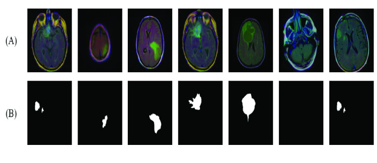
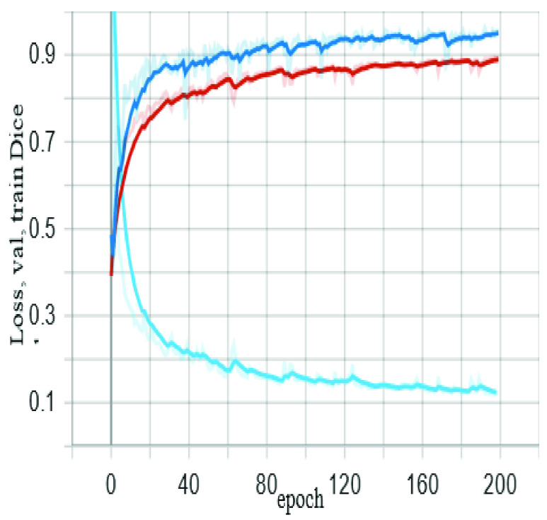
Results: Our experimental results showed high values of the mean dice similarity coefficient (dice = 0.96 during model training and dice = 0.92 for the independent testing dataset). Other evaluation measures were also relatively high, e.g., pixel accuracy = 0.996, F1 score = 0.81, and Matthews Correlation Coefficient, MCC = 0.81. The results and visualization of the DNN-derived tumor masks in the testing dataset showcase the ZNet model's capability to localize and auto-segment brain tumors in MR images. This approach can further be generalized to 3D brain volumes, other pathologies, and a wide range of image modalities.
Conclusion: We can confirm the ability of deep learning methods and the proposed Znet framework to detect and segment tumors in MR images. Furthermore, pixel accuracy evaluation may not be a suitable evaluation measure for semantic segmentation in case of class imbalance in MR images segmentation. This is because the dominant class in ground truth images is the background. Therefore, a high value of pixel accuracy can be misleading in some computer vision applications. On the other hand, alternative evaluation metrics, such as dice and IoU (Intersection over Union), are more factual for semantic segmentation.
Clinical impact: Artificial intelligence (AI) applications in medicine are advancing swiftly, however, there is a lack of deployed techniques in clinical practice. This research demonstrates a practical example of AI applications in medical imaging, which can be deployed as a tool for auto-segmentation of tumors in MR images.
Keywords: Brain tumor; augmentation; deep learning; neural networks; region segmentation.
Figures


References
-
- Ottom M. A., “Convolutional neural network for diagnosing skin cancer,” Int. J. Adv. Comput. Sci. Appl., vol. 10, no. 7, pp. 333–338, 2019.
-
- Dinov I. D., Data Science and Predictive Analytics. Cham, Switzerland: Springer, 2018.
Publication types
MeSH terms
Grants and funding
LinkOut - more resources
Full Text Sources
Medical
