. 2022 Jun 21:2022:3061154.
doi: 10.1155/2022/3061154. eCollection 2022.

Automation of Cephalometrics Using Machine Learning Methods

Affiliations

Automation of Cephalometrics Using Machine Learning Methods

Khalaf Alshamrani et al. Comput Intell Neurosci. .

Retraction in

Abstract

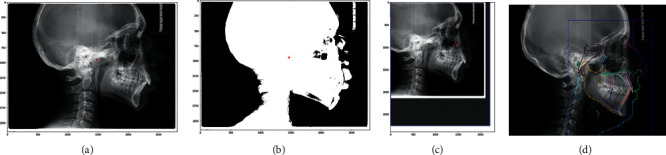
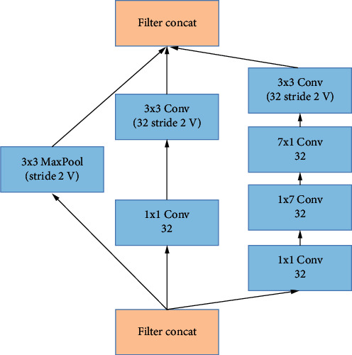
Cephalometry is a medical test that can detect teeth, skeleton, or appearance problems. In this scenario, the patient's lateral radiograph of the face was utilised to construct a tracing from the tracing of lines on the lateral radiograph of the face of the soft and hard structures (skin and bone, respectively). Certain cephalometric locations and characteristic lines and angles are indicated after the tracing is completed to do the real examination. In this unique study, it is proposed that machine learning models be employed to create cephalometry. These models can recognise cephalometric locations in X-ray images, allowing the study's computing procedure to be completed faster. To correlate a probability map with an input image, they combine an Autoencoder architecture with convolutional neural networks and Inception layers. These innovative architectures were demonstrated. When many models were compared, it was observed that they all performed admirably in this task.


Conflict of interest statement

The authors declare that they have no conflicts of interest.

Figures

Figure 1
Figure 1
RX Crop using centroids. (a) Original image, (b) thresholded image, (c) cropped image using average values to fill in the missing information. (d) Original image. In blue, a box is shown that covers all the points of the structures and where they will be cut. Each colored line represents a different structure.
Figure 2
Figure 2
(a) X-ray image extracted from the ISBI 2014 dataset. (b) 2X zoom of the original image; (c) 4X zoom of the original image; ((d), (e)) Two images from the dataset.
Figure 3
Figure 3
Modified InceptionD layer.
Figure 4
Figure 4
Modified InceptionE layer.
Figure 5
Figure 5
Diagram of the parts of an autoencoder.
Figure 6
Figure 6
(a) Architecture of an encoder with Inception layers. (b) Architecture of a decoder with Inception Inter2 layers.
Figure 7
Figure 7
(a) Original image with the Silla point marked in red. (b) Heat map generated with a Gaussian activation centred on the coordinates of the point. (c) Original image with the Gonion point marked in red. (d) Heat map generated with a Gaussian activation centred on the coordinates of the point.
Figure 8
Figure 8
Autoencoder complex model decoder. The number of filters in all convolutional layers doubled.
Figure 9
Figure 9
Encoder architecture in the wider model. The size of the filters and padding have been modified and added one more Inception layer.
Figure 10
Figure 10
Transformation of images to grayscale. (a) Original image. (b) Image after transformation. (c, d) Calculation of box. In red distance between point Silla and PiC X-axis. In blue distance between point Silla and Nasion. (e) Original. (f) Image activation map of the convolutional model. (g) In blue, a rectangle calculated from the training data is observed. In red, a false detection outside the rectangle. (h) Image example with boxes and tan.
Figure 11
Figure 11
Skeleton.

References

    1. Kannan S. K. A simplified overview on clinical cephalometrics PJ Antony, S.karthiga Kanannan, Joby paulose, Manu M Mathew, Charis chandy joseph. Journal of Indian Academy of Oral Medicine and Radiology . 2013;25(3):214–217.
    1. Toman H. A., Nasir A., Hassan R., Hassan R. Skeletal, dentoalveolar, and soft tissue cephalometric measurements of Malay transfusion-dependent thalassaemia patients. The European Journal of Orthodontics . 2011;33(6):700–704. doi: 10.1093/ejo/cjq147. - DOI - PubMed
    1. Agrawal D. Cephalometric analysis for Diagnosis and treatment of orthodontic patients. Journal of Oral Health and Community Dentistry . 2013;7(2):75–79. doi: 10.5005/johcd-7-2-75. - DOI
    1. Lee S. M., Kim H. P., Jeon K., Lee S.-H., Seo J. K. Automatic 3D cephalometric annotation system using shadowed 2D image-based machine learning. Physics in Medicine and Biology . 2019;64(5) doi: 10.1088/1361-6560/ab00c9.055002 - DOI - PubMed
    1. Abdullah Hamad A., Thivagar M. L., Bader Alazzam M., Alassery F., Hajjej F., Shihab A. A. Applying dynamic systems to social media by using controlling stability. Computational Intelligence and Neuroscience . 2022;2022:1–7. doi: 10.1155/2022/4569879. - DOI - PMC - PubMed

Publication types

LinkOut - more resources