

Young Man With Non-hypertensive Ascites of Unexpected Cause: When Ockham's Razor Is Not Sufficient
- PMID: 35774671
- PMCID: PMC9236692
- DOI: 10.7759/cureus.25385
Young Man With Non-hypertensive Ascites of Unexpected Cause: When Ockham's Razor Is Not Sufficient
Abstract
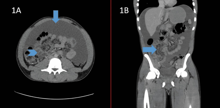
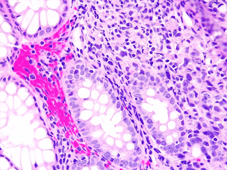
Ascites is defined as the accumulation of fluid in the peritoneal cavity, following an imbalance between production and reabsorption; it is detectable from 50 mL on ultrasound. Three mechanisms have been classically implicated, according to Starling's forces: an increase in the hydrostatic pressure gradient (increased portal venous pressure), a reduction in the oncotic pressure gradient (loss of total proteins, especially albumin), and an increase in peritoneal capillary permeability. This latter mechanism, plus the difference between lymph production and excretion (which favors the accumulation of exudate), explains some of the most notable causes of non-hypertensive ascites (according to the serum albumin in ascites gradient (SAAG)), including peritoneal carcinomatosis and tuberculosis. We present the case of a young man, originally from a tuberculosis endemic area, in whom the study of ascitic fluid guided the workup and the definitive diagnosis, which was unexpected for his age. Finally, a practical approach to non-hypertensive ascites is provided.
Keywords: ascites; cancer; peritoneal carcinomatosis; peritoneum; tuberculosis.
Copyright © 2022, Rondón-Carvajal et al.
Conflict of interest statement
The authors have declared that no competing interests exist.
Figures
Similar articles
-
The role of ascitic fluid viscosity in the differential diagnosis of ascites.Can J Gastroenterol. 2010 Apr;24(4):255-9. doi: 10.1155/2010/896786. Can J Gastroenterol. 2010. PMID: 20431815 Free PMC article.
-
The utility of evaluating low serum albumin gradient ascites in patients with cirrhosis.Am J Gastroenterol. 2009 Jun;104(6):1401-5. doi: 10.1038/ajg.2009.117. Epub 2009 Apr 21. Am J Gastroenterol. 2009. PMID: 19491852
-
Negative Serum Ascites Albumin Gradient (SAAG) in the Setting of Cholangiocarcinoma: A Case Report.Cureus. 2023 Apr 13;15(4):e37528. doi: 10.7759/cureus.37528. eCollection 2023 Apr. Cureus. 2023. PMID: 37193465 Free PMC article.
-
Ascites in Children.Indian J Pediatr. 2016 Nov;83(11):1334-1340. doi: 10.1007/s12098-016-2168-1. Epub 2016 Jun 9. Indian J Pediatr. 2016. PMID: 27278239 Review.
-
Pathogenesis of malignant ascites: Starling's law of capillary hemodynamics revisited.Ann Oncol. 2001 Oct;12(10):1353-7. doi: 10.1023/a:1012504904713. Ann Oncol. 2001. PMID: 11762804 Review.
References
-
- Diagnosis, evaluation, and management of ascites, spontaneous bacterial peritonitis and hepatorenal syndrome: 2021 Practice Guidance by the American Association for the Study of Liver Diseases. Biggins SW, Angeli P, Garcia-Tsao G, et al. Hepatology. 2021;74:1014–1048. - PubMed
-
- The cytologic diagnosis of malignant neoplasms in pleural and peritoneal effusions. Sears D, Hajdu SI. http://pubmed.ncbi.nlm.nih.gov/3469856/#:~:text=The%20most%20common%20pr... Acta Cytol. 1987;31:85–97. - PubMed
-
- Ascitic fluid analysis in malignancy-related ascites. Runyon BA, Hoefs JC, Morgan TR. Hepatology. 1988;8:1104–1109. - PubMed
-
- Biochemical analysis of ascitic (peritoneal) fluid: what should we measure? Tarn AC, Lapworth R. Ann Clin Biochem. 2010;47:397–407. - PubMed
Publication types
LinkOut - more resources
Full Text Sources