

Sufentanil Alleviates Sepsis-Induced Myocardial Injury and Stress Response in Rats through the ERK/GSK-3 β Signaling Axis
- PMID: 35774755
- PMCID: PMC9239792
- DOI: 10.1155/2022/9630716
Sufentanil Alleviates Sepsis-Induced Myocardial Injury and Stress Response in Rats through the ERK/GSK-3 β Signaling Axis
Retraction in
-
Retracted: Sufentanil Alleviates Sepsis-Induced Myocardial Injury and Stress Response in Rats through the ERK/GSK-3β Signaling Axis.Evid Based Complement Alternat Med. 2023 Aug 2;2023:9854369. doi: 10.1155/2023/9854369. eCollection 2023. Evid Based Complement Alternat Med. 2023. PMID: 37565231 Free PMC article.
Abstract
Objective: To explore the effect and possible mechanism of sufentanil on sepsis-induced myocardial injury and stress response in rats.
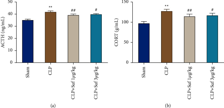
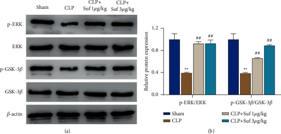
Methods: The cecal ligation and puncture (CLP) method was utilized to establish the sepsis model of rats to explore the effect of sufentanil pretreatment with different concentrations on myocardial injury and oxidative stress in CLP rats. Echocardiogram was applied for detecting cardiac hemodynamic parameters in rats; hematoxylin and eosin (HE) staining as well as TUNEL staining was done for observing pathological changes of myocardial tissue and cardiomyocyte apoptosis in rats, respectively; biochemical testing and enzyme-linked immunosorbent assay (ELISA) were done for determining myocardial injury marker level in serum, oxidative stress substances in myocardial tissue, and neuroendocrine hormone level in serum of rats, respectively; finally, Western blot was performed for checking the expression level of ERK/GSK-3β signaling pathway-related proteins in myocardial tissue of rats.
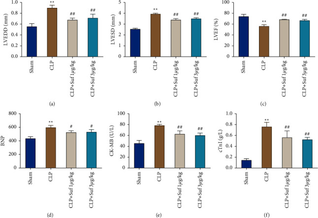
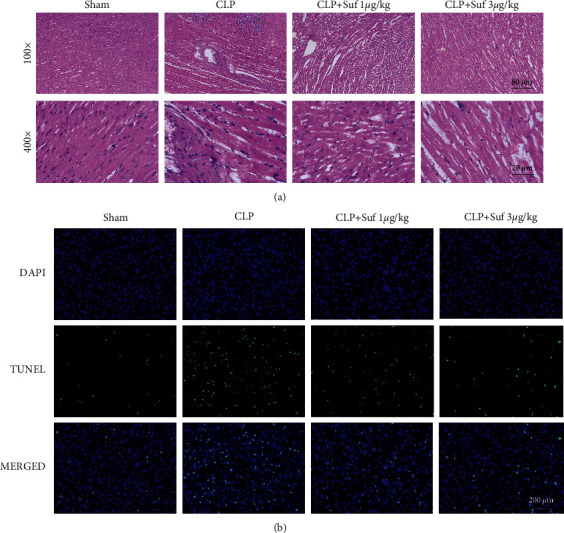
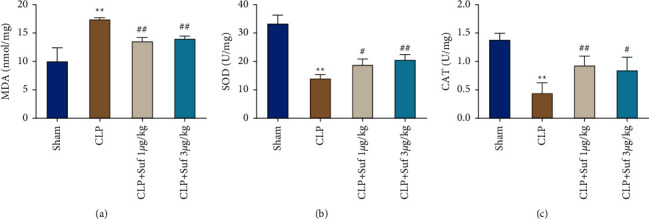
Results: A model of rat with sepsis-induced myocardial injury was constructed with the CLP method. Specifically, this rat model was characterized by obvious cardiac function and tissue damage, cardiomyocyte apoptosis, and oxidative stress response. Sufentanil pretreatment significantly improved cardiac function injury, alleviated pathological injury and oxidative stress response in myocardial tissue, and inhibited cardiomyocyte apoptosis. Specifically, after sufentanil pretreatment, left ventricular end-diastolic dimension (LVEDD) and left ventricular end-systolic dimension (LVESD) were downregulated, and left ventricular ejection fraction (LVEF) was upregulated; the level of B-type natriuretic peptide (BNP) of serum, creatine kinase isoenzyme (CK-MB), and troponin (cTnl) decreased; besides, malondialdehyde (MDA) level was declined, while activities of superoxide dismutase (SOD) and catalase (CAT) were increased. What is more, further mechanism exploration also revealed that sufentanil could reverse the activity of the sepsis-induced ERK/GSK-3β signaling pathway.
Conclusion: Sufentanil has an obvious protective effect on myocardial injury and stress response in CLP rats, and this protective effect may be related to the activation of the ERK/GSK-3β signaling pathway.
Copyright © 2022 Hongcheng Zang et al.
Conflict of interest statement
The authors declare that there are no conflicts of interest.
Figures
References
-
- Driessen R. G. H., van de Poll M. C. G., Mol M. F., van Mook W. N. K. A., Schnabel R. M. The influence of a change in septic shock definitions on intensive care epidemiology and outcome: comparison of sepsis-2 and sepsis-3 definitions. Infectious Diseases . 2018;50(3):207–213. doi: 10.1080/23744235.2017.1383630. - DOI - PubMed
Publication types
LinkOut - more resources
Full Text Sources
Research Materials
Miscellaneous
