. 2022 Jun 14:13:935413.
doi: 10.3389/fpls.2022.935413. eCollection 2022.

Hydroxyproline- O-Galactosyltransferases Synthesizing Type II Arabinogalactans Are Essential for Male Gametophytic Development in Arabidopsis

Affiliations

Hydroxyproline- O-Galactosyltransferases Synthesizing Type II Arabinogalactans Are Essential for Male Gametophytic Development in Arabidopsis

Dasmeet Kaur et al. Front Plant Sci. .

Abstract

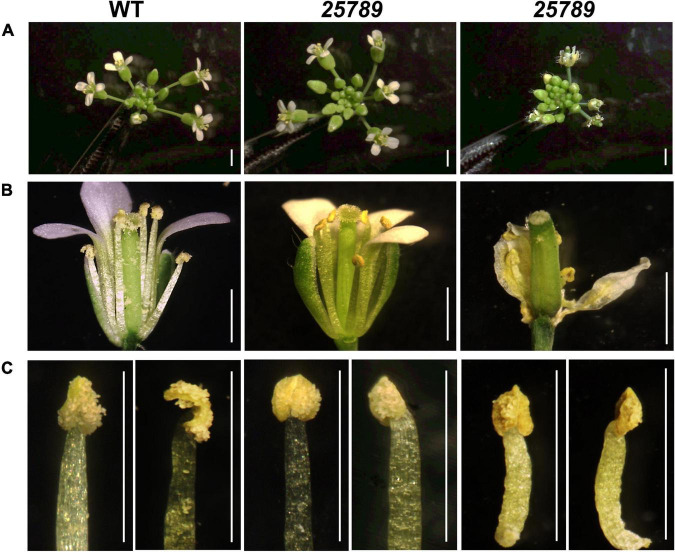
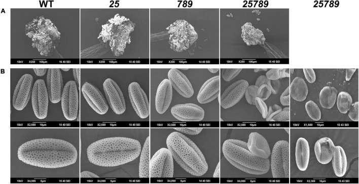
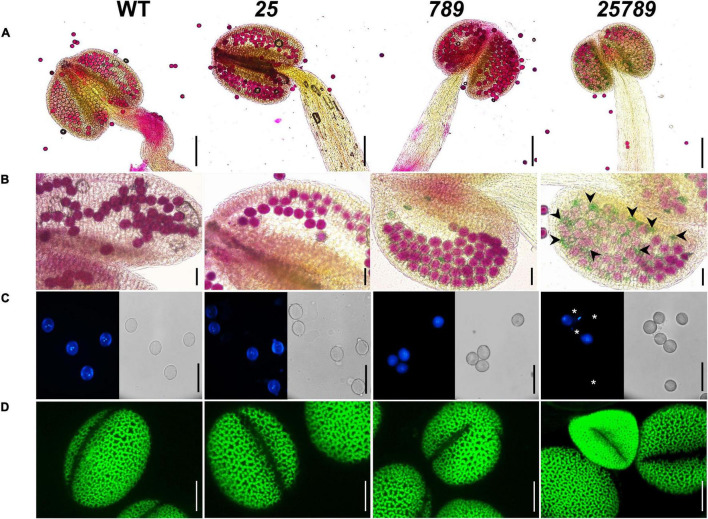
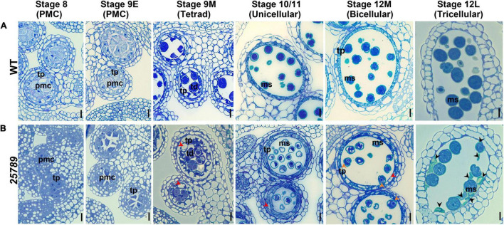
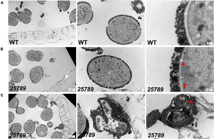
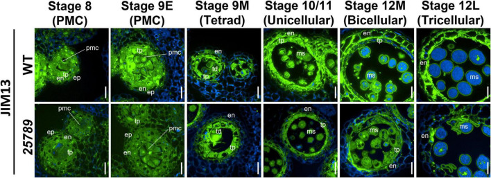
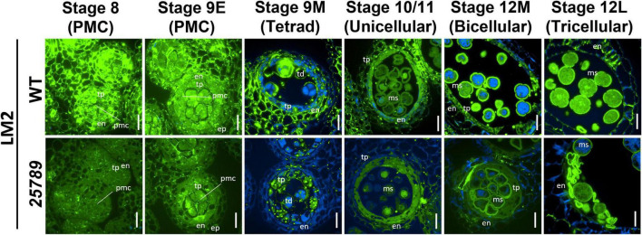
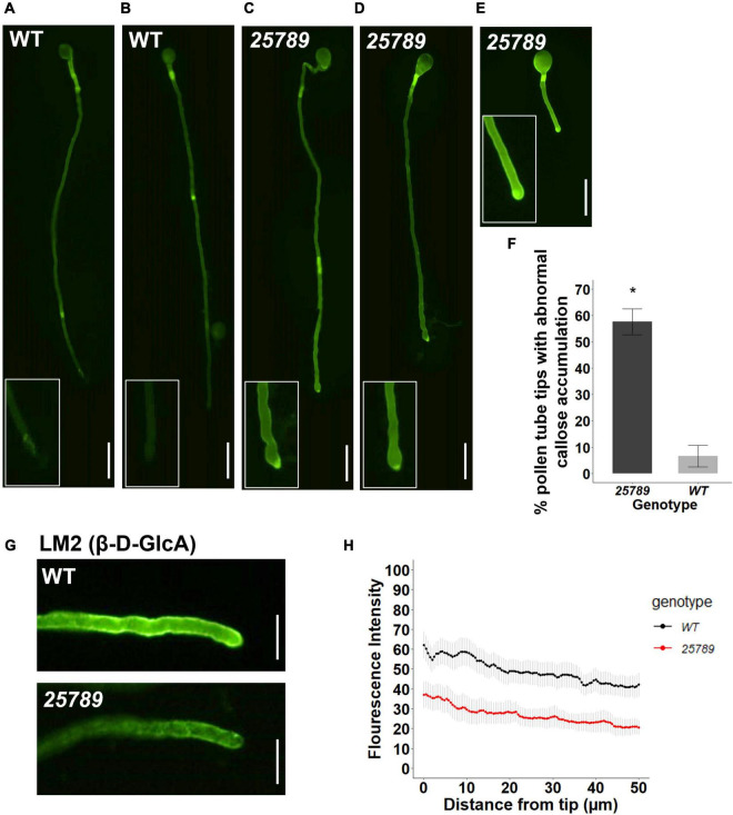
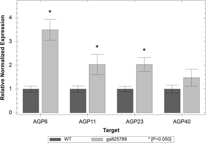
In flowering plants, male reproductive function is determined by successful development and performance of stamens, pollen grains, and pollen tubes. Despite the crucial role of highly glycosylated arabinogalactan-proteins (AGPs) in male gamete formation, pollen grain, and pollen tube cell walls, the underlying mechanisms defining these functions of AGPs have remained elusive. Eight partially redundant Hyp-galactosyltransferases (named GALT2-GALT9) genes/enzymes are known to initiate Hyp-O-galactosylation for Hyp-arabinogalactan (AG) production in Arabidopsis thaliana. To assess the contributions of these Hyp-AGs to male reproductive function, we used a galt2galt5galt7galt8galt9 quintuple Hyp-GALT mutant for this study. Both anther size and pollen viability were compromised in the quintuple mutants. Defects in male gametogenesis were observed in later stages of maturing microspores after meiosis, accompanied by membrane blebbing and numerous lytic vacuoles. Cytological and ultramicroscopic observations revealed that pollen exine reticulate architecture and intine layer development were affected such that non-viable collapsed mature pollen grains were produced, which were devoid of cell content and nuclei, with virtually no intine. AGP immunolabeling demonstrated alterations in cell wall architecture of the anther, pollen grains, and pollen tube. Specifically, the LM2 monoclonal antibody (which recognized β-GlcA epitopes on AGPs) showed a weak signal for the endothecium, microspores, and pollen tube apex. Pollen tube tips also displayed excessive callose deposition. Interestingly, expression patterns of pollen-specific AGPs, namely AGP6, AGP11, AGP23, and AGP40, were determined to be higher in the quintuple mutants. Taken together, our data illustrate the importance of type-II AGs in male reproductive function for successful fertilization.

Keywords: arabinogalactan-proteins; exine; hydroxyproline-galactosyltransferases; intine; microgametogenesis; pollen grains; pollen tube.


Conflict of interest statement

The authors declare that the research was conducted in the absence of any commercial or financial relationships that could be construed as a potential conflict of interest.

Figures

FIGURE 1
FIGURE 1
Comparison of the WT and Hyp-GALT galt25789 quintuple mutant inflorescences and stamen. (A) WT and galt25789 quintuple mutant inflorescences. (B) Comparison of stage 13 flowers of the WT and galt25789 quintuple mutants. Normal inflorescences were accompanied by withered/stunted inflorescences in galt25789 mutants. Note, the galt25789 mutant flowers have reduced amount of pollen sticking to the style and stigma. (C) Anther morphology of stage 13 flowers of the WT and galt25789 quintuple mutants with front view and side view, respectively. Scale bar = 1.0 mm.
FIGURE 2
FIGURE 2
Anther and pollen grains reveal defects in mature pollen grains of galt25789 Hyp-GALT mutants. (A) SEM images of anthers from WT, galt25 and galt789 and galt25789 showed that anther size is reduced in the galt25789 mutants. (B) SEM images of the WT, galt25, galt789 and galt25789 pollen grains. Galt25789 mutant displayed misshaped, collapsed, and defective pollen with abnormal exine patterns in comparison to regular reticulate exine structure in WT. Scale bars = 100 μm in (A), 10 and 5 μm in (B) as indicated.
FIGURE 3
FIGURE 3
Characterization of pollen grain defects by alexander, DAPI, and auramine-O staining of Hyp-GALT mutants compared to WT. (A) Complete view of anther showing pollen viability of WT, galt25, galt789, and galt25789, accessed by alexander staining. (B) High magnification view showing alexander staining of pollen grains. galt25789 had abnormal aborted pollen (indicated by green-colored pollen and black arrowheads in panel). (C) DAPI staining of mature pollen grains released from anthers, under a light microscope and a DAPI filter. Mature pollen grains detected the presence of two sperm nuclei and one vegetative nucleus in WT, galt25 and galt789 and galt25789 except for the aborted pollen grains (indicated by white asterisks) in galt25789 mutant. (D) Confocal images of pollen stained with 0.1% auramine-O in WT, galt25 and galt789 and galt25789. Scale bars = 200 μm in (A); 50 μm in (B,C); 10 μm in panel (D).
FIGURE 4
FIGURE 4
Light and TEM micrographs reveal defects in pollen development in galt25789 Hyp-GALT quintuple mutant. Light micrographs of cross-sections of resin-embedded anthers of WT (A) and galt25789 quintuple (B) plants stained with toluidine blue. Developmental stages 8 to 12L of anthers along with corresponding pollen developmental stages are indicated at top. Red arrowheads in stages indicate vacuolated and thicker tapetal cell layer, as well as vacuolated pollen grains, at binucleate stage of the galt25789 mutant. Orange arrowheads indicate cytoplasmic shrinkage of pollen grains. Black arrowheads indicate cell wall debris of crushed pollen grains. Scale bar = 10 μm. pmc-pollen mother cell; td-tetrad; tp-tapetum; ms-microspore.
FIGURE 5
FIGURE 5
TEM images of WT and galt25789 mutant microspores. (A) Microspores from WT plants. Exine, Intine, and pollen coat of pollen grains (tricellular stage) at stage 12L. (B) Microspores from galt25789 mutant plants. Intine is thin and membrane blebbing is evident in galt25789 mutant normal microspores (indicated by red arrows). (C) Abnormal pollen wall patterning is visible in the aborted mature pollen stage. The mature aborted pollen grain wall structure is aberrant. The pollen grain gets devoid of any content and nuclei. Intine layers are virtually absent (marked by red arrow), and a very dark cytoplasm marks the degradation of the cytoplasm. ba, baculum; ex, exine; in, intine; te, tectum; pc-pollen coat. Scale bars = 5 μm; 3 μm; 2 μm; 500 nm as indicated.
FIGURE 6
FIGURE 6
Distribution of JIM13-epitope labeling of AGPs in microsporocytes and microspores of galt25789 mutants and WT. Cross sections of resin-embedded anthers of WT (Col-0) and galt25789 from stages 8 to 12L were labeled with JIM13 primary antibodies followed by Alexa Fluor 488-labeled secondary antibody labeling subsequently. Fluorescence of Alexa Fluor 488 (green) and autofluorescence (blue) was separately captured by epifluorescence microscopy and merged. Scale bar = 20 μm. en-endothecium; ep-epithelium; ms-microspore; pmc-pollen mother cell; td-tetrad; tp-tapetum.
FIGURE 7
FIGURE 7
Distribution of LM2-epitope labeling of AGPs in microsporocytes and microspores of galt25789 mutants and WT. Cross sections of resin-embedded anthers of WT (Col-0) and galt25789 from stages 8 to 12L were labeled with LM2 primary antibodies followed by Alexa Fluor 488-labeled secondary antibody labeling subsequently. Fluorescence of Alexa Fluor 488 (green) and autofluorescence (blue) was separately captured by epifluorescence microscopy and merged. Scale bar = 20 μm. en-endothecium; ep-epithelium; ms-microspore; pmc-pollen mother cell; td-tetrad; tp-tapetum.
FIGURE 8
FIGURE 8
galt25789 mutant pollen tubes displayed high levels of callose distribution signal at the mutant pollen tube tip and low intensity of LM2 antibody signal during in vitro pollen tube growth. (A,B) Cytochemical staining of β-glucan (callose) with aniline blue shows WT pollen tube with callose signal in the shaft but lacks staining in the pollen tube tip (enlarged in inset). (C–E) displays representative images of abnormal galt25789 mutant pollen tubes with a higher callose signal at the pollen tube tip than the shaft (enlarged in inset). Quantification of abnormal callose deposition of pollen tube tips is shown in (F). Values are expressed as percentage. Error bars denote mean ± SE, asterisks indicate statistically significant, p < 0.005 (n ≥ 45 pollen tubes for each genotype). (G) Immunofluorescence labeling of cell wall epitopes probed with anti-AGP (β-D-GlcA) MAbs LM2, displays strong staining of pollen tube tip and the surface of pollen tubes in WT. LM2 epitope signal is weaker in galt25789 mutant pollen tube tip and shaft. (H) Quantification of signal intensity from the pollen tube tip to 50 μm down the shaft. Graph represents the mean ± SE of the fluorescent intensities for each genotype (n ≥ 25 for each genotype). Scale bars = 50 μm in (A–E), 20 μm in panel (G).
FIGURE 9
FIGURE 9
Real time RT-PCR showing relative expression of pollen-specific AGP genes (AGP6, AGP11, AGP23, and AGP40) in galt25789 mutants compared to their expression in WT inflorescences. The level of the transcripts was normalized according to the reference genes ACT8 and RUB1. Each bar represents an average of the three technical replicas; the * represents a significant result for p < 0.05.

Similar articles

Cited by

References

    1. Ajayi O. O., Held M. A., Showalter A. M. (2021). Glucuronidation of type II arabinogalactan polysaccharides function in sexual reproduction of Arabidopsis. Plant J. 109 164–181. 10.1111/tpj.15562 - DOI - PubMed
    1. Ariizumi T., Steber C. M. (2007). Seed germination of GA-insensitive sleepy1 mutants does not require RGL2 protein disappearance in Arabidopsis. Plant Cell 19 791–804. 10.1105/tpc.106.048009 - DOI - PMC - PubMed
    1. Ariizumi T., Toriyama K. (2011). genetic regulation of sporopollenin synthesis and pollen exine development. Annu. Rev. Plant Biol. 62 437–460. 10.1146/annurev-arplant-042809-112312 - DOI - PubMed
    1. Basu D., Tian L., Wang W., Bobbs S., Herock H., Travers A., et al. (2015a). A small multigene hydroxyproline-O-galactosyltransferase family functions in arabinogalactan-protein glycosylation, growth and development in Arabidopsis. BMC Plant Biol. 15:295. 10.1186/s12870-015-0670-7 - DOI - PMC - PubMed
    1. Basu D., Wang W., Ma S., DeBrosse T., Poirier E., Emch K., et al. (2015b). Two hydroxyproline galactosyltransferases, galt5 and galt2, function in arabinogalactan-protein glycosylation, growth and development in Arabidopsis. PLoS One 10:e0125624. 10.1371/journal.pone.0125624 - DOI - PMC - PubMed

LinkOut - more resources