. 2022 Aug;24(8):1327-1345.
doi: 10.1002/ejhf.2601. Epub 2022 Jul 31.

Exercise testing in heart failure with preserved ejection fraction: an appraisal through diagnosis, pathophysiology and therapy - A clinical consensus statement of the Heart Failure Association and European Association of Preventive Cardiology of the European Society of Cardiology

Affiliations

Exercise testing in heart failure with preserved ejection fraction: an appraisal through diagnosis, pathophysiology and therapy - A clinical consensus statement of the Heart Failure Association and European Association of Preventive Cardiology of the European Society of Cardiology

Marco Guazzi et al. Eur J Heart Fail. 2022 Aug.

Abstract

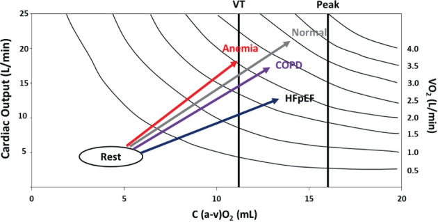
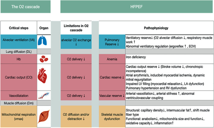
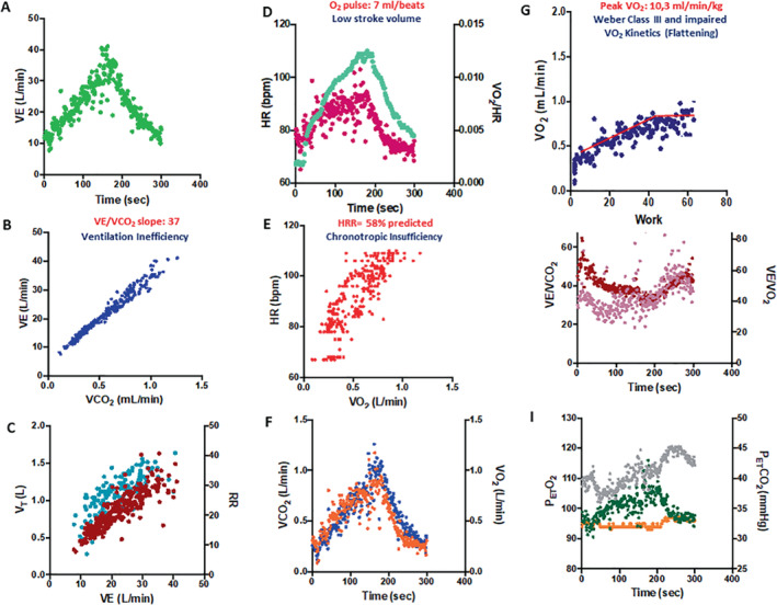
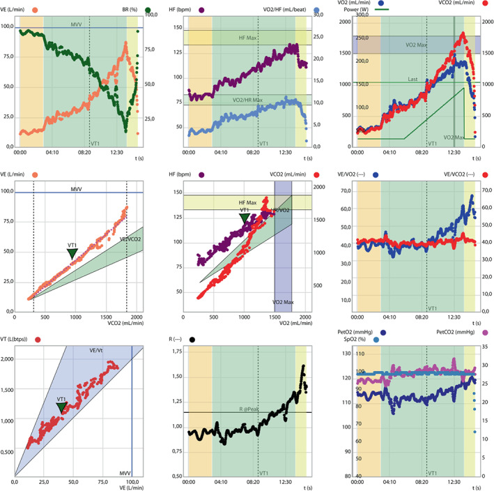
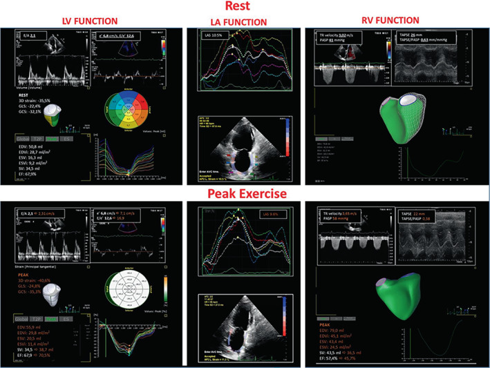
Patients with heart failure with preserved ejection fraction (HFpEF) universally complain of exercise intolerance and dyspnoea as key clinical correlates. Cardiac as well as extracardiac components play a role for the limited exercise capacity, including an impaired cardiac and peripheral vascular reserve, a limitation in mechanical ventilation and/or gas exchange with reduced pulmonary vascular reserve, skeletal muscle dysfunction and iron deficiency/anaemia. Although most of these components can be differentiated and quantified through gas exchange analysis by cardiopulmonary exercise testing (CPET), the information provided by objective measures of exercise performance has not been systematically considered in the recent algorithms/scores for HFpEF diagnosis, by neither European nor US groups. The current clinical consensus statement by the Heart Failure Association (HFA) and European Association of Preventive Cardiology (EAPC) of the European Society of Cardiology (ESC) aims at outlining the role of exercise testing and its pathophysiological, clinical and prognostic insights, addressing the implications of a thorough functional evaluation from the diagnostic algorithm to the pathophysiology and treatment perspectives of HFpEF. Along with these goals, we provide a specific analysis of the evidence that CPET is the standard for assessing, quantifying, and differentiating the origin of dyspnoea and exercise impairment and even more so when combined with echocardiography and/or invasive haemodynamic evaluation. This will lead to improved quality of diagnosis when applying the proposed scores and may also help to implement the progressive characterization of the specific HFpEF phenotypes, a critical step toward the delivery of phenotype-specific treatments.

Keywords: Exercise; Functional limitation; Gas exchange analysis; HFpEF.


Figures

Figure 1
Figure 1
Plot of the Fick principle relating cardiac output to artero‐venous oxygen (a–vO2) difference and isoplets curves of oxygen uptake (VO2). The graph describes the expected relationship of heart failure with normal control pattern along with chronic obstructive pulmonary disease (COPD) and anaemia conditions as the most common comorbidities that affect oxygen (O2) content and delivery and may add on heart failure with preserved ejection fraction (HFpEF) haemodynamic, i.e. cardiac output, limitation. VT, ventilatory threshold.
Figure 2
Figure 2
The oxygen (O2) cascade during exercise. The organ systems and pathways (from air to mitochondria) involved in exercise performance are depicted along with the limiting steps and pathophysiology behind exercise limitation in heart failure with preserved ejection fraction (HFpEF). EOV, exercise oscillatory ventilation; LA, left atrial; RV, right ventricular.
Figure 3
Figure 3
Cascade of the cardiac, haemodynamic and pulmonary maladaptive response under the effects of pulmonary capillary wedge pressure (PCWP) increase. DLco, exercise diffusing lung capacity for carbon monoxide; Dm, membrane diffusion; LVEDP, left ventricular end‐diastolic pressure; Pc, pulmonary circulation; RV, right ventricular; Vc, capillary volume; VD/VT, dead space to tidal volume ratio; VE, minute ventilation; VE/VCO2, minute ventilation to carbon dioxide output. V/Q, ventilation/perfusion
Figure 4
Figure 4
Continuous of mechanisms involved in right ventricular maladaptive response to increased load and pulmonary vascular disease, affecting cardiac output and exercise performance in heart failure with preserved ejection fraction. TR, tricuspid regurgitation.
Figure 5
Figure 5
Nine‐plot analysis (A–I) of a typical cardiopulmonary exercise test response of an old hypertensive female patient with exertional dyspnoea. See text for explanation. HR, heart rate; PetCO2, end‐tidal carbon dioxide tension; PetO2, end‐tidal oxygen tension; VCO2, carbon dioxide output; VE, minute ventilation; VE/VCO2, minute ventilation to carbon dioxide output; VO2, oxygen uptake; VT, ventilatory threshold.
Figure 6
Figure 6
Nine‐plot analysis of a middle aged man with initial exertional dyspnoea presenting with a different cardiopulmonary exercise test phenotype. See text for explanation. HF, heart frequency; HR, heart rate; PetCO2, end‐tidal carbon dioxide tension; PetO2, end‐tidal oxygen tension; VCO2, carbon dioxide ouput; VE, minute ventilation; VO2, oxygen uptake; VT, ventilatory threshold.
Figure 7
Figure 7
Cardiopulmonary exercise test imaging rest to peak exercise analysis of the same case as Figure  5 . Measures obtained by stress echocardiography (rest to peak exercise). The analysis was performed analysing the diastolic (E/e') and systolic (three‐dimensional longitudinal and circumferential strain) left ventricular (LV) function; the adaptive left atrial (LA) dynamics by LA strain (LAS); right ventricular (RV) function (RV ejection fraction 3D analysis) and its coupling with the pulmonary circulation by the tricuspid annular plane systolic excursion/systolic pulmonary artery pressure (TAPSE/PASP) ratio. Data are reported at rest (white) and at peak exercise (orange) with the changes occurring in the main variables from rest to peak. TR, tricuspid regurgitation.

References

    1. Pieske B, Tschope C, de Boer RA, Fraser AG, Anker SD, Donal E, et al. How to diagnose heart failure with preserved ejection fraction: the HFA‐PEFF diagnostic algorithm: a consensus recommendation from the Heart Failure Association (HFA) of the European Society of Cardiology (ESC). Eur Heart J. 2019;40:3297–317. - PubMed
    1. Reddy YNV, Carter RE, Obokata M, Redfield MM, Borlaug BA. A simple, evidence‐based approach to help guide diagnosis of heart failure with preserved ejection fraction. Circulation. 2018;138:861–70. - PMC - PubMed
    1. Verbrugge FH, Omote K, Reddy YNV, Sorimachi H, Obokata M, Borlaug BA. Heart failure with preserved ejection fraction in patients with normal natriuretic peptide levels is associated with increased morbidity and mortality. Eur Heart J. 2022;43:1941–51. - PMC - PubMed
    1. Solomon SD, Rizkala AR, Lefkowitz MP, Shi VC, Gong J, Anavekar N, et al. Baseline characteristics of patients with heart failure and preserved ejection fraction in the PARAGON‐HF trial. Circ Heart Fail. 2018;11:e004962. - PubMed
    1. Houstis NE, Eisman AS, Pappagianopoulos PP, Wooster L, Bailey CS, Wagner PD, et al. Exercise intolerance in heart failure with preserved ejection fraction: diagnosing and ranking its causes using personalized O2 pathway analysis. Circulation. 2018;137:148–61. - PMC - PubMed

Publication types