

Recent developments in the use of gold and silver nanoparticles in biomedicine
- PMID: 35775611
- PMCID: PMC9539467
- DOI: 10.1002/wnan.1817
Recent developments in the use of gold and silver nanoparticles in biomedicine
Abstract
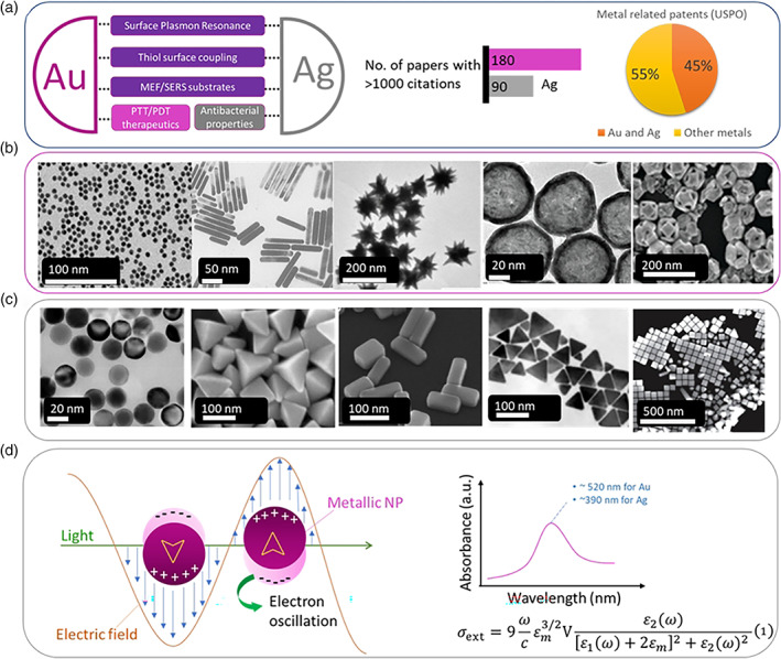
Gold and silver nanoparticles (NPs) are widely used in the biomedical research both in the therapeutic and the sensing/diagnostics fronts. Both metals share some common optical properties with surface plasmon resonance being the most widely exploited property in therapeutics and diagnostics. Au NPs exhibit excellent light-to-heat conversion efficiencies and hence have found applications primarily in precision oncology, while Ag NPs have excellent antibacterial properties which can be harnessed in biomaterials' design. Both metals constitute excellent biosensing platforms owing to their plasmonic properties and are now routinely used in various optical platforms. The utilization of Au and Ag NPs in the COVID-19 pandemic was rapidly expanded mostly in biosensing and point-of-care platforms and to some extent in therapeutics. In this review article, the main physicochemical properties of Au and Ag NPs are discussed with selective examples from the recent literature. This article is categorized under: Therapeutic Approaches and Drug Discovery > Nanomedicine for Oncologic Disease Diagnostic Tools > In Vitro Nanoparticle-Based Sensing Nanotechnology Approaches to Biology > Nanoscale Systems in Biology.
Keywords: COVID-19; diagnostics; gold nanoparticles; nanomedicine; silver nanoparticles.
© 2022 The Author. WIREs Nanomedicine and Nanobiotechnology published by Wiley Periodicals LLC.
Conflict of interest statement
The author has declared no conflicts of interest for this article.
Figures

Similar articles
-
Silver-Based Plasmonic Nanoparticles for and Their Use in Biosensing.Biosensors (Basel). 2019 Jun 10;9(2):78. doi: 10.3390/bios9020078. Biosensors (Basel). 2019. PMID: 31185689 Free PMC article. Review.
-
Engineering gold nanoparticles for molecular diagnostics and biosensing.Wiley Interdiscip Rev Nanomed Nanobiotechnol. 2023 Jan;15(1):e1836. doi: 10.1002/wnan.1836. Epub 2022 Aug 5. Wiley Interdiscip Rev Nanomed Nanobiotechnol. 2023. PMID: 35932114 Review.
-
Ag and Au nanoparticles/reduced graphene oxide composite materials: Synthesis and application in diagnostics and therapeutics.Adv Colloid Interface Sci. 2019 Sep;271:101991. doi: 10.1016/j.cis.2019.101991. Epub 2019 Jul 17. Adv Colloid Interface Sci. 2019. PMID: 31376639 Review.
-
Noble metals on the nanoscale: optical and photothermal properties and some applications in imaging, sensing, biology, and medicine.Acc Chem Res. 2008 Dec;41(12):1578-86. doi: 10.1021/ar7002804. Acc Chem Res. 2008. PMID: 18447366
-
Recent Advances in Silver and Gold Nanoparticles-Based Colorimetric Sensors for Heavy Metal Ions Detection: A Review.Crit Rev Anal Chem. 2023;53(3):718-750. doi: 10.1080/10408347.2021.1973886. Epub 2021 Sep 12. Crit Rev Anal Chem. 2023. PMID: 34510976 Review.
Cited by
-
Impacts of loading thymoquinone to gold or silver nanoparticles on the efficacy of anti-tumor treatments in breast cancer with or without chemotherapeutic cisplatin.BMC Biotechnol. 2025 Apr 10;25(1):26. doi: 10.1186/s12896-025-00958-6. BMC Biotechnol. 2025. PMID: 40211258 Free PMC article.
-
Capped Plasmonic Gold and Silver Nanoparticles with Porphyrins for Potential Use as Anticancer Agents-A Review.Pharmaceutics. 2024 Sep 28;16(10):1268. doi: 10.3390/pharmaceutics16101268. Pharmaceutics. 2024. PMID: 39458600 Free PMC article. Review.
-
α-Amino Acids as Reducing and Capping Agents in Gold Nanoparticles Synthesis Using the Turkevich Method.Langmuir. 2023 Jun 27;39(25):8646-8657. doi: 10.1021/acs.langmuir.3c00507. Epub 2023 Jun 14. Langmuir. 2023. PMID: 37314886 Free PMC article.
-
The quest for nanoparticle-powered vaccines in cancer immunotherapy.J Nanobiotechnology. 2024 Feb 14;22(1):61. doi: 10.1186/s12951-024-02311-z. J Nanobiotechnology. 2024. PMID: 38355548 Free PMC article. Review.
-
SERS Determination of Oxidative Stress Markers in Saliva Using Substrates with Silver Nanoparticle-Decorated Silicon Nanowires.Biosensors (Basel). 2023 Feb 14;13(2):273. doi: 10.3390/bios13020273. Biosensors (Basel). 2023. PMID: 36832039 Free PMC article.
References
-
- Abnous, K. , Danesh, N. M. , Ramezani, M. , Alibolandi, M. , Nameghi, M. A. , Zavvar, T. S. , & Taghdisi, S. M. (2021). A novel colorimetric aptasensor for ultrasensitive detection of aflatoxin M1 based on the combination of CRISPR‐Cas12a, rolling circle amplification and catalytic activity of gold nanoparticles. Analytica Chimica Acta, 1165, 338549. 10.1016/j.aca.2021.338549 - DOI - PubMed
-
- Adams, S. , Thai, D. , Mascona, X. , Schwartzberg, A. M. , & Zhang, J. Z. (2014). Key factors affecting the reproducibility of synthesis and growth mechanism of near‐infrared absorbing hollow gold nanospheres. Chemistry of Materials, 26(23), 6805–6810. 10.1021/CM5033892 - DOI
-
- Adnan, N. N. M. , Cheng, Y. Y. , Ong, N. M. N. , Kamaruddin, T. T. , Rozlan, E. , Schmidt, T. W. , Duong, H. T. T. , & Boyer, C. (2016). Effect of gold nanoparticle shapes for phototherapy and drug delivery. Polymer Chemistry, 7, 2888–2903. 10.1039/c6py00465b - DOI
-
- Alberti, G. , Zanoni, C. , Magnaghi, L. R. , & Biesuz, R. (2021). Gold and silver nanoparticle‐based colorimetric sensors: New trends and applications. Chemosensors, 9(11), 305. 10.3390/CHEMOSENSORS9110305 - DOI
Publication types
MeSH terms
Substances
LinkOut - more resources
Full Text Sources
Medical
