. 2023 Jan;72(1):151-168.
doi: 10.1007/s00262-022-03239-9. Epub 2022 Jul 1.

Cancer-targeted photoimmunotherapy induces antitumor immunity and can be augmented by anti-PD-1 therapy for durable anticancer responses in an immunologically active murine tumor model

Affiliations

Cancer-targeted photoimmunotherapy induces antitumor immunity and can be augmented by anti-PD-1 therapy for durable anticancer responses in an immunologically active murine tumor model

Michelle A Hsu et al. Cancer Immunol Immunother. 2023 Jan.

Abstract

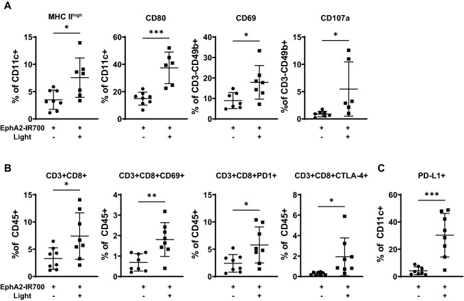
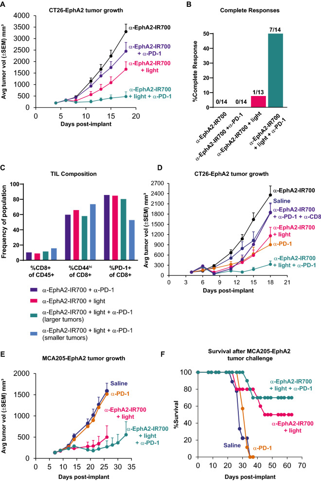
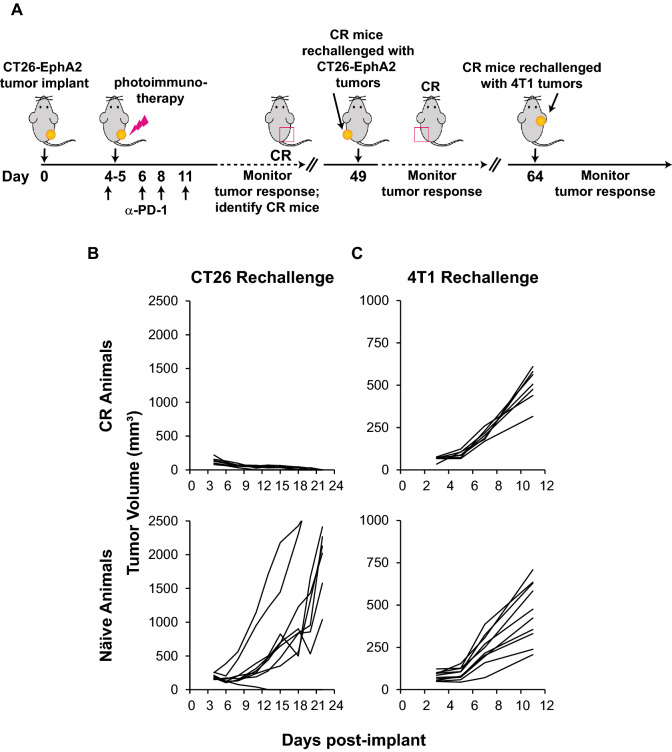
The complex immunosuppressive nature of solid tumor microenvironments poses a significant challenge to generating efficacious and durable anticancer responses. Photoimmunotherapy is a cancer treatment strategy by which an antibody is conjugated with a non-toxic light-activatable dye. Following administration of the conjugate and binding to the target tumor, subsequent local laser illumination activates the dye, resulting in highly specific target cell membrane disruption. Here we demonstrate that photoimmunotherapy treatment elicited tumor necrosis, thus inducing immunogenic cell death characterized by the release of damage-associated molecular patterns (DAMPs). Photoimmunotherapy-killed tumor cells activated dendritic cells (DC), leading to the production of proinflammatory cytokines, T cell stimulation, priming antigen-specific T cells, and durable memory T cell responses, which led complete responder mice to effectively reject new tumors upon rechallenge. PD-1 blockade in combination with photoimmunotherapy enhanced overall anticancer efficacy, including against anti-PD-1-resistant tumors. The combination treatment also elicited abscopal anticancer activity, as observed by reduction of distal, non-illuminated tumors, further demonstrating the ability of photoimmunotherapy to harness local and peripheral T cell responses. With this work we therefore delineate the immune mechanisms of action for photoimmunotherapy and demonstrate the potential for cancer-targeted photoimmunotherapy to be combined with other immunotherapy approaches for augmented, durable anticancer efficacy. Moreover, we demonstrate responses utilizing various immunocompetent mouse models, as well as in vitro data from human cells, suggesting broad translational potential.

Keywords: Cancer; Immuno-oncology; Immunology; Photoimmunotherapy.


Conflict of interest statement

This research was funded by Rakuten Medical, Inc (RMI). All authors are currently, or were employed by RMI, at the time of research. SMO, DMB, JJF, RH, MGG, MW, CDMF, and DY hold stock options at RMI. MGG, JJF, RH, CDMF, and DY hold IP interests at RMI. MW participates in biopharma consulting activities outside of RMI. MGG serves a leadership role at Rakuten Medical and holds IP interests at UC Irvine and RMI.

Figures

Fig. 1
Fig. 1
Characterization of an immunocompetent mouse tumor model for cancer-targeted photoimmunotherapy. Flow cytometry analysis demonstrated stable expression of mouse EphA2 on the surface of CT26 mouse colon carcinoma cells after viral transduction (A). CT26-EphA2 cells were treated with increasing concentrations of anti-EphA2-IR700 followed by illumination at indicated doses, demonstrating that EphA2 photoimmunotherapy results in cell killing in a light- and conjugate- dose-dependent manner in vitro (B). Anti-EphA2-IR700 accumulates in CT26-EphA2 tumors in mice after systemic administration, as detected by fluorescence imaging (C). Mice were implanted with CT26-EphA2 tumors, then treated with anti-EphA2-IR700 and light or only anti-EphA2-IR700 without light, as reference. Tumor growth was inhibited in the anti-EphA2-IR700 plus light group (n = 15 per group). Final tumor measurements: saline vs EphA2 photoimmunotherapy, p < 0.01; EphA2 vs EphA2 photoimmunotherapy, p < 0.0001 based on two-way ANOVA and Tukey test. The tumor growth results shown are representative of several replicate experiments performed by different personnel. (D). The ability of EphA2 photoimmunotherapy to elicit vaccinal effects as compared to known ICD-inducing or non-ICD-inducing conditions was determined. Cells killed with cisplatin, doxorubicin, or anti-EphA2-IR700 plus light were allowed to achieve 50% cell death, then were implanted into the right hind flank of naïve mice. Seven days after implant with dying cells, mice were challenged with viable CT26-EphA2 cells in the opposite hind flank and tumor growth was measured for 21 days (E)
Fig. 2
Fig. 2
Photoimmunotherapy elicits intratumoral immune cell activation. Mice were implanted with CT26-EphA2 tumors, then treated with anti-EphA2-IR700 without illumination or EphA2 photoimmunotherapy. Activation markers for intratumoral innate and adaptive immune cells were measured by flow cytometry one day (A) or 7 to 8 days (B & C) after photoimmunotherapy treatment. *p < 0.05, **p < 0.01, ***p < 0.001 as measured by unpaired t-test
Fig. 3
Fig. 3
Photoimmunotherapy enhances anticancer activity of tumor-specific lymphocytes. Mice were implanted with CT26-EphA2 cells, then CD40L blocking antibody (or saline control) was administered at days 0, 1, 2 and 3 (A) or 6, 7, and 8. (B). Anti-EphA2-IR700 was delivered at day 6, followed by illumination 24 ± 2 h later for photoimmunotherapy groups, and tumor volume was measured to determine tumor growth inhibition (A & B). Final tumor measurements: EphA2 photoimmunotherapy vs EphA2 photoimmunotherapy + anti-CD40L, p < 0.01 (A) and n.s. (B) based on two-way ANOVA and Tukey test. To evaluate the memory potential against syngeneic tumors, mice identified as complete responders after photoimmunotherapy were rechallenged with syngeneic tumor cells in the opposite hind flank 49 days after the original tumor challenge, and 2 to 3 weeks after complete responses were achieved. Tumor growth was measured over 21 days as compared to growth in naïve mice (C). The tumor growth curves shown for both efficacy and tumor rechallenge are representative of at least two replicate experiments performed by different personnel
Fig. 4
Fig. 4
Combining photoimmunotherapy with checkpoint inhibition elicits enhanced anticancer responses. Mice were implanted with CT26-EphA2 tumors, then treated with anti-EphA2-IR700 alone, or EphA2 photoimmunotherapy. Anti-PD-1 was administered to indicated groups at days 4, 6, 8, and 11 post-inoculation, and tumor growth was measured from days 18 to 22, and graphed here through day 18 (A). Final tumor measurements: anti-PD-1 vs EphA2 photoimmunotherapy + anti-PD-1, p = 0.0001; EphA2 photoimmunotherapy vs EphA2 photoimmunotherapy + anti-PD-1, p < 0.05 based on two-way ANOVA and Tukey test. The percent of complete responses (calculated by number of CRs/total animals in treatment group) as defined as undetectable tumors at study end were determined (B). Activation patterns among intratumoral lymphocytes were determined by flow cytometry at day 21, among mice that had palpable tumors. Tumors with limiting volume were pooled to achieve sufficient cell yield for analysis (C). Mice were implanted with tumors (n = 15 per group) and received saline, anti-EphA2-IR700 without illumination, anti-PD-1 monotherapy (days 4, 6, 8, and 11 post implant), or anti-CD8 (days 4 and 7 for CD8 T cell depletion) and evaluated for tumor growth through day 18 post implant. Tumor-bearing mice also received photoimmunotherapy treatment (as described above, n = 12), photoimmunotherapy treatment with anti-PD-1 (n = 11), or photoimmunotherapy treatment with anti-PD-1 and anti-CD8 (n = 9), and evaluated for tumor growth through day 18. Final tumor measurements: EphA2 photoimmunotherapy + anti-PD-1 vs. EphA2 photoimmunotherapy + anti-PD-1 + anti-CD8, p < 0.01 based on two-way ANOVA and Tukey test (D). MCA205-EphA2 cells were inoculated into the hind flanks of mice. Mice were then treated with saline, anti-PD-1 alone, EphA2 photoimmunotherapy, or EphA2 photoimmunotherapy combined with anti-PD-1 antibodies, as indicated (n = 10 mice per group). Anti-PD-1 was administered beginning on day 7 and continuing 3 times per weeks until study end. Tumor growth (E) and survival (F) were measured through days 33 and 63, respectively. Final tumor measurements: anti-PD-1 vs EphA2 photoimmunotherapy + anti-PD-1, p < 0.0001; EphA2 photoimmunotherapy vs EphA2 photoimmunotherapy + anti-PD-1, n.s. based on two-way ANOVA and Tukey test (E). The tumor growth curves shown are representative of at least two replicate experiments performed by different personnel
Fig. 5
Fig. 5
Complete responder mice, previously treated with anti-PD-1 and photoimmunotherapy, mount tumor-specific memory immune responses. Mice that had achieved a complete response (n = 7) after dual treatment with anti-PD-1 and photoimmunotherapy were re-challenged with syngeneic CT26-EphA2 cells in the contralateral hind flank, followed by 4T1 cells in the axilla (A). Naïve mice (n = 10) were inoculated as control. Tumor growth was measured to determine rejection (B & C). The resulting tumor growth curves after rechallenge are representative of at least two replicate experiments performed by different personnel
Fig. 6
Fig. 6
Photoimmunotherapy plus anti-PD-1 treatment expands peripheral tumor-specific CD8 + T cells. At day 14 after photoimmunotherapy, splenocytes from mice bearing CT26-EphA2 tumors that had been treated with either anti-EphA2-IR700, anti-EphA2-IR700 plus anti-PD-1, EphA2 photoimmunotherapy, or EphA2 photoimmunotherapy plus anti-PD-1, were harvested and expanded in the presence of the antigen AH1, which is expressed on the surface of CT26 cells, and co-cultured with CT26 cell monolayers in vitro at the indicated effector:target ratios. The AH1 antigen-specific cytotoxic potential of primed splenocytes was measured by Lactic Acid Dehydrogenase (LDH) release assay
Fig. 7
Fig. 7
Photoimmunotherapy plus anti-PD-1 enhances abscopal anticancer activity. CT26-EphA2 cells were implanted into both hind flanks of mice. 3 × 106 cells were implanted to establish primary tumors, whereas 3 × 105 cells were delivered to establish distal tumors (A). Mice were treated with either anti-EphA2-IR700, anti-EphA2-IR700 plus anti-PD-1, EphA2 photoimmunotherapy, or EphA2 photoimmunotherapy plus anti-PD-1. Anti-PD-1 was delivered on days 4, 6, 8, and 11. Only target tumors, but not distal tumors were illuminated in groups receiving applied light. Tumor growth was measured through day 25 (B) and complete responses were determined at study end (C). The tumor growth curves shown are representative of at least two replicate experiments performed by different personnel

References

    1. Sharma P, Allison JP. The future of immune checkpoint therapy. Science. 2015;348:56–61. doi: 10.1126/science.aaa8172. - DOI - PubMed
    1. Topalian SL, Drake CG, Pardoll DM. Immune checkpoint blockade: a common denominator approach to cancer therapy. Cancer Cell. 2015;27:450–461. doi: 10.1016/j.ccell.2015.03.001. - DOI - PMC - PubMed
    1. Anderson KG, Stromnes IM, Greenberg PD. Obstacles Posed by the Tumor Microenvironment to T cell Activity: A Case for Synergistic Therapies. Cancer Cell. 2017;31:311–325. doi: 10.1016/j.ccell.2017.02.008. - DOI - PMC - PubMed
    1. Galluzzi L, Chan TA, Kroemer G, Wolchok JD, Lopez-Soto A. The hallmarks of successful anticancer immunotherapy. Sci Transl Med. 2018 doi: 10.1126/scitranslmed.aat7807. - DOI - PubMed
    1. Kroemer G, Senovilla L, Galluzzi L, Andre F, Zitvogel L. Natural and therapy-induced immunosurveillance in breast cancer. Nat Med. 2015;21:1128–1138. doi: 10.1038/nm.3944. - DOI - PubMed