Review
. 2022 Jul 1;79(7):390.
doi: 10.1007/s00018-022-04394-z.

-Omics potential of in vitro skin models for radiation exposure

Affiliations
Review

-Omics potential of in vitro skin models for radiation exposure

Leyla A Akh et al. Cell Mol Life Sci. .

Abstract

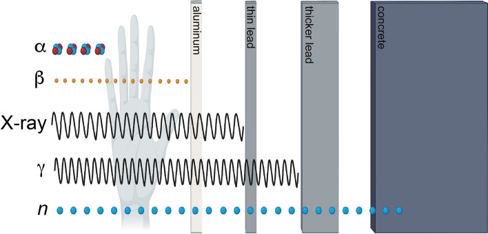
There is a growing need to uncover biomarkers of ionizing radiation exposure that leads to a better understanding of how exposures take place, including dose type, rate, and time since exposure. As one of the first organs to be exposed to external sources of ionizing radiation, skin is uniquely positioned in terms of model systems for radiation exposure study. The simultaneous evolution of both MS-based -omics studies, as well as in vitro 3D skin models, has created the ability to develop a far more holistic understanding of how ionizing radiation affects the many interconnected biomolecular processes that occur in human skin. However, there are a limited number of studies describing the biomolecular consequences of low-dose ionizing radiation to the skin. This review will seek to explore the current state-of-the-art technology in terms of in vitro 3D skin models, as well as track the trajectory of MS-based -omics techniques and their application to ionizing radiation research, specifically, the search for biomarkers within the low-dose range.

Keywords: 3D tissue model; Biomarker; Ionizing radiation; Low dose; Multi-omics; Skin.


Conflict of interest statement

The authors have no relevant financial or non-financial interests to disclose.

Figures

Fig. 1
Fig. 1
Common types of ionizing radiation, along with their associated depths of penetration. While alpha-particles have a high LET they are stopped by the first few layers of skin or thin paper. High-energy beta particles can penetrate through several cm of human skin but can be shielded by thin aluminum or plastic. X-ray and gamma-rays have much higher penetration depths being stopped by lead or iron shielding. Neutrons have the highest penetration depth only being attenuated by thick concrete or water
Fig. 2
Fig. 2
Simplified 3D model of skin, along with most cell types commonly found in each layer. Immune cells are marked with blue text. The layer of skin immediately exposed to the environment is the stratum corneum with the epidermis underneath. The dermis has the greatest number of different cell types responsible for protecting the body. The hypodermis is largely composed of adipose (fat) tissue
Fig. 3
Fig. 3
General categories of skin models cultured at the air–liquid interface. The models discussed in this paper all fall into these general categories with some novel variations. a Reconstructed human epidermis (RHE), composed of stratified keratinocytes on an inert porous membrane cultured at the air–liquid interface. b A full-thickness model, where a RHE is cultured atop a dermal equivalent composed of fibroblasts embedded in a collagen matrix. c Re-epidermized dermis, where the epidermis is removed from human skin leaving a de-epidermized dermis. Then, a RHE is cultured on the de-epidermized dermis to produce a model with both epidermal and dermal components. d A skin explant model where human skin is cultured at the air–liquid interface directly from the donor
Fig. 4
Fig. 4
Sample workflow for MS-based -omics analysis. Beginning with sample radiation exposure, this workflow includes both targeted (analyte(s) are known) and untargeted (analyte(s) are unknown) mass spectrometry and highlights the components involved in each step. The ontological tools listed here are not exhaustive but represent the bulk of available options

References

    1. Braganza MZ, Kitahara CM, Berrington de González A, Inskip PD, Johnson KJ, Rajaraman P. Ionizing radiation and the risk of brain and central nervous system tumors: a systematic review. Neuro Oncol. 2012;14:1316–1324. - PMC - PubMed
    1. Lang S, Servomaa K, Kosma VM, Rytömaa T. Biokinetics of nuclear fuel compounds and biological effects of nonuniform radiation. Environ Health Perspect. 1995;103:920–934. - PMC - PubMed
    1. Mettler FA (1985) Medical effects of ionizing radiation. Saunders/Elsevier
    1. Prasad KN, Cole WC, Hasse GM. Health risks of low dose ionizing radiation in humans: a review. Exp Biol Med. 2004;229:378–382. - PubMed
    1. Puukila S, Lemon JA, Lees SJ, Tai TC, Boreham DR, Khaper N. Impact of ionizing radiation on the cardiovascular system: a review. Radiat Res. 2017;188:539–546. - PubMed