Review
. 2023 Feb;96(1142):20220463.
doi: 10.1259/bjr.20220463. Epub 2022 Jul 13.

Non-oncologic incidental uptake on FAPI PET/CT imaging

Affiliations
Review

Non-oncologic incidental uptake on FAPI PET/CT imaging

Masatoshi Hotta et al. Br J Radiol. 2023 Feb.

Abstract

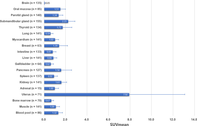
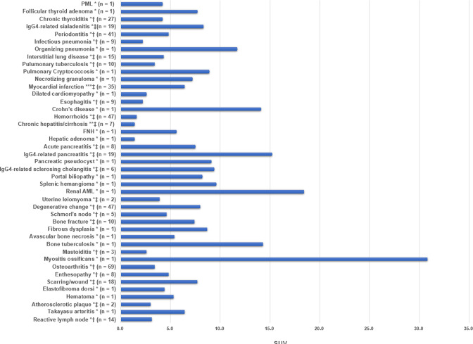
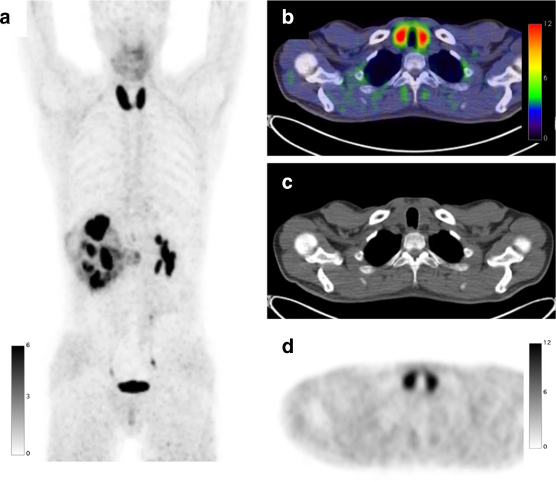
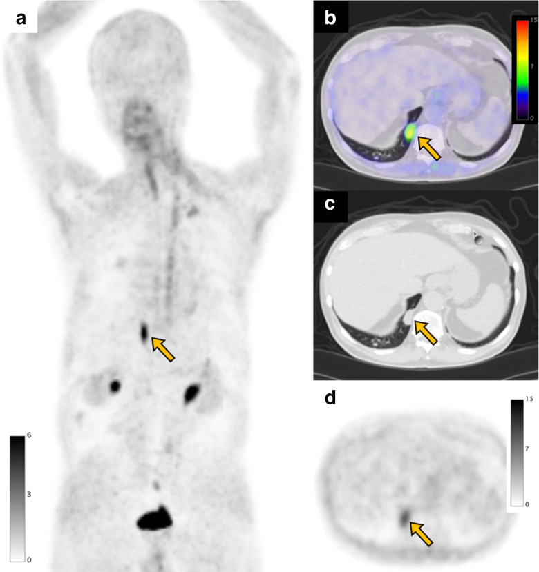
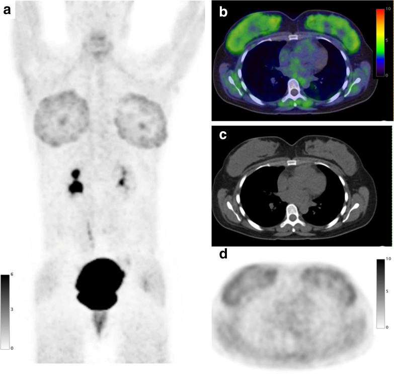
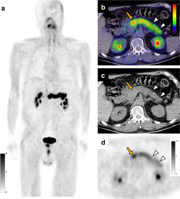
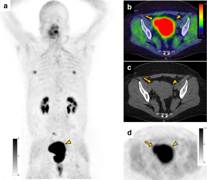
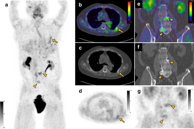
Fibroblast-activation protein (FAP) is a serine protease classified in the dipeptidyl peptidase 4 (DPP4) family. FAP is predominantly expressed in activated fibroblasts such as the cancer-associated fibroblasts (CAFs). FAP expression in CAFs is associated with tumor progression and poor prognosis in solid cancers. Recently, radiolabeled FAP inhibitors (FAPI) has been developed, which enables positron emission tomography (PET) imaging of FAP. FAPI PET/CT can provide a higher tumor-to-background ratio (TBR) than 18F-fludeoxyglucose PET/CT in various cancers, and thus has attracted substantial attention. As studies on FAPI PET grow in number and size, incidental findings related to non-oncologic conditions have been increasingly reported. FAPI PET uptake has been reported in various conditions such as benign tumors, fibrotic, granulomatosis, scarring/wound, degenerative diseases, and inflammatory diseases.The knowledge of physiological and non-oncologic causes of FAPI uptake is indispensable for accurate FAPI PET/CT interpretation and can help appropriate management of incidental findings on FAPI PET/CT in patients referred for cancer staging indications. In this review article, we describe for each organ system (Brain, Oral mucosa, Salivary Glands, Thyroid, Lung, Myocardium, Breast, Esophagus, Stomach, Intestine, Liver, Gallbladder, Pancreas, Spleen, Kidney, , Uterus, Bone marrow, Joints, Muscle, Vessels, Lymph nodes), the patterns of physiological FAPI uptake and the main causes of non-oncological uptake reported from the literature with FAPI-02, FAPI-04 and FAPI-46. We also illustrate some examples from our institutional database at UCLA.


Conflict of interest statement

CONFLICTS OF INTEREST: Jeremie Calais was the recipient of grants from the ERF-SNMMI (2019–2021 Molecular Imaging Research Grant for Junior Academic Faculty) and the Prostate Cancer Foundation (grant 20YOUN05 and 19CHAL02). Jeremie Calais reported receiving personal fees from Advanced Accelerator Applications, Astellas, Blue Earth Diagnostics, Curium Pharma, DS pharma, EXINI, GE Healthcare, IBA RadioPharma, Isoray, Janssen Pharmaceuticals, Lantheus, Lightpointmedical, Novartis, POINT Biopharma, RadioMedix, Progenics, Telix Pharmaceuticals, outside the submitted work.

Figures

Figure 1.
Figure 1.
Pooled SUVmean and SD of each organ system. Error bars show SD, SD, standard deviation; SUV, standardized uptake value.
Figure 2.
Figure 2.
Reported SUVs of non-oncologic diseases. (* SUVmax, ** SUVmean, *** SUVpeak, † median, ‡ average). SUV, standardized uptake value.
Figure 3.
Figure 3.
A 45-year-old male with right clear cell renal cell carcinoma treated with neoadjuvant immune checkpoint inhibitors underwent a FAPI PET/CT scan before surgery. FAPI PET MIP image (a) FAPI PET/CT (b), CT (c), and FAPI PET (d) demonstrate diffuse intense increased uptake (SUVmax: 23.5) in the enlarged thyroid indicating thyroiditis induced by immune checkpoint inhibitors. FAPI, fibroblast-activation protein inhibitor; PET, positron emission tomography; SUV, standardized uptake value.
Figure 4.
Figure 4.
A 56-year-old female with colon cancer underwent a FAPI PET/CT scan before surgery. FAPI PET MIP image (a) FAPI PET/CT (b), CT (c), and FAPI PET (d) demonstrate focal uptake (SUVmax: 5.0, arrow) in the tooth root in the maxillary bone representing periodontitis. Note that FAPI uptake is seen in the multiple joints such as shoulders (SUVmax: 6.3) and hips (SUVmax: 2.4), indicating osteoarthritis. FAPI, fibroblast-activation protein inhibitor; maximum intensity projection; PET, positron emission tomography; SUV, standardized uptake value.
Figure 5.
Figure 5.
A 75-year-old male with a subcutaneous lipomatous tumor underwent a FAPI PET/CT scan before surgery. FAPI PET MIP image (a) FAPI PET/CT (b), CT (c), and FAPI PET (d) demonstrate focal uptake (SUVmax: 7.2, arrow) in a pulmonary nodule in the right lower lobe, which was diagnosed as necrotizing granuloma by biopsy. FAPI, fibroblast-activation protein inhibitor; maximum intensity projection; PET, positron emission tomography; SUV, standardized uptake value.
Figure 6.
Figure 6.
A 36-year-old female with invasive squamous cell carcinoma of the cervix after hormonal stimulation with gonadotropin injections underwent a FAPI PET/CT scan before surgery. FAPI PET MIP image (a) FAPI PET/CT (b), CT (c), and FAPI PET (d) demonstrate diffuse bilateral uptake (SUVmax: 4.0) in the breasts suggesting fibroglandular tissue composition after hormonal stimulation. FAPI, fibroblast-activation protein inhibitor; MIP, maximum intensity projection; PET, positron emission tomography; SUV, standardized uptake value.
Figure 7.
Figure 7.
A 73-year-old male with pancreatic cancer underwent a FAPI PET/CT scan before surgery. FAPI PET MIP image (a) shows diffuse intense uptake in the pancreas. FAPI PET/CT (b), CT (c), and FAPI PET (d) demonstrate intense uptake in the enlarged pancreas head (arrow, SUVmax: 13.4) indicating pancreatic cancer. FAPI accumulation in the pancreas body to tail is slightly lower than primary tumor (arrowheads, SUVmax: 9.7) suggesting tumor-induced pancreatitis. FAPI, fibroblast-activation protein inhibitor; PET, positron emission tomography; SUV, standardized uptake value.
Figure 8.
Figure 8.
A 47-year-old female with gonadal vein tumor concerning for sarcoma underwent a FAPI PET/CT scan before surgery. FAPI PET MIP (a), FAPI PET/CT (b), CT (c), and FAPI PET (d) images show intense uptake in the uterus (arrowhead, SUVmax: 29.7). FAPI uptake is also shown in the uterine leiomyoma (arrow, SUVmax: 14.1) demonstrated as a slightly high-density mass on CT. FAPI uptake (SUVmax: 5.2) in the bilateral nipple (SUVmax: 5.2) is also seen. FAPI, fibroblast-activation protein inhibitor; PET, positron emission tomography; SUV, standardized uptake value.
Figure 9.
Figure 9.
A 78-year-old male with right clear cell renal cell carcinoma underwent a FAPI PET/CT scan before surgery. FAPI PET MIP (a), FAPI PET/CT (b), CT (c), and FAPI PET (d) images show uptake in a bone fracture of the left rib (arrow, SUVmax: 4.1). Also, coronal FAPI PET/CT (e), CT (f), and FAPI PET (g) images demonstrate FAPI uptake (arrowheads, SUVmax: 5.4) in the lumbar spines with osteophytes representing degenerative changes. FAPI, fibroblast-activation protein inhibitor; MIP, maximum intensity projection; PET, positron emission tomography; SUV, standardized uptake value.

References

    1. Kalluri R. The biology and function of fibroblasts in cancer. Nat Rev Cancer 2016; 16: 582–98. doi: 10.1038/nrc.2016.73 - DOI - PubMed
    1. Koczorowska MM, Tholen S, Bucher F, Lutz L, Kizhakkedathu JN, De Wever O, et al. . Fibroblast activation protein-α, a stromal cell surface protease, shapes key features of cancer associated fibroblasts through proteome and degradome alterations. Mol Oncol 2016; 10: 40–58. doi: 10.1016/j.molonc.2015.08.001 - DOI - PMC - PubMed
    1. Liu F, Qi L, Liu B, Liu J, Zhang H, Che D, et al. . Fibroblast activation protein overexpression and clinical implications in solid tumors: a meta-analysis. PLoS ONE 2015; 10. doi: 10.1371/journal.pone.0116683 - DOI - PMC - PubMed
    1. Mona CE, Benz MR, Hikmat F, Grogan TR, Lueckerath K, Razmaria A, et al. . Correlation of 68ga-fapi-46 PET biodistribution with FAP expression by immunohistochemistry in patients with solid cancers: interim analysis of a prospective translational exploratory study. J Nucl Med 2022; 63: 1021–26. doi: 10.2967/jnumed.121.262426 - DOI - PMC - PubMed
    1. Kratochwil C, Flechsig P, Lindner T, Abderrahim L, Altmann A, Mier W, et al. . 68)ga-FAPI PET/CT: tracer uptake in 28 different kinds of cancer. J Nucl Med 2019; 60: 801–5. doi: 10.2967/jnumed.119.227967 - DOI - PMC - PubMed

Substances