. 2022 Jul 1;17(7):e0270915.
doi: 10.1371/journal.pone.0270915. eCollection 2022.

Cardiac glycoside-mediated turnover of Na, K-ATPases as a rational approach to reducing cell surface levels of the cellular prion protein

Affiliations

Cardiac glycoside-mediated turnover of Na, K-ATPases as a rational approach to reducing cell surface levels of the cellular prion protein

Mohadeseh Mehrabian et al. PLoS One. .

Abstract

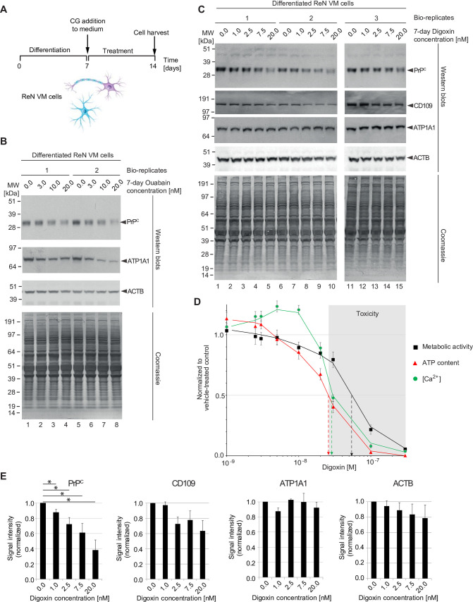
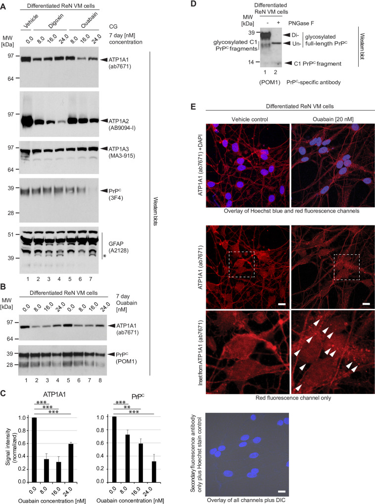
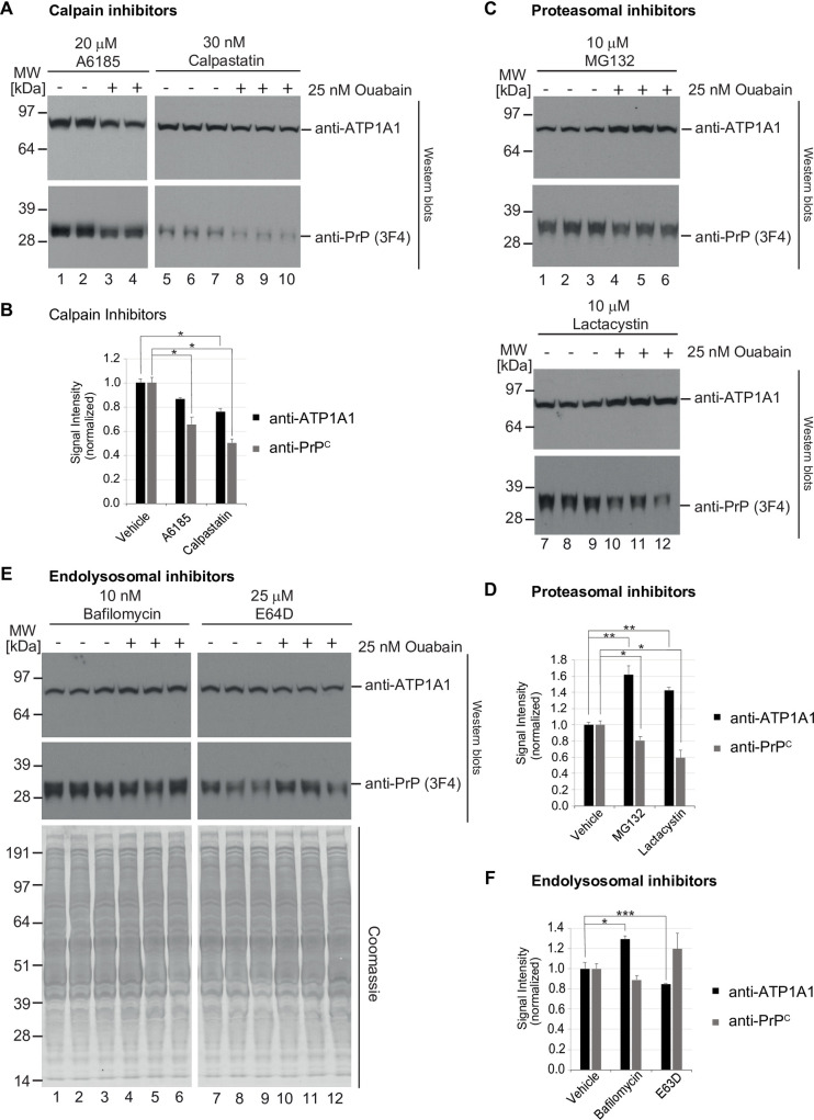
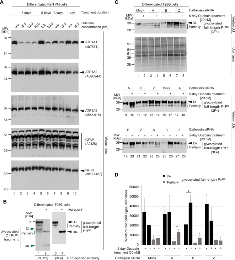
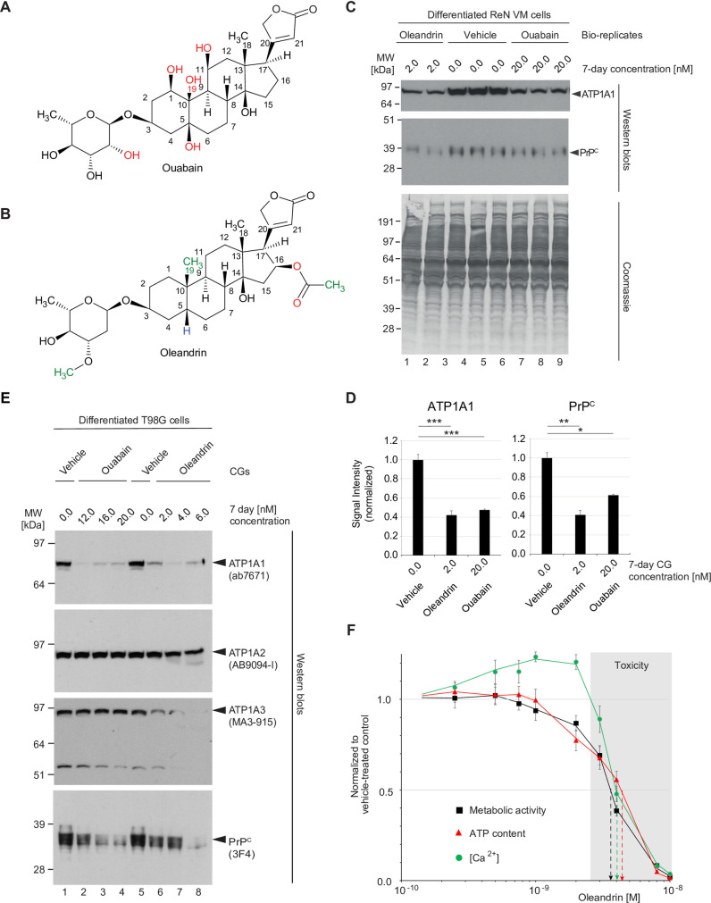
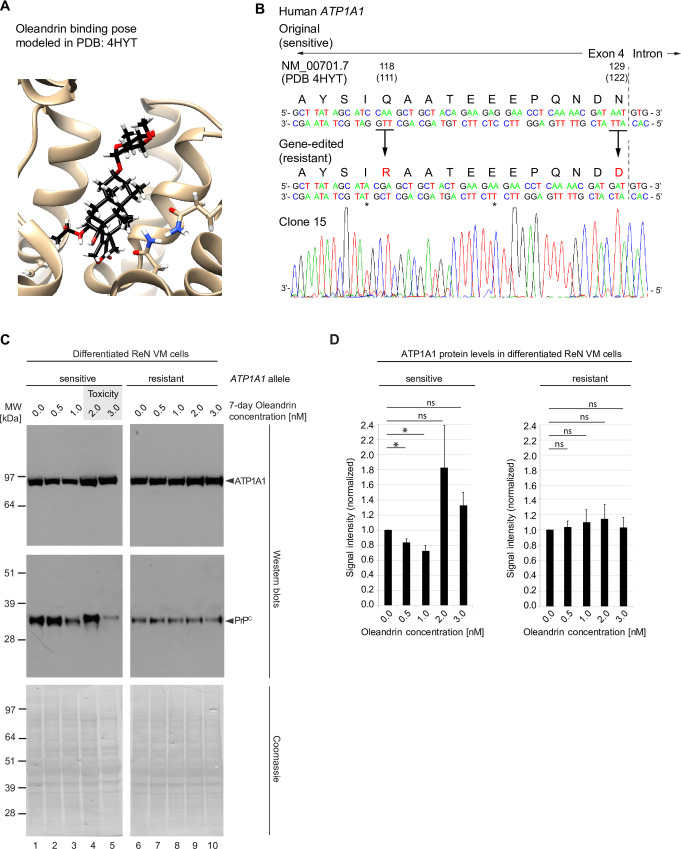
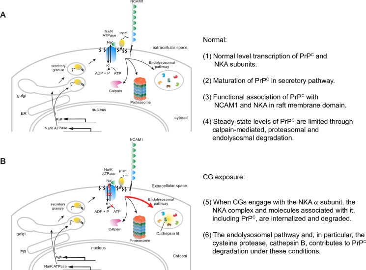
It is widely anticipated that a reduction of brain levels of the cellular prion protein (PrPC) can prolong survival in a group of neurodegenerative diseases known as prion diseases. To date, efforts to decrease steady-state PrPC levels by targeting this protein directly with small molecule drug-like compounds have largely been unsuccessful. Recently, we reported Na,K-ATPases to reside in immediate proximity to PrPC in the brain, unlocking an opportunity for an indirect PrPC targeting approach that capitalizes on the availability of potent cardiac glycosides (CGs). Here, we report that exposure of human co-cultures of neurons and astrocytes to non-toxic nanomolar levels of CGs causes profound reductions in PrPC levels. The mechanism of action underpinning this outcome relies primarily on a subset of CGs engaging the ATP1A1 isoform, one of three α subunits of Na,K-ATPases expressed in brain cells. Upon CG docking to ATP1A1, the ligand receptor complex, and PrPC along with it, is internalized by the cell. Subsequently, PrPC is channeled to the lysosomal compartment where it is digested in a manner that can be rescued by silencing the cysteine protease cathepsin B. These data signify that the repurposing of CGs may be beneficial for the treatment of prion disorders.


Conflict of interest statement

GS, MM and DW declare that they are co-inventors on a provisional patent (United States Provisional Application No. 63/159,289) named ‘Compounds and methods to treat prion and related diseases’ describing the use of cardiac glycosides for the purpose of reducing steady-state levels of PrPC. No other competing interests exist, and this provisional patent does not alter the adherence of these authors to all PLOS ONE policies on sharing data and materials.

Figures

Fig 1
Fig 1. Prolonged non-toxic CG exposure of human ReN VM cells causes concentration-dependent reduction in NKA α subunit and PrPC levels.
(A) Workflow of in vitro ReN VM cell-based CG exposure analyses. (B) Exposure of differentiated ReN VM cells to ouabain at low nanomolar concentrations (up to 20 nM) caused a concentration-dependent reduction in PrPC and ATP1A1 levels that was already apparent at 3 nM ouabain levels. Levels of actin or other proteins in the cell visualized by Coomassie staining were not similarly affected by ouabain. (C) Western blot documenting that extended exposure of differentiated neurons to low levels of digoxin (up to 20 nM) caused a dose-dependent reduction in PrPC levels—and to a lesser extent steady-state protein levels of CD109 and actin—but did not affect the levels of ATP1A1 or many other cellular proteins, as evidenced by Coomassie staining. (D) 7-day ReN VM cell treatments with digoxin are toxic at concentrations exceeding 25 nM. Effects of a range of digoxin concentrations (0–500 nM) on cell viability (black squares), cellular ATP content (red triangles) and intracellular Ca2+ concentration ([Ca2+]i, green circles). Toxic concentrations of digoxin are shaded in grey. (E) Graphs depicting quantitation of steady-state levels of PrPC and CD109 in the presence of non-toxic low nanomolar levels of digoxin.
Fig 2
Fig 2. Digoxin and ouabain affect steady-state levels of NKA α subunits differently, and CG-dependent changes to PrPC levels are more closely associated with ATP1A1 than ATP1A2 protein levels.
(A) Digoxin exerts a lesser effect on steady-state ATP1A1 than ATP1A2 levels. The opposite is observed for ouabain exposed cells. Cellular extracts of 7-day differentiated, then 7-day CG-treated ReN VM cells were adjusted in their protein concentration and analyzed by western blotting with probes directed against NKA α subunits, PrPC and GFAP. Note that 24 nM levels of digoxin (Lane 4) diminished ATP1A2 protein levels but had relatively little effect on ATP1A1 protein levels. In contrast, 16 nM ouabain dramatically depletes ATP1A1 protein levels, with little changes to ATP1A2 protein levels (Lane 6). Levels of 3F4-reactive PrPC signals were more profoundly reduced in cells exposed to ouabain than digoxin (compare Lanes 4 and 7). GFAP levels were mostly unchanged. However, consistent with results we had reported before [17], a GFAP-antibody reactive band (see asterisk), migrating with an apparent molecular weight of ~39 kDa, became more pronounced in CG exposed cells. (B) Whereas steady-state levels of ATP1A1 recover at borderline toxic ouabain concentration of 24 nM, steady-state levels of PrPC remain inversely correlated with ouabain levels within the window of ouabain concentrations tested in this experiment. Note that this western blot was probed with the PrPC-reactive antibody POM1, which detects both full-length and endoproteolytically digested C1 fragments of PrPC. (C) Quantitation of ATP1A1 and PrPC western blot signals following 7-day ouabain treatment. (D) Following 7 days of differentiation, ReN VM cell cultures primarily express diglycosylated full-length PrPC. Cellular extracts of ReN VM cells were analyzed without or with prior PNGase F digestion and western blot detection of POM1-reactive signals. (E) Ouabain administration elicits internalization of ATP1A1 in 7-day differentiated ReN VM cells. Under basal conditions, ATP1A1 localizes primarily to the plasma membrane. Following treatment with 20 nM ouabain, ATP1A1 is increasingly internalized by cells into punctate structures (white arrowheads). Scale bar = 10 μm.
Fig 3
Fig 3. CG-dependent reduction of PrPC levels relies predominantly on endo/lysosomal degradation pathway.
(A,B) Inhibition of calpain proteases with A6185 or calpastatin did not rescue the ouabain-dependent reduction in steady-state ATP1A1 or PrPC levels in ReN VM cells. (C,D) Proteasomal inhibition with MG132 or lactacystin caused an increase in steady-state ATP1A1 levels yet did not rescue the reduction in PrPC levels that is observed in ouabain-treated cells. (E,F) In contrast, inhibition of endo/lysosomal degradation pathways with bafilomycin A1 or E64D blocked the ouabain-dependent PrPC reduction. Quantitative graphs in this figure depict the means of signal intensities and their standard deviations. Asterisks reflect significance thresholds with one asterisk shown if t-tests returned p<0.05 and two asterisks indicating p<0.005.
Fig 4
Fig 4. Cathepsin B is the main cathepsin involved in the ouabain-induced reduction in PrPC levels.
(A) Treatment with 25 nM ouabain over 3 to 7 days is sufficient to induce a profound reduction in ATP1A1 protein levels in T98G cells. (B) PrPC in T98G cell extracts consists primarily of diglycosylated full-length PrPC, with partially N-glycosylated and endoproteolytically cleaved PrPC representing a minor sub pool. (C) Western blot and Coomassie SDS-PAGE analyses of cell extracts from T98G cells silenced for individual cathepsins (as indicated) and treated with 25 nM ouabain or vehicle in the cell culture medium for a duration of four days. Probing of the western blot with the anti-human PrP antibody 3F4 led to three bands per lane, with the slowest migrating band representing diglycosylated full-length PrPC and faster migrating bands representing partially N-glycosylated full-length PrPC. As expected, mock silencing did not prevent the ouabain-induced reduction in full-length PrPC levels (compare Lanes 1 and 2). The same was observed after silencing of cells with a cathepsin Z-directed siRNA pool (compare Lanes 7 and 8). Although silencing of cathepsin A increased PrPC levels of all sizes, it had only a minor effect on stabilizing levels of full-length PrPC (compare Lanes 3 and 4). Finally, silencing of cathepsin B rescued the ouabain-dependent diminution of full-length PrPC levels (compare Lanes 5 and 6). The amounts of total proteins analyzed was equal for all samples following adjustment by bicinchoninic acid (BCA) assay analysis. (D) Graph depicting results of densitometry analyses of western blot signals shown in Panel B. The analyses were undertaken separately for the slowest migrating 3F4-reactive bands, which were interpreted to represent N-glycosylated full-length PrPC (black color), and for slower migrating 3F4-reactive PrPC bands (grey color). Bars indicate the means of signal intensities and their standard deviations. Were indicated with asterisks, sample cohort comparisons met the significance threshold of p<0.05.
Fig 5
Fig 5. Oleandrin is a potent yet toxic CG for the NKA-mediated reduction of PrPC levels.
Comparison of (A) ouabain, a cardiotonic steroid sourced from the Strophanthus gratus plant native to eastern Africa, and (B) oleandrin, found in Nerium oleander, an ornamental shrub of uncertain origin. A lack of hydroxyl groups in the steroid core and the presence of additional methyl groups that are absent in ouabain account for the more hydrophobic characteristics of oleandrin. (C) Tenfold lower levels of oleandrin than ouabain cause a similar reduction in steady-state PrPC levels during 7-day treatment of differentiated ReN VM cells. (D) Graphs comparing the means of western blot signal intensities of ATP1A1 and PrPC levels and their standard deviations in 2 nM oleandrin- versus 20 nM ouabain-treated ReN VM cells. The number of asterisks shown reflect significance thresholds as follows: one: p<0.05; two: p<0.005; three: p<0.0005. (E) The potency of oleandrin translates to T98G cells and may reflect an ability of this CG to reduce not only levels of ATP1A1 but also ATP1A3. (F) Influence on cell viability (black squares), cellular ATP content (red triangles) and intracellular Ca2+ concentration ([Ca2+]i, green circles) of 7-day exposure of ReN VM cells to a range of oleandrin concentrations (10 pM to 10 nM). Toxic concentrations of oleandrin (exceeding 3 nM) are shaded in grey.
Fig 6
Fig 6. CG-dependent reduction of PrPC levels depends on CG-ATP1A1 ligand-receptor engagement.
(A) Predicted oleandrin binding pose modeled in ATP1A1 structure reported in PDB entry 4HYT. (B) Genetic sequencing results confirm successful amino acid substitutions Q118R and N129D, two changes required to render the human ATP1A1 resistant to CGs, in a ReN VM clone. (C) Comparison of ATP1A1 levels (top panel) and PrPC levels (bottom panel) in response to oleandrin treatment in ReN VM cells that express either the CG sensitive wild-type or the CRISPR-Cas9-engineered CG resistant ATP1A1 alleles. In wild-type cells levels of ATP1A1 and PrPC decreased as oleandrin concentration increased from 0 to 1 nM. When oleandrin was administered at a concentration of 2 nM, levels of ATP1A1 and PrPC rebounded, and toxicity was observed (grey shaded box). In contrast, steady-state protein levels of ATP1A1 and PrPC remained stable at all oleandrin concentrations tested in ReN VM cells expressing the resistant form of ATP1A1, and no toxicity was observed. Coomassie staining documents that protein loading was adjusted across lanes. (D) Graphs depicting quantitation of steady-state levels of ATP1A1 in the presence of oleandrin concentrations up to 3 nM in CG-sensitive wild-type and CG-resistant ReN VM cells.
Fig 7
Fig 7. Model of CG-dependent reduction of PrPC.
(A) Normal cell homeostasis. (B) Enhanced lysosomal degradation of PrPC upon exposure to non-toxic levels of CGs.

References

    1. Sailer A, Bueler H, Fischer M, Aguzzi A, Weissmann C. No propagation of prions in mice devoid of PrP. Cell. 1994;77(7):967–8. doi: 10.1016/0092-8674(94)90436-7 - DOI - PubMed
    1. Bueler H, Raeber A, Sailer A, Fischer M, Aguzzi A, Weissmann C. High prion and PrPSc levels but delayed onset of disease in scrapie-inoculated mice heterozygous for a disrupted PrP gene. Molecular Medicine (Cambridge, Mass). 1994;1(1):19–30. - PMC - PubMed
    1. Minikel EV, Vallabh SM, Lek M, Estrada K, Samocha KE, Sathirapongsasuti JF, et al. Quantifying prion disease penetrance using large population control cohorts. Sci Transl Med. 2016;8(322):322ra9. doi: 10.1126/scitranslmed.aad5169 - DOI - PMC - PubMed
    1. Richt JA, Kasinathan P, Hamir AN, Castilla J, Sathiyaseelan T, Vargas F, et al. Production of cattle lacking prion protein. Nat Biotechnol. 2007;25(1):132–8. doi: 10.1038/nbt1271 - DOI - PMC - PubMed
    1. Benestad SL, Austbø L, Tranulis MA, Espenes A, Olsaker I. Healthy goats naturally devoid of prion protein. Veterinary Research. 2012;43(1):87. doi: 10.1186/1297-9716-43-87 - DOI - PMC - PubMed

Publication types