. 2022 Jul;8(26):eabn3868.
doi: 10.1126/sciadv.abn3868. Epub 2022 Jul 1.

Molecular mechanism of S-adenosylmethionine sensing by SAMTOR in mTORC1 signaling

Affiliations

Molecular mechanism of S-adenosylmethionine sensing by SAMTOR in mTORC1 signaling

Xin Tang et al. Sci Adv. 2022 Jul.

Abstract

The mechanistic target of rapamycin-mLST8-raptor complex (mTORC1) functions as a central regulator of cell growth and metabolism in response to changes in nutrient signals such as amino acids. SAMTOR is an S-adenosylmethionine (SAM) sensor, which regulates the mTORC1 activity through its interaction with the GTPase-activating protein activity toward Rags-1 (GATOR1)-KPTN, ITFG2, C12orf66 and SZT2-containing regulator (KICSTOR) complex. In this work, we report the crystal structures of Drosophila melanogaster SAMTOR in apo form and in complex with SAM. SAMTOR comprises an N-terminal helical domain and a C-terminal SAM-dependent methyltransferase (MTase) domain. The MTase domain contains the SAM-binding site and the potential GATOR1-KICSTOR-binding site. The helical domain functions as a molecular switch, which undergoes conformational change upon SAM binding and thereby modulates the interaction of SAMTOR with GATOR1-KICSTOR. The functional roles of the key residues and the helical domain are validated by functional assays. Our structural and functional data together reveal the molecular mechanism of the SAM sensing of SAMTOR and its functional role in mTORC1 signaling.


Figures
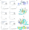
Fig. 1.
Fig. 1.. SAM binds exclusively to the MTase domain of dSAMTOR.
(A) ITC measurements for the ligand-binding affinity of the WT and the Δ1-64 mutant dSAMTOR. ND, not detected. The experiments were performed three times for those with measurable binding and two times for those with undetectable binding, and the repeated experiments yielded similar results; for each case, only the result of one representative experiment is shown. (B) Composite simulated annealing FoFc omit map (contoured at 1.5 σ) for the bound SAM and several surrounding residues. (C) Overall structure of the SAM-bound MTase domain of dSAMTOR in two different views. The α helices, major β sheet, and minor β sheet are colored in cyan, yellow, and blue, respectively. The bound SAM is shown with a stick model in green. The topology of the secondary structure elements of the MTase domain is shown below. The linker between β8 and α4 (residues 232 to 238) is disordered and thus is shown with a dashed line.
Fig. 2.
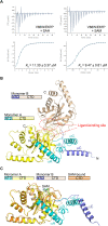
Fig. 2.. The N-terminal domain of the dSAMTOR adopts multiple conformations.
(A) ITC measurements for the ligand-binding affinity of the V66W/E67P mutant dSAMTOR. The experiments were performed three times, which yielded similar results, and for each case, only the result of one representative experiment is shown. (B) Overall structure of the homodimer of the V66W/E67P mutant dSAMTOR in apo form. The two monomers are designated as monomer A and monomer B. The N-terminal helical domain (NTD) and the C-terminal MTase domain (CTD) of monomer A are colored in cyan and yellow, and those of monomer B are in blue and wheat, respectively. The ligand-binding site is indicated by dashed ovals. (C) Superposition of monomer A and monomer B in the V66W/E67P mutant dSAMTOR structure onto the SAM-bound MTase domain of dSAMTOR. The color coding of each structure is shown above.
Fig. 3.
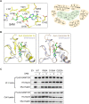
Fig. 3.. The key residues at the SAM-binding site play a critical role in SAM binding and the interaction with GATOR1-KICSTOR.
(A) Interactions between SAM and the surrounding residues are shown in a ball-and-stick model (left) and a schematic diagram (right) in the structure of the SAM-bound MTase domain of dSAMTOR. (B) Structural comparison of the ligand-binding site of monomer A (left) and monomer B (right) in the V66W/E67P mutant dSAMTOR and the SAM-bound MTase domain of dSAMTOR. The color coding of each structure is shown above. (C) Co-IP assay to examine the interactions of Flag-hSAMTOR (WT and mutants) with HA-Nprl3 and Myc-Kaptin in the presence and absence of SAM. EV, empty vector. The assay was performed three times, which yielded similar results, and only the result of one representative experiment is shown.
Fig. 4.
Fig. 4.. The N-terminal domain functions as a molecular switch in mTORC1 signaling.
(A) Co-IP assay to examine the interactions of the full-length (FL), the N-terminal helical domain (NTD), and the C-terminal MTase domain (CTD) of hSAMTOR with HA-Nprl3 and Myc-Kaptin in the presence and absence of SAM in the HEK 293T cells. The assays in (A), (C), and (D) were performed three times, which yielded similar results, and for each case, only the result of one representative experiment is shown. (B) Structural comparison of the SAM-bound MTase domain of dSAMTOR (left) and the apo V66W/E67P mutant dSAMTOR (monomer A) (right) with the predicted dSAMTOR structure from the AlphaFold Protein Structure Database. The color coding of each structure is shown above. (C) Co-IP assay to examine the interactions of the WT and F175A mutant of the full-length (FL) and the C-terminal MTase domain (CTD) of hSAMTOR with HA-Nprl3 and Myc-Kaptin in the HEK 293T cells. (D) Immunoblotting assay to examine the mTORC1 kinase activity in the HEK 293T cells transiently overexpressing the WT and F175A mutant hSAMTOR at three different levels. The cell lysates were analyzed by immunoblotting of the phosphorylation level (p-T389) of S6K1 and the expression levels of the indicated proteins. (E) A schematic diagram illustrating the proposed molecular mechanism for SAMTOR to sense and bind SAM and then function as a molecular switch through conformational change to regulate its interaction with GATOR1-KICSTOR in mTORC1 signaling.

Similar articles

Cited by

References

    1. Cornu M., Albert V., Hall M. N., mTOR in aging, metabolism, and cancer. Curr. Opin. Genet. Dev. 23, 53–62 (2013). - PubMed
    1. Liu G. Y., Sabatini D. M., mTOR at the nexus of nutrition, growth, ageing and disease. Nat. Rev. Mol. Cell Biol. 21, 183–203 (2020). - PMC - PubMed
    1. Yang H., Rudge D. G., Koos J. D., Vaidialingam B., Yang H. J., Pavletich N. P., mTOR kinase structure, mechanism and regulation. Nature 497, 217–223 (2013). - PMC - PubMed
    1. Yang H., Jiang X., Li B., Yang H. J., Miller M., Yang A., Dhar A., Pavletich N. P., Mechanisms of mTORC1 activation by RHEB and inhibition by PRAS40. Nature 552, 368–373 (2017). - PMC - PubMed
    1. Wullschleger S., Loewith R., Hall M. N., TOR signaling in growth and metabolism. Cell 124, 471–484 (2006). - PubMed

MeSH terms

Substances