. 2022 Jul 13;144(27):12431-12442.
doi: 10.1021/jacs.2c04287. Epub 2022 Jul 1.

Solid-State NMR Reveals Asymmetric ATP Hydrolysis in the Multidrug ABC Transporter BmrA

Affiliations

Solid-State NMR Reveals Asymmetric ATP Hydrolysis in the Multidrug ABC Transporter BmrA

Denis Lacabanne et al. J Am Chem Soc. .

Abstract

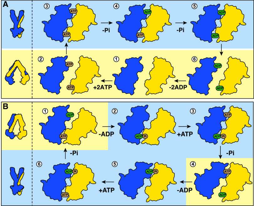
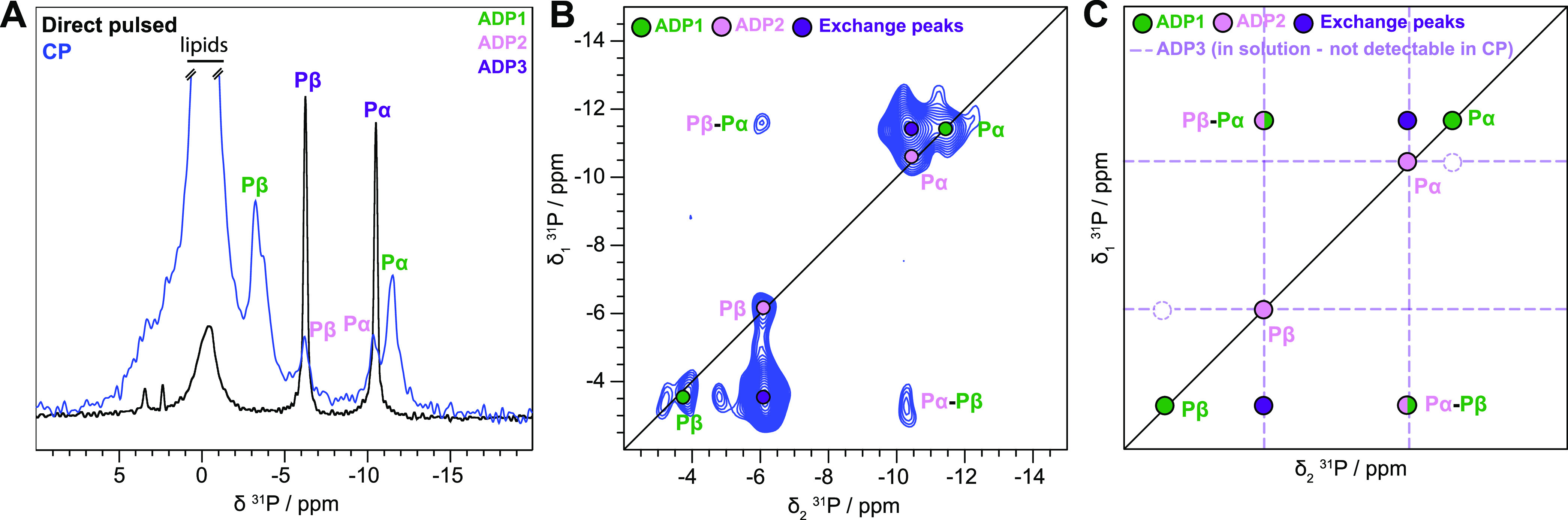
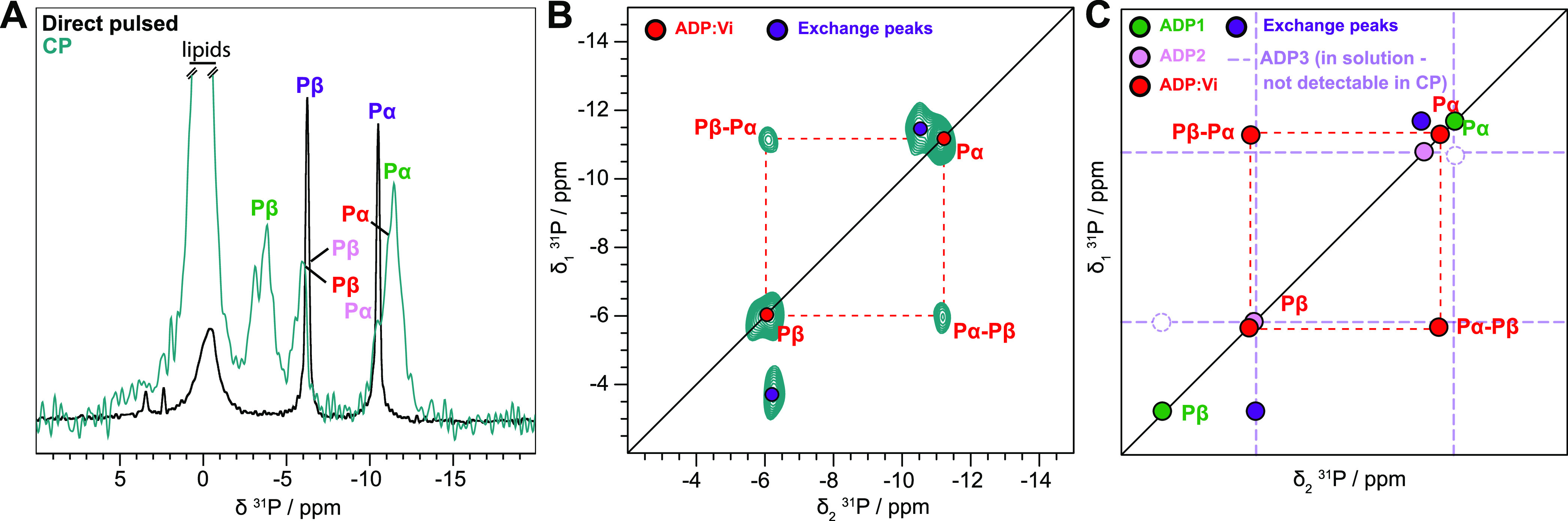
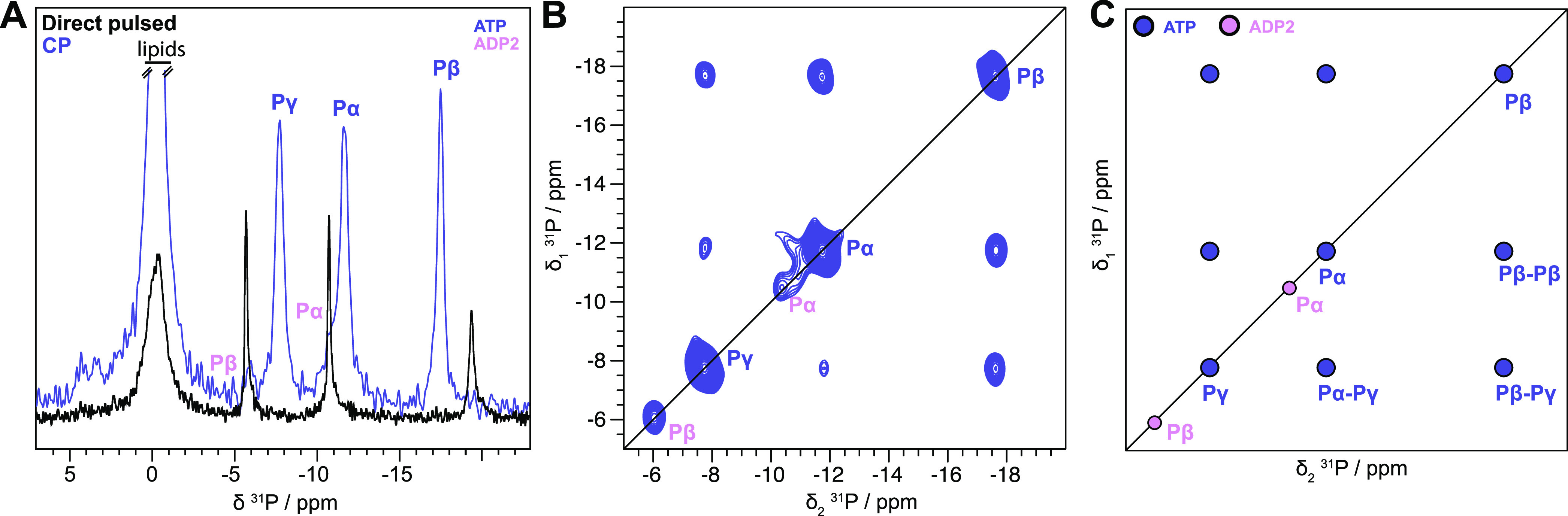
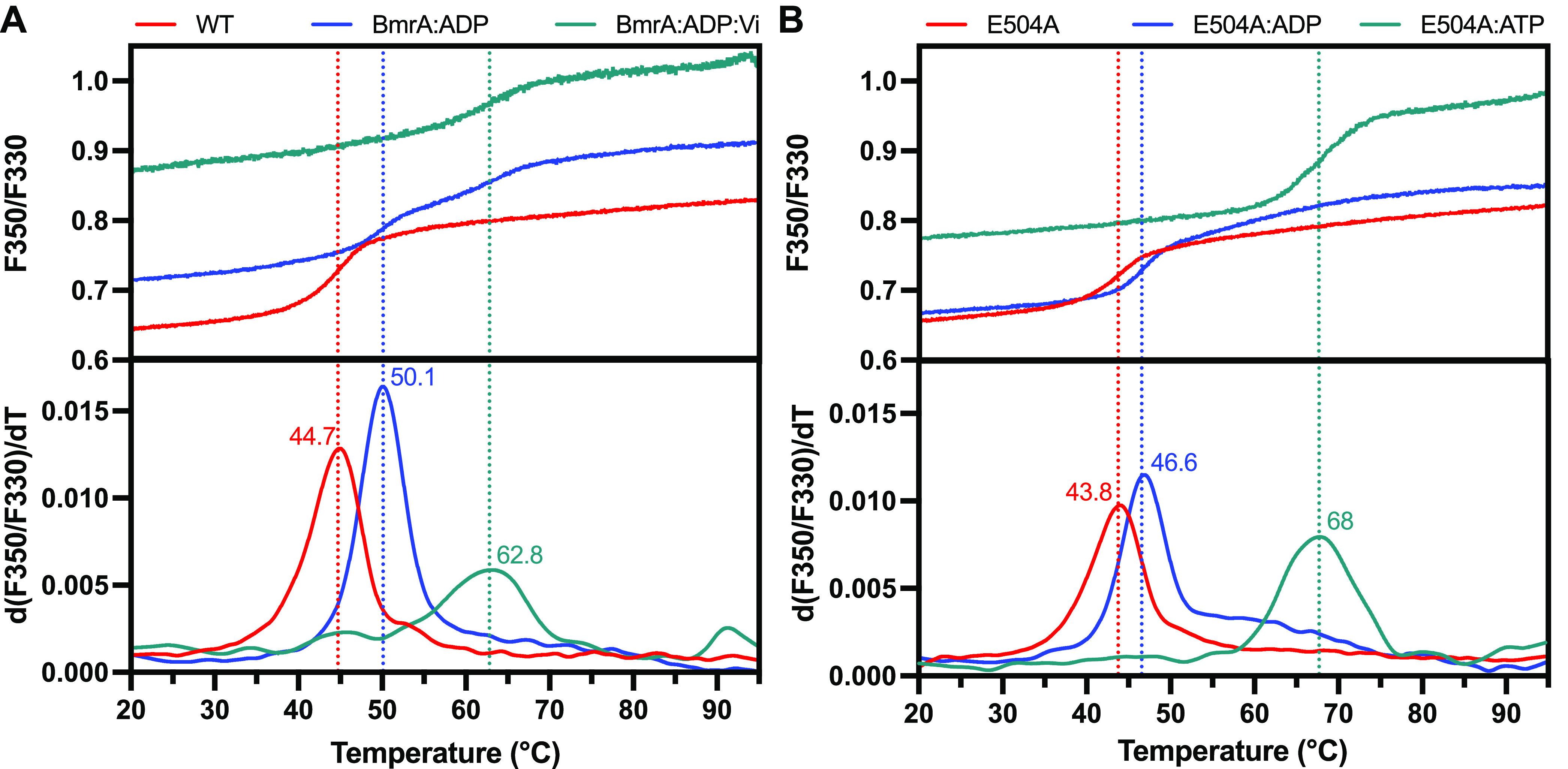
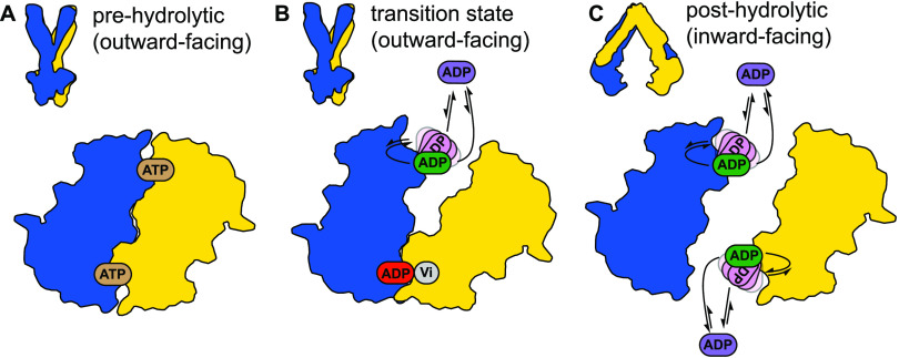
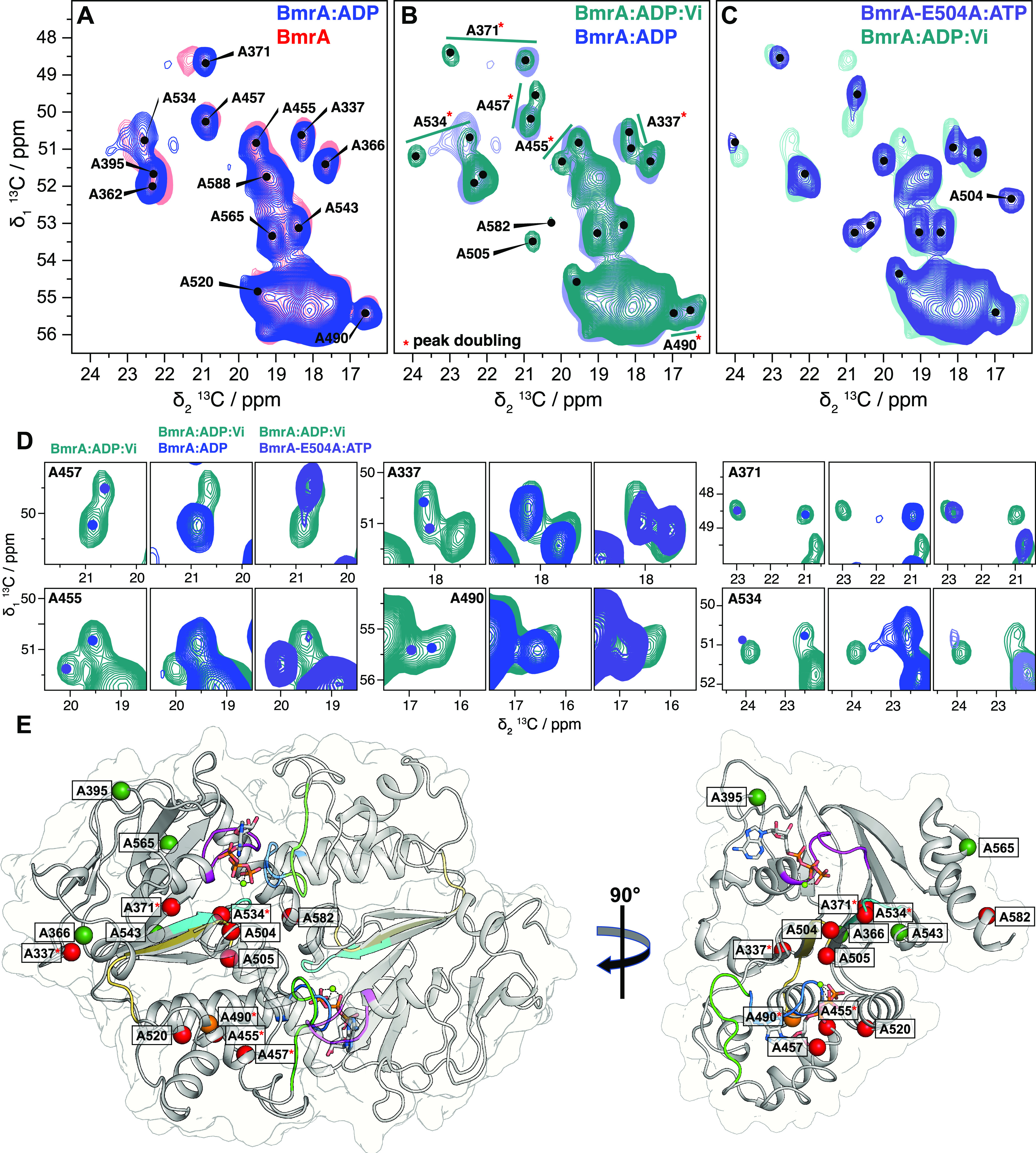
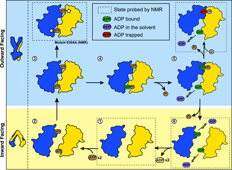
The detailed mechanism of ATP hydrolysis in ATP-binding cassette (ABC) transporters is still not fully understood. Here, we employed 31P solid-state NMR to probe the conformational changes and dynamics during the catalytic cycle by locking the multidrug ABC transporter BmrA in prehydrolytic, transition, and posthydrolytic states, using a combination of mutants and ATP analogues. The 31P spectra reveal that ATP binds strongly in the prehydrolytic state to both ATP-binding sites as inferred from the analysis of the nonhydrolytic E504A mutant. In the transition state of wild-type BmrA, the symmetry of the dimer is broken and only a single site is tightly bound to ADP:Mg2+:vanadate, while the second site is more 'open' allowing exchange with the nucleotides in the solvent. In the posthydrolytic state, weak binding, as characterized by chemical exchange with free ADP and by asymmetric 31P-31P two-dimensional (2D) correlation spectra, is observed for both sites. Revisiting the 13C spectra in light of these findings confirms the conformational nonequivalence of the two nucleotide-binding sites in the transition state. Our results show that following ATP binding, the symmetry of the ATP-binding sites of BmrA is lost in the ATP-hydrolysis step, but is then recovered in the posthydrolytic ADP-bound state.


Conflict of interest statement

The authors declare no competing financial interest.

Figures

Figure 1
Figure 1
Schematic representation of the (A) ATP switch model or processive clamp model (sequential ATP hydrolysis) and the (B) constant contact model or alternating site model., In the blue background, the conformation of the transporter is presumably in an outward-facing conformation, and in the yellow background, it is in a putative inward-facing conformation.
Figure 2
Figure 2
Different ADP species detected in BmrA:ADP. (A) 31P cross-polarization spectrum (blue) and direct-pulsed spectrum (black) of BmrA:ADP, showing bound and free ADP species, respectively. The one-dimensional (1D) CP spectrum of BmrA:ADP was taken from Lacabanne et al.-Copyright 2020 Lacabanne http://creativecommons.org/licenses/by/4.0/. (B) 31P–31P 150 ms DARR correlation spectrum of BmrA:ADP. (C) Schematic representation of the experimental spectrum shown in panel (B).
Figure 3
Figure 3
ADP:Vi is tightly bound to BmrA. (A) 31P cross-polarization spectrum (green) and direct-pulsed spectrum (black) of BmrA:ADP:Vi. The 1D CP spectrum of BmrA:ADP:Vi was taken from Lacabanne et al.-Copyright 2020 Lacabanne http://creativecommons.org/licenses/by/4.0/. (B) 31P–31P 150 ms DARR correlation spectrum of BmrA:ADP:Vi. (C) Schematic representation of the experimental spectrum shown in panel (B).
Figure 4
Figure 4
Hydrolytic-deficient E504A mutant binds ATP in the two NBDs. (A) 31P cross-polarization spectrum (pale purple) and direct-pulsed spectrum (black) of BmrA-E504A:ATP. The 1D CP spectrum of BmrA-E504A:ATP was taken from Lacabanne et al.—Copyright 2020 Lacabanne http://creativecommons.org/licenses/by/4.0/. (B) 31P–31P 150 ms DARR correlation spectrum of BmrA-E504A:ATP revealing no chemical exchange of bound ATP (pale purple). A small fraction of hydrolyzed ADP can be observed (ADP2, pink). (C) Schematic representation of the experimental spectrum shown in panel (B).
Figure 5
Figure 5
Thermostability of BmrA probed by nanoDSF measurements. Unfolding curves (top panel) and derivatives of the unfolding curves with the apparent melting temperatures (bottom panel) of (A) BmrA and (B) BmrA-E504A in the apo state (red lines) or in the presence of ADP (blue lines), ADP:Vi (green line, panel A), or ATP (green line, panel B).
Figure 6
Figure 6
Nucleotide-binding models as deduced from the NMR data and assuming that ADP is in three different states. (A) Symmetric ATP binding as revealed by the E504A mutant in the presence of ATP, (B) asymmetric ADP binding as observed for the ADP:Vi-bound state (transition state) of the wild-type transporter, and (C) symmetric ADP binding observed for BmrA:ADP (posthydrolytic state).
Figure 7
Figure 7
BmrA conformation probed by 13C-detected 2D DARR experiments. Alanine region spectral fingerprint of (A) ADP-bound state of the WT protein overlaid with the apo state, (B) transition state of the WT:ADP:Vi overlaid with the ADP-bound state, and (C) prehydrolytic state using the E504A mutant overlaid with the WT:ADP:Vi. (D) Zooms of peak splitting from WT:ADP:Vi compared with WT:ADP and E504A:ATP. Examples of peak splitting from other regions of the spectrum are presented in Figure S4. BmrA, BmrA:ADP:Vi, and BmrA-E504A:ATP spectra were adapted from data previously recorded (Lacabanne et al.—Copyright 2019 Lacabanne http://creativecommons.org/licenses/by/4.0/). The colored dots indicate peak maxima in the BmrA:ADP (light blue) and BmrA-E504A:ATP spectra (gray). Resonance assignments were transferred from solution-state NMR assignments obtained on the isolated NBD. (E) View of the NBDs of BmrA-E504A:ATP in the full-length structure (pdb 7OW8); the different motifs are highlighted in different colors: the X-loop (470-TEVGERG-476) in green, the Walker A motif (374-GPSGGKT-381) in magenta, the Walker B motif (496-ILMLDE-504) in dark yellow, the ABC signature (477-LSGGQ-483) in blue, and the H-loop (532-AHR-536) in cyan. Alanine residues resolved in the 2D NMR spectra are shown as colored spheres according to the chemical-shift perturbations determined between BmrA-apo and BmrA-E504A:ATP: red CSP > 0.6 ppm (or appearing peaks: A504, A505, A582), orange CSP > 0.2 ppm, and dark green CSP < 0.2 ppm. Resonances displaying peak doubling are indicated by a red star.
Figure 8
Figure 8
Scheme of ATP hydrolysis by the multidrug ABC transporter BmrA. The two BmrA monomers are shown in yellow and blue and the NBDs are shown in contours. Dashed squares indicated the states that were studied by NMR. The E504A mutation is indicated by a white star. For a description of the cycle, please see the text. One can see that the process adopted by BmrA results in a mixture of the two alternative models shown in Figure 1.

Similar articles

Cited by

References

    1. Dean M.; Rzhetsky A.; Allikmets R. The human ATP-binding cassette (ABC) transporter superfamily. Genome Res. 2001, 11, 1156–1166. 10.1101/gr.184901. - DOI - PubMed
    2. Schneider E.; Hunke S. ATP-binding-cassette (ABC) transport systems: functional and structural aspects of the ATP-hydrolyzing subunits/domains. FEMS Microbiol. Rev. 1998, 22, 1–20. 10.1111/j.1574-6976.1998.tb00358.x. - DOI - PubMed
    1. Kerr I. D. Structure and association of ATP-binding cassette transporter nucleotide-binding domains. Biochim. Biophys. Acta, Biomembr. 2002, 1561, 47–64. 10.1016/S0304-4157(01)00008-9. - DOI - PubMed
    2. Wilkens S. Structure and mechanism of ABC transporters. F1000Prime Rep. 2015, 7, 14.10.12703/p7-14. - DOI - PMC - PubMed
    3. Rees D. C.; Johnson E.; Lewinson O. ABC transporters: the power to change. Nat Rev Mol Cell Biol 2009, 10, 218–227. 10.1038/nrm2646. - DOI - PMC - PubMed
    1. Thomas C.; Tampé R. Structural and Mechanistic Principles of ABC Transporters. Annu. Rev. Biochem. 2020, 89, 605–636. 10.1146/annurev-biochem-011520-105201. - DOI - PubMed
    1. Choi Y. H.; Yu A. M. ABC transporters in multidrug resistance and pharmacokinetics, and strategies for drug development. Curr. Pharm. Des. 2014, 20, 793–807. 10.2174/138161282005140214165212. - DOI - PMC - PubMed
    1. Robey R. W.; Pluchino K. M.; Hall M. D.; Fojo A. T.; Bates S. E.; Gottesman M. M. Revisiting the role of ABC transporters in multidrug-resistant cancer. Nat. Rev. Cancer 2018, 18, 452–464. 10.1038/s41568-018-0005-8. - DOI - PMC - PubMed

Publication types

MeSH terms

Substances