

Regulatory T Cells Require CCR6 for Skin Migration and Local Suppression of Vitiligo
- PMID: 35777498
- PMCID: PMC10198248
- DOI: 10.1016/j.jid.2022.05.1090
Regulatory T Cells Require CCR6 for Skin Migration and Local Suppression of Vitiligo
Abstract
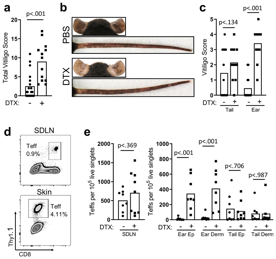
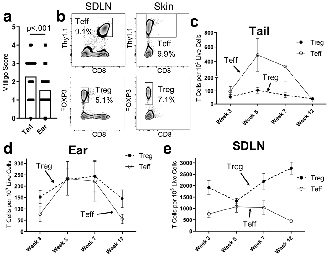
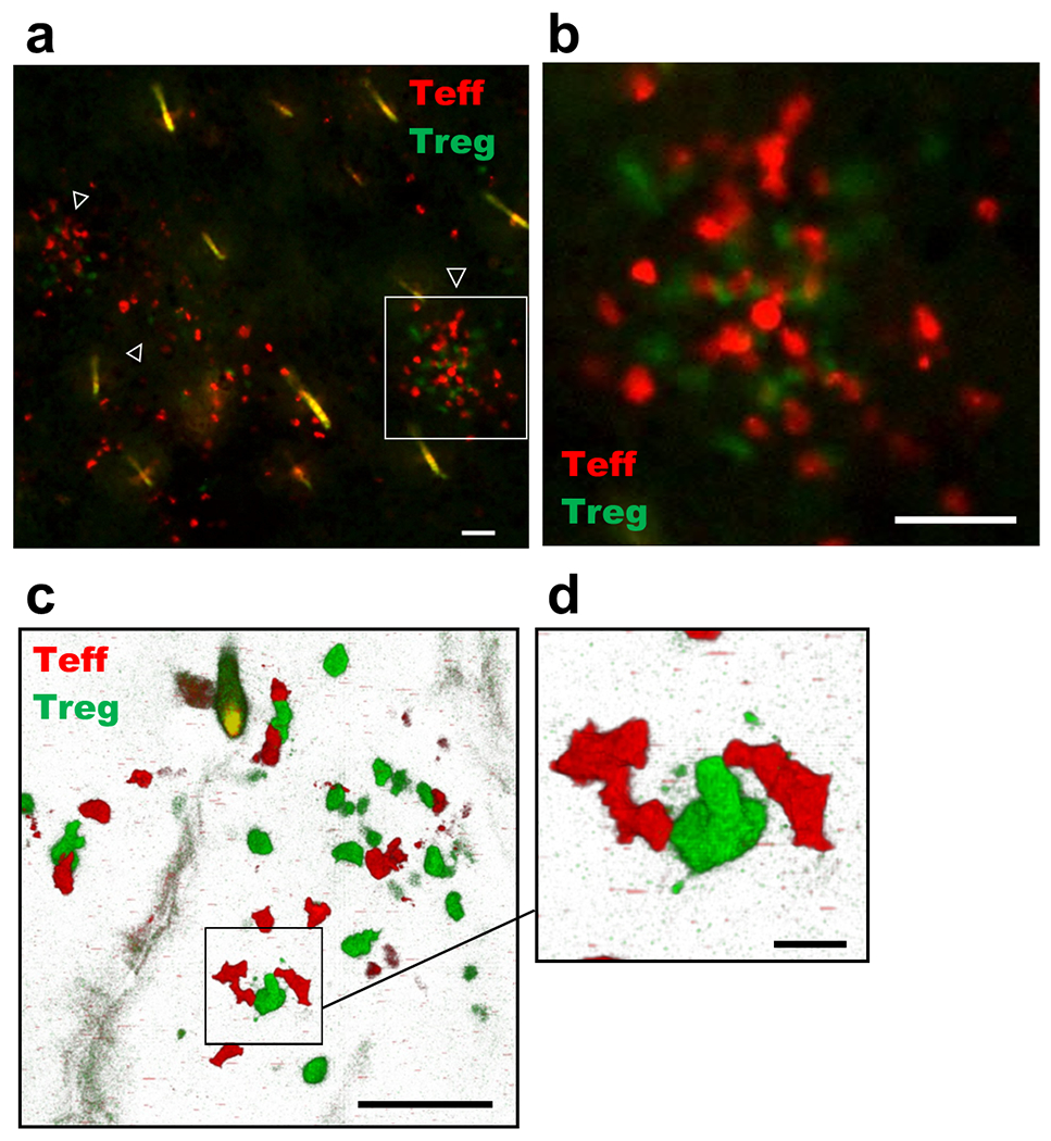
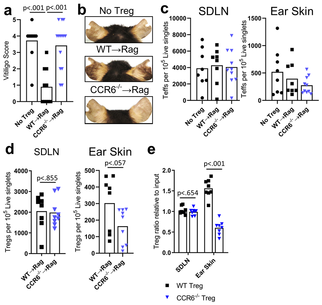
Vitiligo is an autoimmune skin disease caused by melanocyte-targeting autoreactive CD8+ T cells. Regulatory T cells (Tregs) have been implicated in restraining vitiligo severity in both mouse models and human patients; however, whether they must be present in the skin for their suppressive function is still unclear. We observed uneven distribution of Tregs within different anatomical locations of mouse skin, which correlated with reduced depigmentation after vitiligo induction. We specifically depleted Tregs in our mouse model of vitiligo and observed increased disease. Next, we found that Tregs contact CD8+ T effector cells in vitiligo lesional skin and that Treg recruitment to the skin inversely correlated with disease severity, suggesting a critical role for Treg suppression within the skin. When we investigated the signals facilitating Treg migration to the skin, we found that although CXCR3 was dispensable for Treg migration and function in vitiligo, Tregs lacking CCR6 exhibited a reduced capacity to migrate to the skin and suppress depigmentation, despite normal systemic numbers in the skin-draining lymph nodes. Our observations highlight a key role for cutaneous Tregs in disease suppression during vitiligo and identify CCR6 as a chemokine receptor that contributes to Treg migration to the skin.
Copyright © 2022 The Authors. Published by Elsevier Inc. All rights reserved.
Conflict of interest statement
CONFLICT OF INTEREST STATEMENT
JEH is an inventor on patent application #15/851,651, “Anti-human CXCR3 antibodies for the Treatment of Vitiligo” which covers targeting CXCR3 for the treatment of vitiligo. J.E.H. is a scientific founder of Villaris Therapeutics Inc., which develops therapeutic treatments for vitiligo.
Figures
References
-
- Abdallah M, Lotfi R, Othman W, and Galal R. 2014. ‘Assessment of tissue FoxP3+, CD4+ and CD8+ T-cells in active and stable nonsegmental vitiligo’, Int J Dermatol, 53: 940–6. - PubMed
-
- Antony PA, Piccirillo CA, Akpinarli A, Finkelstein SE, Speiss PJ, Surman DR, Palmer DC, Chan CC, Klebanoff CA, Overwijk WW, Rosenberg SA, and Restifo NP. 2005. ‘CD8+ T cell immunity against a tumor/self-antigen is augmented by CD4+ T helper cells and hindered by naturally occurring T regulatory cells’, J Immunol, 174: 2591–601. - PMC - PubMed
-
- Bassiouny DA, and Shaker O. 2011. ‘Role of interleukin-17 in the pathogenesis of vitiligo’, Clin Exp Dermatol, 36: 292–7. - PubMed
-
- Ben Ahmed M, Zaraa I, Rekik R, Elbeldi-Ferchiou A, Kourda N, Belhadj Hmida N, Abdeladhim M, Karoui O, Ben Osman A, Mokni M, and Louzir H. 2012. ‘Functional defects of peripheral regulatory T lymphocytes in patients with progressive vitiligo’, Pigment Cell Melanoma Res, 25: 99–109. - PubMed
Publication types
MeSH terms
Substances
Grants and funding
LinkOut - more resources
Full Text Sources
Medical
Research Materials
