

Quantification of lung ventilation defects on hyperpolarized MRI: The Multi-Ethnic Study of Atherosclerosis (MESA) COPD study
- PMID: 35777684
- PMCID: PMC9957614
- DOI: 10.1016/j.mri.2022.06.016
Quantification of lung ventilation defects on hyperpolarized MRI: The Multi-Ethnic Study of Atherosclerosis (MESA) COPD study
Abstract
Purpose: To develop an end-to-end deep learning (DL) framework to segment ventilation defects on pulmonary hyperpolarized MRI.
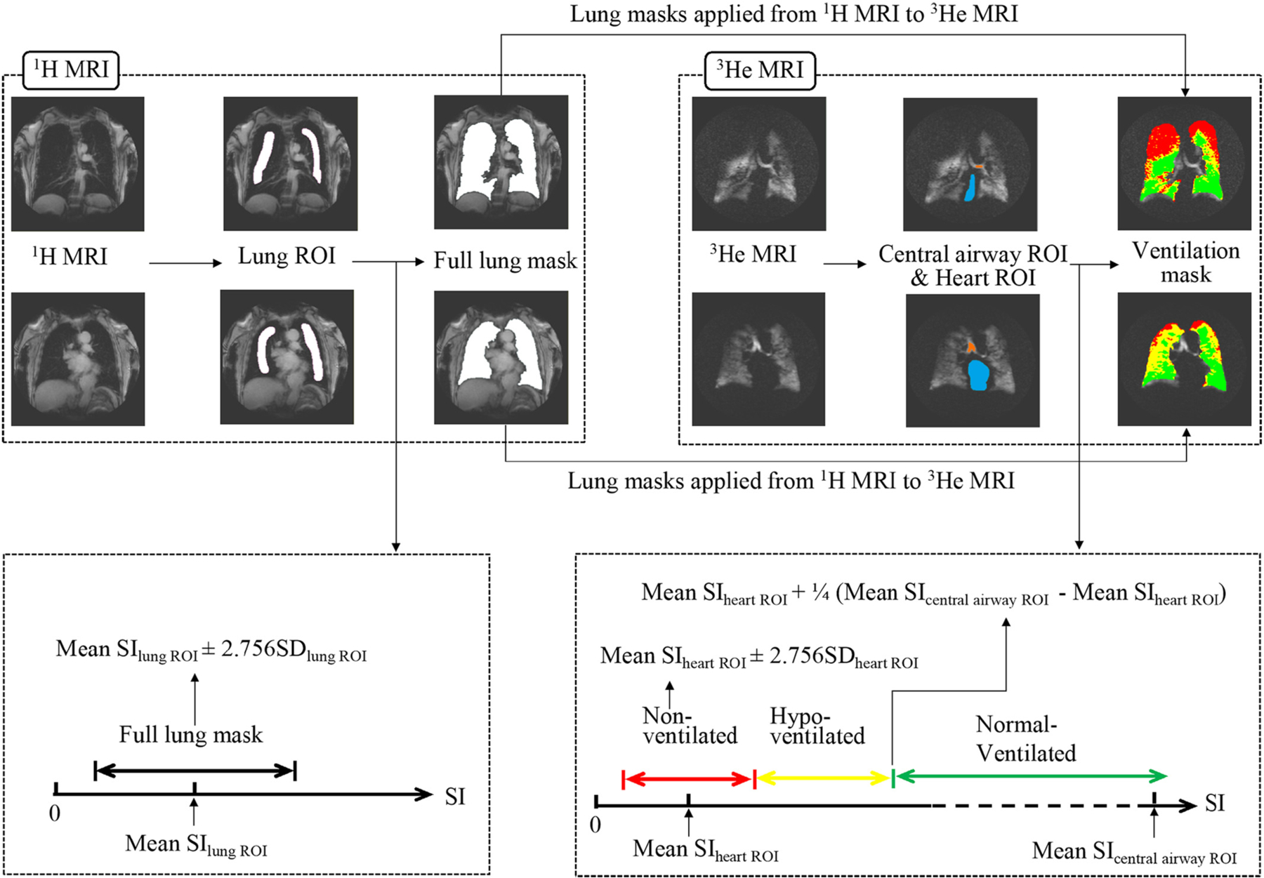
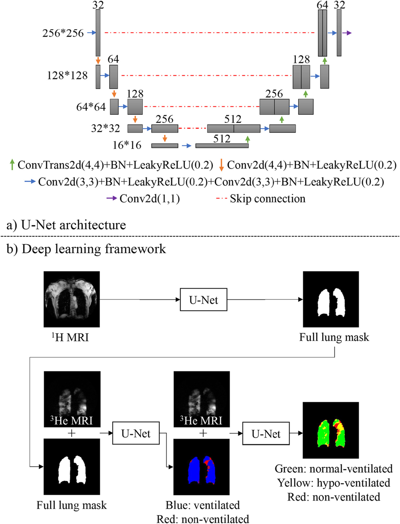
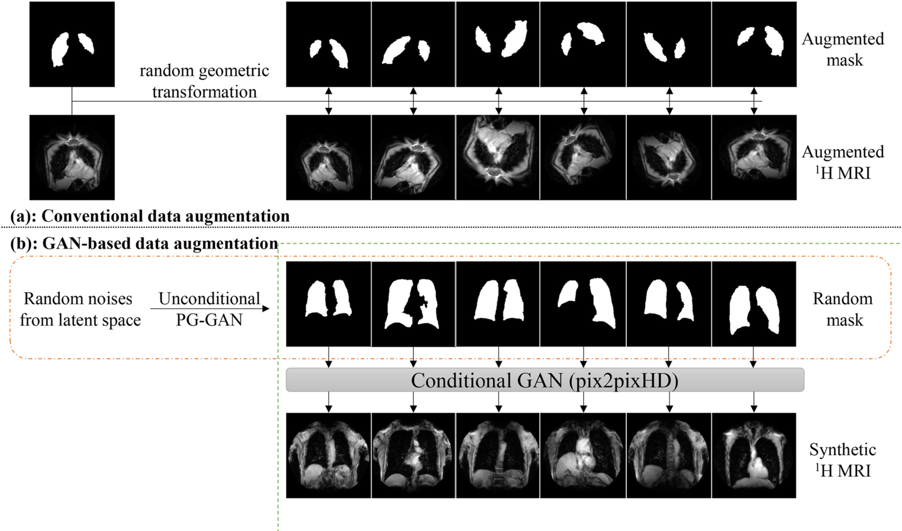
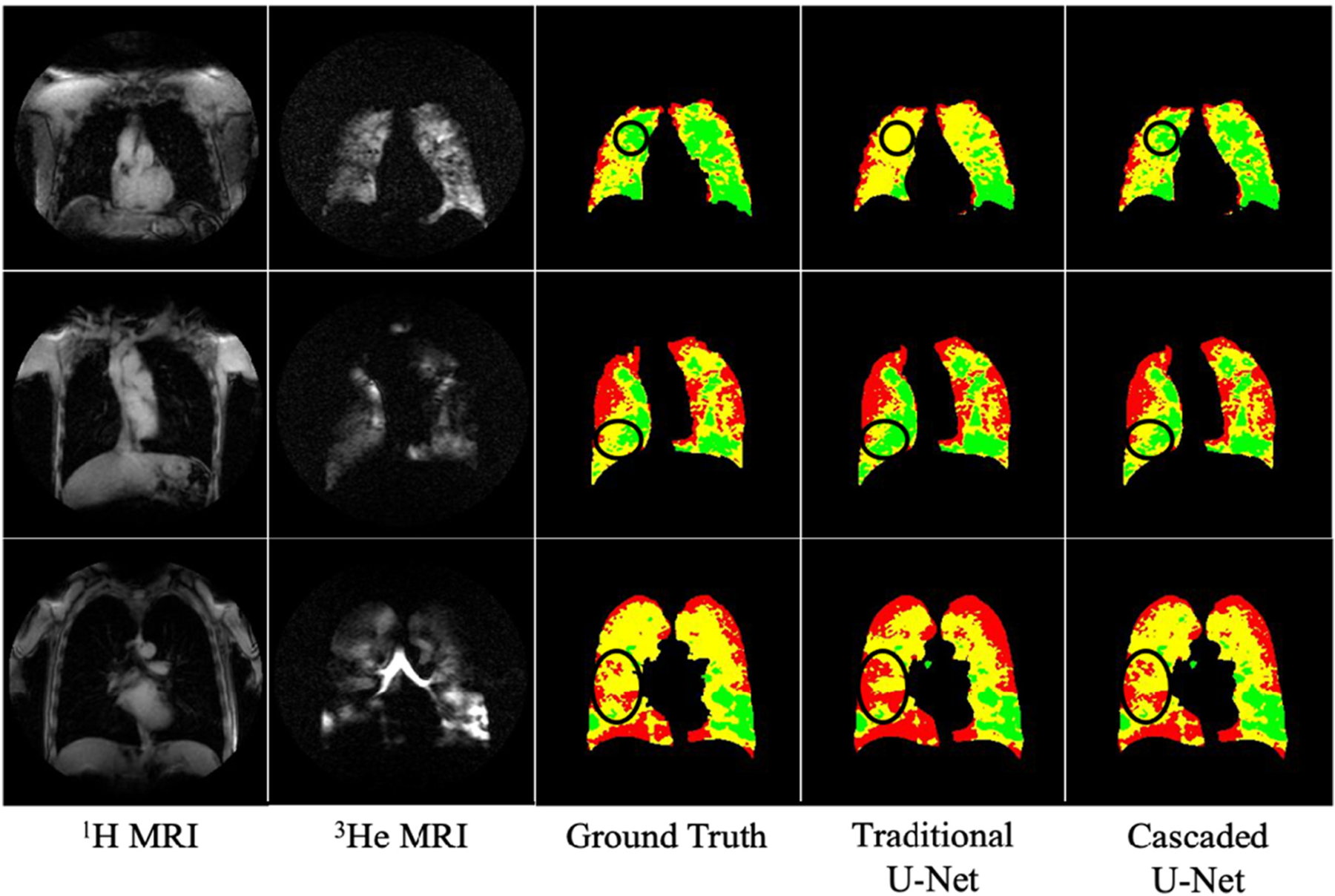
Materials and methods: The Multi-Ethnic Study of Atherosclerosis Chronic Obstructive Pulmonary Disease (COPD) study is a nested longitudinal case-control study in older smokers. Between February 2016 and July 2017, 56 participants (age, mean ± SD, 74 ± 8 years; 34 men) underwent same breath-hold proton (1H) and helium (3He) MRI, which were annotated for non-ventilated, hypo-ventilated, and normal-ventilated lungs. In this retrospective DL study, 820 1H and 3He slices from 42/56 (75%) participants were randomly selected for training, with the remaining 14/56 (25%) for test. Full lung masks were segmented using a traditional U-Net on 1H MRI and were imported into a cascaded U-Net, which were used to segment ventilation defects on 3He MRI. Models were trained with conventional data augmentation (DA) and generative adversarial networks (GAN)-DA.
Results: Conventional-DA improved 1H and 3He MRI segmentation over the non-DA model (P = 0.007 to 0.03) but GAN-DA did not yield further improvement. The cascaded U-Net improved non-ventilated lung segmentation (P < 0.005). Dice similarity coefficients (DSC) between manually and DL-segmented full lung, non-ventilated, hypo-ventilated, and normal-ventilated regions were 0.965 ± 0.010, 0.840 ± 0.057, 0.715 ± 0.175, and 0.883 ± 0.060, respectively. We observed no statistically significant difference in DCSs between participants with and without COPD (P = 0.41, 0.06, and 0.18 for non-ventilated, hypo-ventilated, and normal-ventilated regions, respectively).
Conclusion: The proposed cascaded U-Net framework generated fully-automated segmentation of ventilation defects on 3He MRI among older smokers with and without COPD that is consistent with our reference method.
Keywords: COPD; Deep learning; Hyperpolarized gas; MRI; Ventilation defects.
Copyright © 2022. Published by Elsevier Inc.
Figures
References
-
- de Lange EE, Altes TA, Patrie JT, Gaare JD, Knake JJ, Mugler JP 3rd, et al. Evaluation of asthma with hyperpolarized helium-3 MRI: correlation with clinical severity and spirometry. Chest 2006;130(4):1055–62. - PubMed
-
- Kirby M, Pike D, Coxson HO, McCormack DG, Parraga G. Hyperpolarized (3)He ventilation defects used to predict pulmonary exacerbations in mild to moderate chronic obstructive pulmonary disease. Radiology 2014;273(3):887–96. - PubMed
-
- Woodhouse N, Wild JM, Paley MN, Fichele S, Said Z, Swift AJ, et al. Combined helium-3/proton magnetic resonance imaging measurement of ventilated lung volumes in smokers compared to never-smokers. J Magn Reson Imaging 2005;21 (4):365–9. - PubMed
-
- Kirby M, Heydarian M, Svenningsen S, Wheatley A, McCormack DG, Etemad-Rezai R, et al. Hyperpolarized 3He magnetic resonance functional imaging semiautomated segmentation. Acad Radiol 2012;19(2):141–52. - PubMed
Publication types
MeSH terms
Substances
Grants and funding
- N01 HC095168/HL/NHLBI NIH HHS/United States
- 75N92020D00001/HL/NHLBI NIH HHS/United States
- R01 HL077612/HL/NHLBI NIH HHS/United States
- N01 HC095167/HL/NHLBI NIH HHS/United States
- R01 HL093081/HL/NHLBI NIH HHS/United States
- SP/14/6/31350/BHF_/British Heart Foundation/United Kingdom
- R01 HL121270/HL/NHLBI NIH HHS/United States
- HHSN268201500003I/HL/NHLBI NIH HHS/United States
- N01 HC095166/HL/NHLBI NIH HHS/United States
- N01 HC095160/HL/NHLBI NIH HHS/United States
- 75N92020D00002/HL/NHLBI NIH HHS/United States
- HHSN268201500003C/HL/NHLBI NIH HHS/United States
- N01 HC095161/HL/NHLBI NIH HHS/United States
- 75N92020D00005/HL/NHLBI NIH HHS/United States
- UL1 TR001079/TR/NCATS NIH HHS/United States
- N01 HC095169/HL/NHLBI NIH HHS/United States
- N01 HC095159/HL/NHLBI NIH HHS/United States
- 75N92020D00003/HL/NHLBI NIH HHS/United States
- UL1 TR001420/TR/NCATS NIH HHS/United States
- 75N92020D00004/HL/NHLBI NIH HHS/United States
- N01 HC095163/HL/NHLBI NIH HHS/United States
- 75N92020D00007/HL/NHLBI NIH HHS/United States
- P30 DK026687/DK/NIDDK NIH HHS/United States
- UL1 TR000040/TR/NCATS NIH HHS/United States
- N01 HC095162/HL/NHLBI NIH HHS/United States
- 75N92020D00006/HL/NHLBI NIH HHS/United States
- N01 HC095165/HL/NHLBI NIH HHS/United States
- MR/M008894/1/MRC_/Medical Research Council/United Kingdom
- N01 HC095164/HL/NHLBI NIH HHS/United States
LinkOut - more resources
Full Text Sources
Medical
