Review
. 2023 Jan-Feb;13(1):71-81.
doi: 10.1016/j.prro.2022.06.009. Epub 2022 Jun 29.

One Year of Clinic-Wide Cherenkov Imaging for Discovery of Quality Improvement Opportunities in Radiation Therapy

Affiliations
Review

One Year of Clinic-Wide Cherenkov Imaging for Discovery of Quality Improvement Opportunities in Radiation Therapy

Daniel A Alexander et al. Pract Radiat Oncol. 2023 Jan-Feb.

Abstract

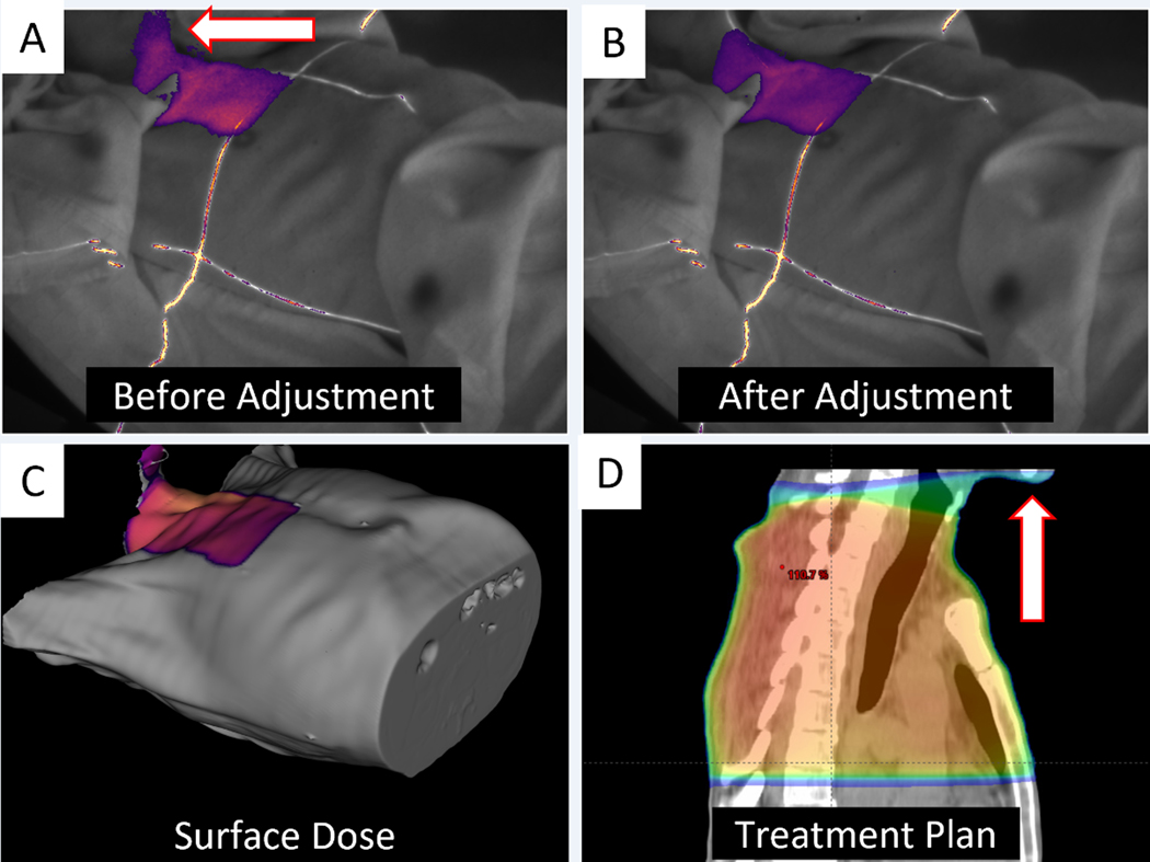
Purpose: Cherenkov imaging is clinically available as a radiation therapy treatment verification tool. The aim of this work was to discover the benefits of always-on Cherenkov imaging as a novel incident detection and quality improvement system through review of all imaging at our center.

Methods and materials: Multicamera Cherenkov imaging systems were permanently installed in 3 treatment bunkers, imaging continuously over a year. Images were acquired as part of normal treatment procedures and reviewed for potential treatment delivery anomalies.

Results: In total, 622 unique patients were evaluated for this study. We identified 9 patients with treatment anomalies occurring over their course of treatment, which were only detected with Cherenkov imaging. Categorizing each event indicated issues arising in simulation, planning, pretreatment review, and treatment delivery, and none of the incidents were detected before this review by conventional measures. The incidents identified in this study included dose to unintended areas in planning, dose to unintended areas due to positioning at treatment, and nonideal bolus placement during setup.

Conclusions: Cherenkov imaging was shown to provide a unique method of detecting radiation therapy incidents that would have otherwise gone undetected. Although none of the events detected in this study reached the threshold of reporting, they identified opportunities for practice improvement and demonstrated added value of Cherenkov imaging in quality assurance programs.


Conflict of interest statement

Conflict of Interest Statement for All Authors:

DAA, and SMD are consultants of, PB and MJ are employees of, and BWP is president and co-founder of, and LAJ has financial interests in, DoseOptics, LLC.

Figures

Figure 1.
Figure 1.
(A) Position of the three permanently mounted BeamSite cameras installed in Bunker A denoted by blue arrows. (B) Position of the two permanently mounted BeamSite cameras installed in Bunker B (blue arrows) and the three custom-gated AlignRT pods (red arrows) installed alongside the clinical pods. (C) Schematic of the 10 Gbps dedicated local area network used to transfer high volumes of Cherenkov image data, and the interface with the hospital network. (D) Example Cherenkov image of a left breast treatment generated by BeamSite. (E) Bar graph highlighting the number of patients treated per disease site along with the corresponding number of patients imaged with BeamSite, for the date range defined by the study (October 1, 2020 – October 1, 2021). In total, 622 patients were imaged out of 1088 treated.
Figure 2.
Figure 2.
Flow chart highlighting the Cherenkov image data pipeline from acquisition to review. Images are acquired, transferred, and processed automatically as shown in the red and green boxes. Subsequently, image review, flagging, and interpretation for events occur manually on a retrospective basis.
Figure 3.
Figure 3.
Case 2 showing dose to the patient’s chin during a palliative thoracic spine treatment, which was noticed live by a radiation therapist, prompting intervention to position the chin slightly further back. Cherenkov image frames accumulated from the beginning of treatment to beam interruption are shown in (A), while Cherenkov image frames accumulated from beam continuation to the end of the relevant field are shown in (B). The surface dose from the relevant field as extracted from the TPS projected on the patient surface extracted from the simulation CT scan is shown in (C), visualized from the camera perspective. The dose to the chin as shown in (C) was verified by checking the treatment plan (D).
Figure 4.
Figure 4.
Cases 3, 5, and 6 showing dose to unintended areas. Case 3 (A-C) shows improper arm placement (7/10 fractions) leading to 4.5 Gy exposure from an exit beam. Case 5 (D-F) shows non-ideal hand position leading to minor exposure (1/10 fractions). Case 6 (G-I) highlights extraneous dose to the left armpit from small deviation in left arm position (1/16 fractions). All surface dose renderings were extracted from the treatment plan and overlaid on the patient CT surface, while red arrows highlight event regions.
Figure 5.
Figure 5.
Cases 7 and 8 showing imperfect bolus coverage leading to uncovered irradiated regions of chest wall. Case 7 shows proper coverage using two adjacent pieces of bolus on fraction 4 (B), and insufficient coverage leading to potential underdose from the exit side on fraction 5 (C). Panels A-C show surface dose and Cherenkov images from only the relevant LPO field. Case 8 (D-F) shows a similar but less extreme bolus placement issue on fraction 22 (F) compared with proper coverage shown on fraction 1 (E). Panels D-F correspond to the entire treatment fraction. All surface dose renderings were extracted from the treatment plan and overlaid on the patient CT surface, while red arrows highlight event regions.

Similar articles

Cited by

References

    1. Otto K. Volumetric modulated arc therapy: IMRT in a single gantry arc. Med Phys. 2008;35(1):310–317. doi:10.1118/1.2818738 - DOI - PubMed
    1. Kamperis E, Kodona C, Hatziioannou K, Giannouzakos V. Complexity in Radiation Therapy: It’s Complicated. Int J Radiat Oncol. 2020;106(1):182–184. doi:10.1016/j.ijrobp.2019.09.003 - DOI - PubMed
    1. Huq MS, Fraass BA, Dunscombe PB, et al. The report of Task Group 100 of the AAPM: Application of risk analysis methods to radiation therapy quality management. Med Phys. 2016;43(7):4209–4262. doi:10.1118/1.4947547 - DOI - PMC - PubMed
    1. Marks LB, Pawlicki TA, Hayman JA. Learning to Appreciate Swiss Cheese and Other Industrial Engineering Concepts. Pract Radiat Oncol. 2015;5(5):277–281. doi:10.1016/j.prro.2015.07.004 - DOI - PubMed
    1. Ford E, Conroy L, Dong L, et al. Strategies for effective physics plan and chart review in radiation therapy: Report of AAPM Task Group 275. Med Phys. 2020;47(6):e236–e272. doi:10.1002/mp.14030 - DOI - PMC - PubMed