

Review
doi: 10.1101/cshperspect.a041061.
Inflammasomes and Other Caspase-Activation Platforms
Affiliations
- PMID: 35777830
- PMCID: PMC9248825
- DOI: 10.1101/cshperspect.a041061
Item in Clipboard
Review
Inflammasomes and Other Caspase-Activation Platforms
Cold Spring Harb Perspect Biol.
.
No abstract available
Figures
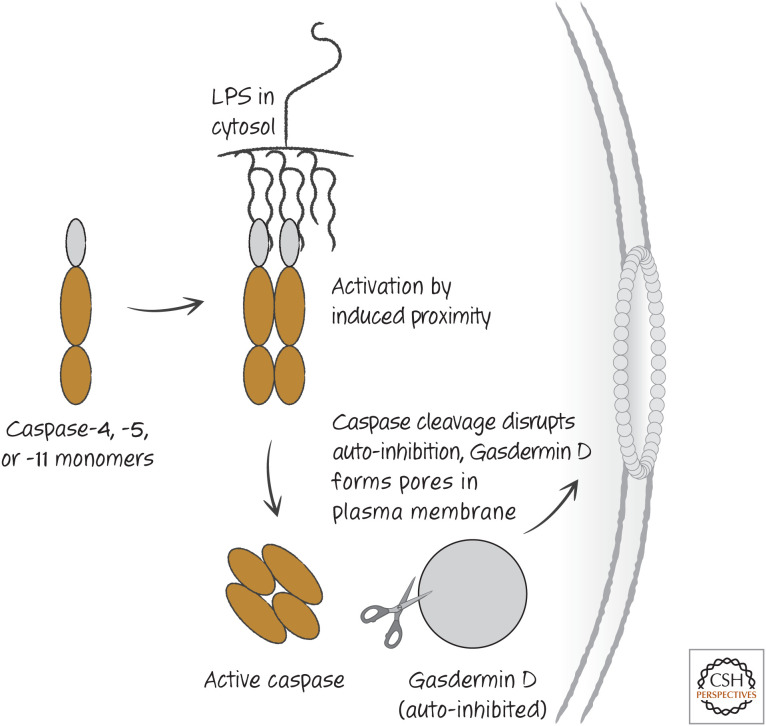
Cytosolic lipopolysaccharide (LPS) directly induces caspases and pyroptosis. This appears to be restricted to caspase-4 and caspase-5 in humans and caspase-11 in mice. Other caspases we have discussed are not activated in this direct manner.
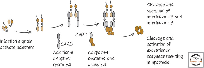
The basic inflammasome and activation of caspase-1.
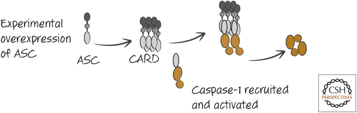
Overexpression of ASC can activate caspase-1. This is not a physiological mechanism of caspase-1 activation, but the observation is informative.
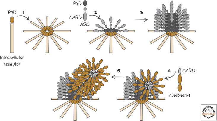
General structure of inflammasomes. 1. The intracellular receptor, containing a pyrin domain (PYD), on interaction with its ligand forms a disc-like structure. 2. The center of this structure interacts with ASC through PYD–PYD interactions. 3. The PYD domains of ASC interact with additional ASC PYD domains to form a cylindrical structure. 4. The CARD domains of caspase-1 monomers bind to the CARD of ASC. 5. Bound caspase-1 recruits additional caspase-1 molecules through CARD–CARD interactions, forming a fibril. A “mature” inflammasome is shown in Figure 5.
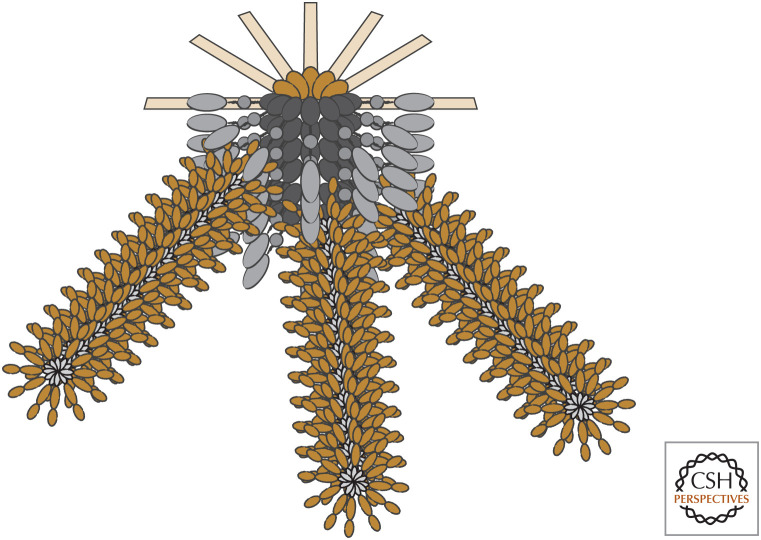
Inflammasomes have fibrils of caspase-1.
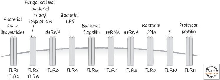
Some TLRs and the PAMPs that activate them.
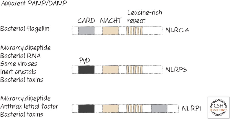
Some NLRs that function in inflammasomes. The PAMPs and DAMPs that induce them are listed. Toxins from bacteria can also alter potassium levels, thereby indirectly facilitating inflammasome formation.
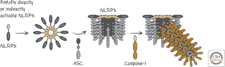
Activation of the NLRP3 inflammasome.
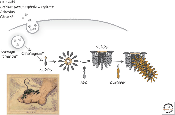
Inert crystals induce the NLRP3 inflammasome in gout and other diseases. (Bottom left, Reprinted from Gillray 1799.)
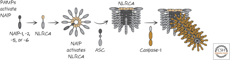
Activation of the NLRC4 inflammasome.
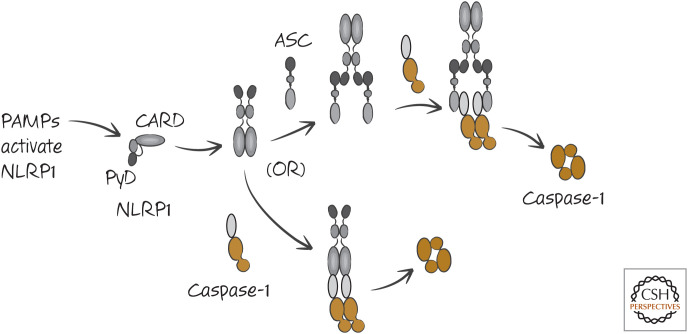
Two models of the NLRP1 inflammasome. Both may be correct in different settings.
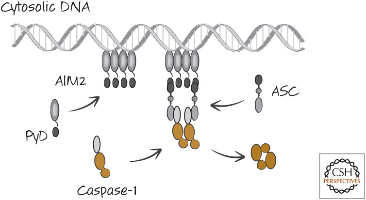
The AIM2 inflammasome. This is a simplified scheme, and AIM2 facilitates the formation of caspase-1 fibrils, as we saw for other inflammasomes.
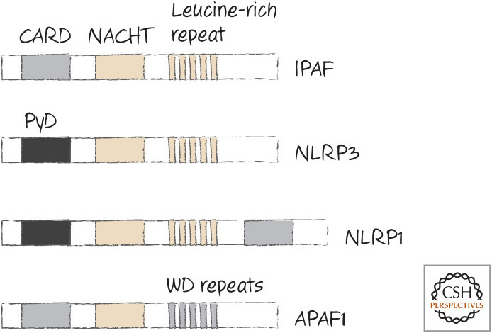
APAF1 is in the Nod-like receptor (NLR) family.
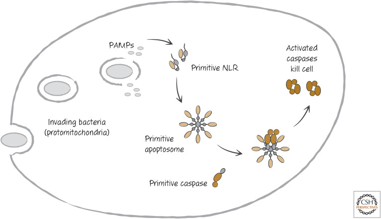
Return of the just-so story.
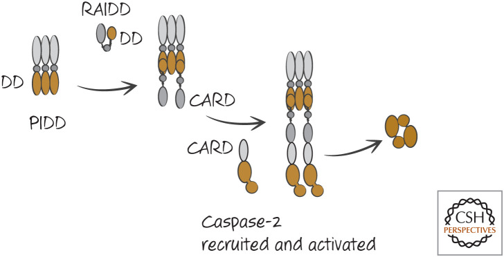
The PIDDosome activates caspase-2. DD, death domain.
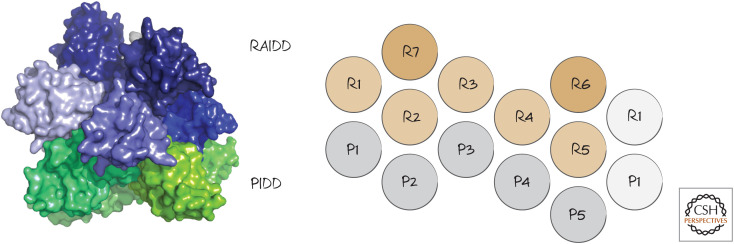
PIDDosome structure. The interactions of PIDD (P) and RAIDD (R) DDs are shown on the right. (Left, PDB 2OF5 [Park et al. 2007]; right, reprinted from Park et al. 2007, ©2007 with permission from Elsevier.)
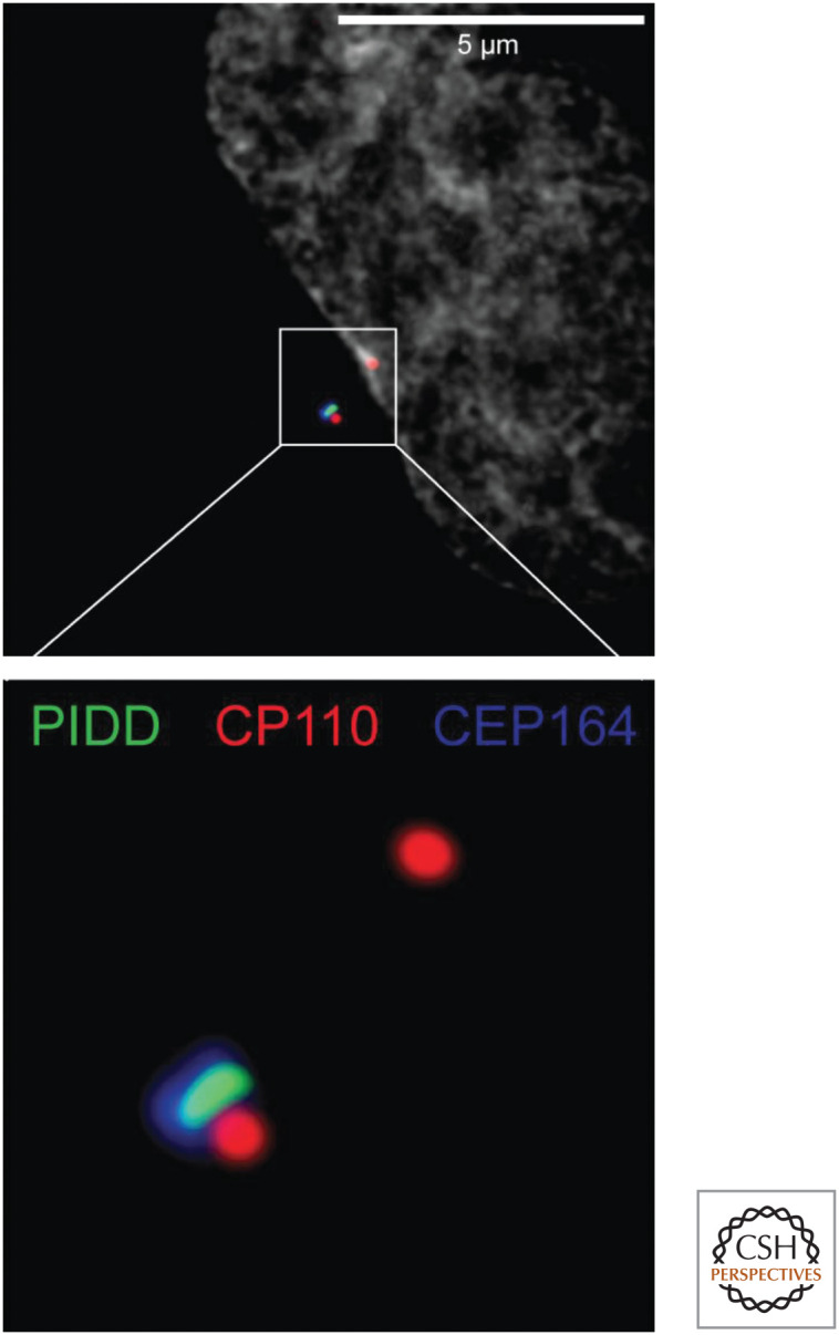
Fluorescence micrograph showing how PIDD localizes to mature centrosomes. PIDD (green) associates with the mature centrosome (which also contains CEP164, blue). The nascent (upper red spot) and mature centrosomes (lower complex of spots) both contain CP110 (red). The lower image is a magnified view of the area enclosed by the white square in the upper image. (Photo courtesy of Dr. Luca Fava and Dr. Andreas Villunger, University of Innsbruck.)
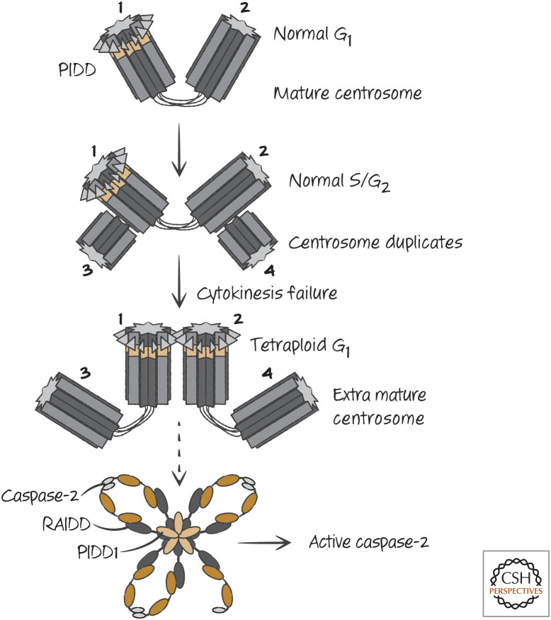
Aberrant extra mature centrosomes activate caspase-2. During the S/G2 phases of the cell cycle, the centrosome duplicates, but PIDD remains associated with only the mature centrosome. If cytokinesis fails, leading to the formation of a tetraploid cell in the following G1 phase, two mature centrosomes appear in the cell, leading to PIDD–PIDD interaction, recruitment of RAIDD, and activation of caspase-2.
Similar articles
-
Inflammatory Caspases: Toward a Unified Model for Caspase Activation by Inflammasomes.Annu Rev Immunol. 2022 Apr 26;40:249-269. doi: 10.1146/annurev-immunol-101220-030653. Epub 2022 Jan 26. Annu Rev Immunol. 2022. PMID: 35080918 Review.
-
Death and survival from executioner caspase activation.Semin Cell Dev Biol. 2024 Mar 15;156:66-73. doi: 10.1016/j.semcdb.2023.07.005. Epub 2023 Jul 18. Semin Cell Dev Biol. 2024. PMID: 37468421 Review.
-
Inflammasomes as polyvalent cell death platforms.Cell Mol Life Sci. 2016 Jun;73(11-12):2335-47. doi: 10.1007/s00018-016-2204-3. Epub 2016 Apr 5. Cell Mol Life Sci. 2016. PMID: 27048821 Free PMC article. Review.
-
Discovery of a caspase cleavage motif antibody reveals insights into noncanonical inflammasome function.Proc Natl Acad Sci U S A. 2021 Mar 23;118(12):e2018024118. doi: 10.1073/pnas.2018024118. Proc Natl Acad Sci U S A. 2021. PMID: 33723046 Free PMC article.
-
Caspase-11: the driving factor for noncanonical inflammasomes.Eur J Immunol. 2013 Sep;43(9):2240-5. doi: 10.1002/eji.201343800. Eur J Immunol. 2013. PMID: 24037676 Review.
Cited by
-
Caspase-2 kills cells with extra centrosomes.Sci Adv. 2024 Nov;10(44):eado6607. doi: 10.1126/sciadv.ado6607. Epub 2024 Oct 30. Sci Adv. 2024. PMID: 39475598 Free PMC article.
-
The Future of Death.Cold Spring Harb Perspect Biol. 2022 Dec 1;14(12):a041111. doi: 10.1101/cshperspect.a041111. Cold Spring Harb Perspect Biol. 2022. PMID: 36456104 Free PMC article. Review. No abstract available.
-
Caspase-5: Structure, Pro-Inflammatory Activity and Evolution.Biomolecules. 2024 Apr 26;14(5):520. doi: 10.3390/biom14050520. Biomolecules. 2024. PMID: 38785927 Free PMC article. Review.
-
The Burial: Clearance and Consequences.Cold Spring Harb Perspect Biol. 2022 Oct 3;14(10):a041087. doi: 10.1101/cshperspect.a041087. Cold Spring Harb Perspect Biol. 2022. PMID: 36192119 Free PMC article. Review. No abstract available.
-
Stepwise phosphorylation and SUMOylation of PIDD1 drive PIDDosome assembly in response to DNA repair failure.Nat Commun. 2024 Oct 24;15(1):9195. doi: 10.1038/s41467-024-53412-0. Nat Commun. 2024. PMID: 39448602 Free PMC article.
References
Publication types
MeSH terms
Substances
LinkOut - more resources
Full Text Sources