Comment
. 2023 Feb;130(2):e370-e380.
doi: 10.1016/j.bja.2022.05.025. Epub 2022 Jun 29.

Protective effects of omega-3 fatty acids in a blood-brain barrier-on-chip model and on postoperative delirium-like behaviour in mice

Affiliations
Comment

Protective effects of omega-3 fatty acids in a blood-brain barrier-on-chip model and on postoperative delirium-like behaviour in mice

Ting Yang et al. Br J Anaesth. 2023 Feb.

Abstract

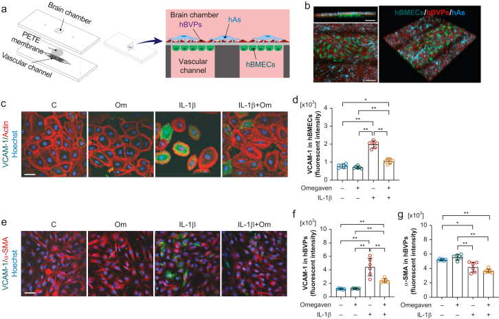
Background: Peripheral surgical trauma can trigger neuroinflammation and ensuing neurological complications, such as delirium. The mechanisms whereby surgery contributes to postoperative neuroinflammation remain unclear and without effective therapies. Here, we developed a microfluidic-assisted blood-brain barrier (BBB) device and tested the effects of omega-3 fatty acids on neuroimmune interactions after orthopaedic surgery.

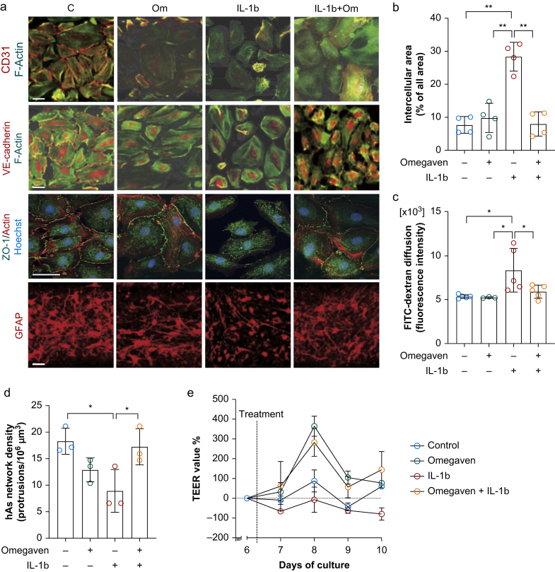
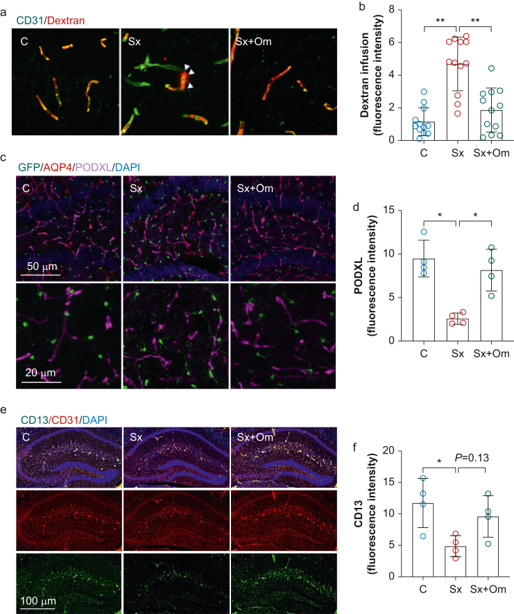
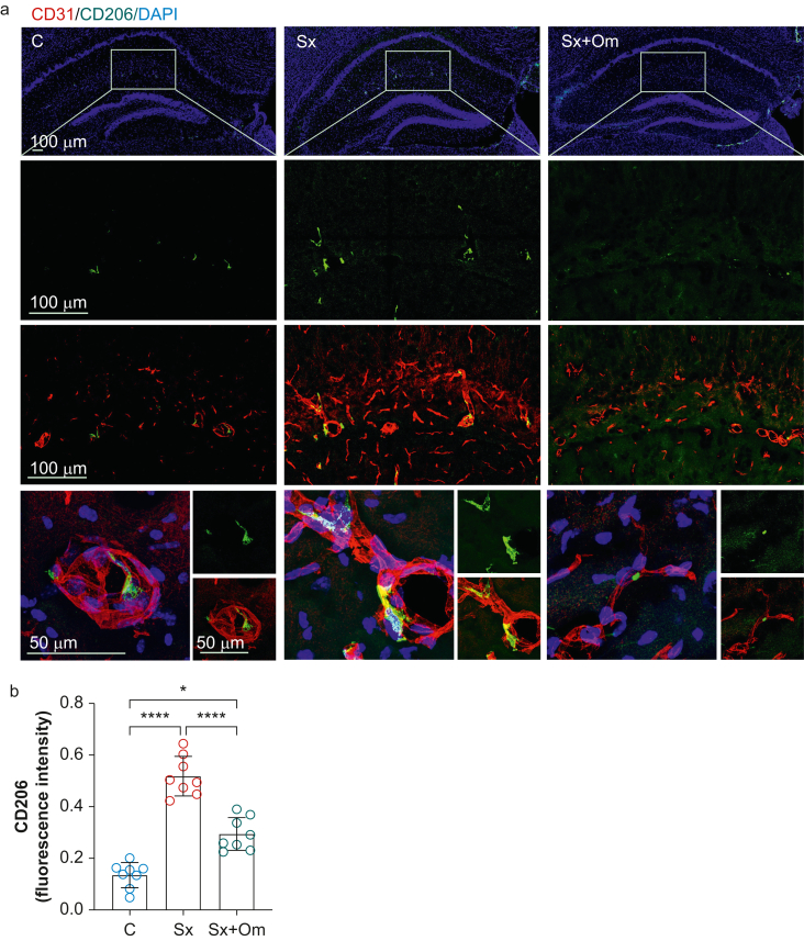
Methods: A microfluidic-assisted BBB device was established using primary human cells. Tight junction proteins, vascular cell adhesion molecule 1 (VCAM-1), BBB permeability, and astrocytic networks were assessed after stimulation with interleukin (IL)-1β and in the presence or absence of a clinically available omega-3 fatty acid emulsion (Omegaven®; Fresenius Kabi, Bad Homburg, Germany). Mice were treated 1 h before orthopaedic surgery with 10 μl g-1 body weight of omega-3 fatty acid emulsion i.v. or equal volumes of saline. Changes in pericytes, perivascular macrophages, BBB opening, microglial activation, and inattention were evaluated.

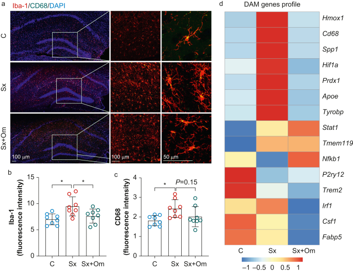
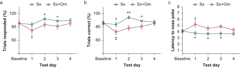
Results: Omega-3 fatty acids protected barrier permeability, endothelial tight junctions, and VCAM-1 after exposure to IL-1β in the BBB model. In vivo studies confirmed that omega-3 fatty acid treatment inhibited surgery-induced BBB impairment, microglial activation, and delirium-like behaviour. We identified a novel role for pericyte loss and perivascular macrophage activation in mice after surgery, which were rescued by prophylaxis with i.v. omega-3 fatty acids.

Conclusions: We present a new approach to study neuroimmune interactions relevant to perioperative recovery using a microphysiological BBB platform. Changes in barrier function, including dysregulation of pericytes and perivascular macrophages, provide new targets to reduce postoperative delirium.

Keywords: blood–brain barrier; delirium; microglia; neurovascular unit; omega-3 fatty acid; organ-on-chip; pericyte; perivascular macrophage.


Figures

Fig 1
Fig 1
Blood–brain barrier (BBB)-on-chip model and interleukin (IL)-1β induced cell activation. (a) Illustration of the microphysiological platform for brain cells co-culture to generate model BBB tissue. The BBB-on-chip device consists of two layers of polydimethylsiloxane substrate separated by the polyester track etched (PETE) membrane, which acts as a basal membrane. The bottom layer has a vascular channel dividing into eight branches of 300 μm width each to culture human brain microvascular endothelial cells (hBMECs), and the top layer has a 5 mm × 5 mm square well that forms the brain chamber. The brain chamber is located on top of the vascular channels to co-culture brain cells (human brain vascular pericytes [hBVPs] and human astrocytes [hAs]) with a brain microvascular endothelial cells separated by the PETE membrane. (b) Representative fluorescence image of live brain microvascular endothelial cells, brain vascular pericytes, and human astrocytes in the BBB device. In counterclockwise order, each image represents a cross-sectional view, top view, 3D view, respectively (scale bar: 100 μm; green: hBMECs; red: hBVPs; cyan: hAs). Omega-3 fatty acids inhibit IL-1β-induced vascular cell adhesion molecule 1 (VCAM-1) overexpression in both (c and d) hBMECs (e and f) and hBVPs (e and g). IL-1β reduced alpha-smooth muscle actin (α-SMA) expression in hBVPs, which was not affected by omega-3 fatty acid treatment. Data expressed as mean (standard deviation); ∗P<0.01; ∗∗P<0.001; n=6 per group. All experiments were repeated three times. C, control; hAs, human astrocytes; hBMECs, human brain microvascular endothelial cells; hBVPs, human brain vascular pericytes; Om, omega-3 fatty acids.
Fig 2
Fig 2
Omega-3 fatty acids inhibit interleukin (IL)-1β-induced blood–brain barrier (BBB) permeability dysfunction by increasing cell–cell tightness. (a) Representative images of immunofluorescence staining of CD31, vascular endothelial cadherin, ZO-1, and GFAP (scale bar: 100 μm). (b) Quantification of cell–cell tightness by measuring the percentage of intercellular area. Omega-3 fatty acids abolished IL-1β-induced intercellular area increase. (c) 70 kDa fluorescein isothiocyanate (FITC)–dextran diffusion across the BBB was significantly increased in response to IL-1β. This effect is prevented by omega-3 fatty acid pretreatment. (d) IL-1β induced significant reduction in the network density of astrocyte protrusions, which is rescued by omega-3 fatty acid pretreatment. (e) Relative changes in trans-endothelial electrical resistance (TEER) values compared with pretreatment values as a function of time for different experimental conditions. Data expressed as mean (standard deviation); ∗P<0.05; ∗∗P<0.01; ∗∗∗P<0.001; n=4 per group for Panel B; n=3–5 per group for Panel C; n=3 per group for Panel D; n=2 per group for panel e. All experiments were repeated three times. C, control; GFAP, glial fibrillary acidic protein; hAs, human astrocytes; hBMECs, human brain microvascular endothelial cells; hBVPs, human brain vascular pericytes; Om, omega-3 fatty acids; ZO-1, zonula occludens-1.
Fig 3
Fig 3
Omega-3 fatty acids prevent surgery-induced blood–brain barrier disruption. Surgery induced significant dextran diffusion (a and b), a reduction of PODXL (c and d), and reduction of a pericyte marker (CD13) into the hippocampus (e and f). These changes were prevented by omega-3 fatty acid pretreatment. Data expressed as mean (standard deviation); ∗P<0.05; ∗∗P<0.01; ∗∗∗P<0.001; n=4 per group (for panel b, the quantification was performed in three different regions in the hippocampus per mouse). AQP4, aquaporin 4; C, control; DAPI, 4′,6-diamidino-2-phenylindole; PODXL, podocalyxin; Sx, surgery; Sx+Om, surgery+omega-3 fatty acids.
Fig 4
Fig 4
Omega-3 fatty acids inhibit surgery-induced perivascular macrophage activation. (a) Representative images of the immunofluorescence staining of a perivascular macrovascular marker, CD206, in the hippocampus. (b) Surgery enhanced expression of CD206 in the hippocampus, which was blocked by omega-3 fatty acid pretreatment. Data expressed as mean (standard deviation); ∗P<0.001; n=8 per group. C, control; DAPI, 4′,6-diamidino-2-phenylindole; Sx, surgery; Sx+Om, surgery+omega-3 fatty acids.
Fig 5
Fig 5
Omega-3 fatty acids reduce surgery-induced microglia activation. (a) Representative images of immunofluorescence staining of microglia (Iba-1) and CD68 in the hippocampus. Surgery-induced microgliosis, with higher expression of Iba-1 (b) and CD68 (c) in the hippocampus. These effects were reduced by omega-3 fatty acids. (d) NanoString gene expression of hippocampal lysate shows changes in multiple microglia activation-associated genes, which were in part restored by omega-3 fatty acid treatment. Data expressed as mean (standard deviation); ∗P<0.05; ∗∗P<0.01; ∗∗∗P<0.001; n=4 or 8 per group. C, control; DAM, disease-associated microglia; DAPI, 4′,6-diamidino-2-phenylindole; Iba-1, ionised calcium binding adaptor molecule-1; Sx, surgery; Sx+Om, surgery+omega-3 fatty acids.
Fig 6
Fig 6
Postoperative inattention behaviour was prevented by omega-3 fatty acid pretreatment. Omega-3 fatty acid-treated mice showed better attention function after surgery in the five-choice serial reaction time task test as evaluated by a higher percentage of responded trials (a), more correct responses in all responded trials (b), and shorter response latency compared with placebo-treated mice (c). ∗P<0.05; ∗∗P<0.01; ∗∗∗P<0.001 vs Sx on the same testing day; P<0.05; P<0.01 vs Sx baseline; n=9 per group. Sx, surgery; Sx+Om, surgery+omega-3 fatty acids.

Comment on

References

    1. Daneman R. The blood–brain barrier in health and disease. Ann Neurol. 2012;72:648–672. - PubMed
    1. Yang T., Velagapudi R., Terrando N. Neuroinflammation after surgery: from mechanisms to therapeutic targets. Nat Immunol. 2020;21:1319–1326. - PMC - PubMed
    1. Wilson J.E., Mart M.F., Cunningham C., et al. Delirium Nat Rev Dis Primers. 2020;6:90. - PMC - PubMed
    1. Leslie D.L., Marcantonio E.R., Zhang Y., Leo-Summers L., Inouye S.K. One-year health care costs associated with delirium in the elderly population. Arch Intern Med. 2008;168:27–32. - PMC - PubMed
    1. Jin Z., Hu J., Ma D. Postoperative delirium: perioperative assessment, risk reduction, and management. Br J Anaesth. 2020;125:492–504. - PubMed