

RF-CNN-F: random forest with convolutional neural network features for coronary artery disease diagnosis based on cardiac magnetic resonance
- PMID: 35778476
- PMCID: PMC9249743
- DOI: 10.1038/s41598-022-15374-5
RF-CNN-F: random forest with convolutional neural network features for coronary artery disease diagnosis based on cardiac magnetic resonance
Abstract
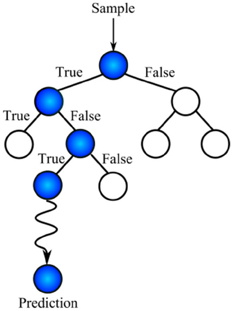
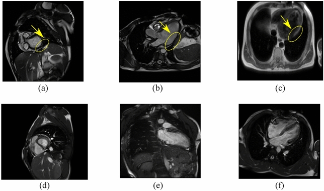
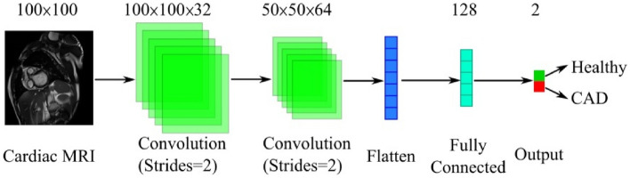
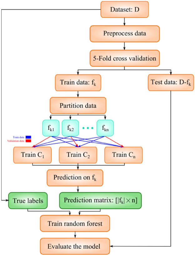
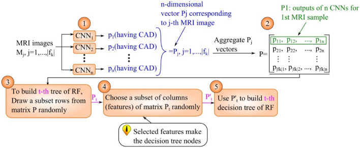
Coronary artery disease (CAD) is a prevalent disease with high morbidity and mortality rates. Invasive coronary angiography is the reference standard for diagnosing CAD but is costly and associated with risks. Noninvasive imaging like cardiac magnetic resonance (CMR) facilitates CAD assessment and can serve as a gatekeeper to downstream invasive testing. Machine learning methods are increasingly applied for automated interpretation of imaging and other clinical results for medical diagnosis. In this study, we proposed a novel CAD detection method based on CMR images by utilizing the feature extraction ability of deep neural networks and combining the features with the aid of a random forest for the very first time. It is necessary to convert image data to numeric features so that they can be used in the nodes of the decision trees. To this end, the predictions of multiple stand-alone convolutional neural networks (CNNs) were considered as input features for the decision trees. The capability of CNNs in representing image data renders our method a generic classification approach applicable to any image dataset. We named our method RF-CNN-F, which stands for Random Forest with CNN Features. We conducted experiments on a large CMR dataset that we have collected and made publicly accessible. Our method achieved excellent accuracy (99.18%) using Adam optimizer compared to a stand-alone CNN trained using fivefold cross validation (93.92%) tested on the same dataset.
© 2022. The Author(s).
Conflict of interest statement
The authors declare no competing interests.
Figures

References
-
- Śpiewak M. Imaging in coronary artery disease cardiac magnetic resonance. Cor et Vasa. 2015;57(6):e453–e461. doi: 10.1016/j.crvasa.2015.09.009. - DOI
MeSH terms
LinkOut - more resources
Full Text Sources
Medical
Miscellaneous
