Review
. 2023 Jan;44(1):1-7.
doi: 10.1038/s41401-022-00938-y. Epub 2022 Jul 1.

AlphaFold2 versus experimental structures: evaluation on G protein-coupled receptors

Affiliations
Review

AlphaFold2 versus experimental structures: evaluation on G protein-coupled receptors

Xin-Heng He et al. Acta Pharmacol Sin. 2023 Jan.

Abstract

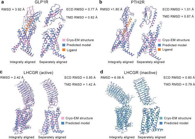
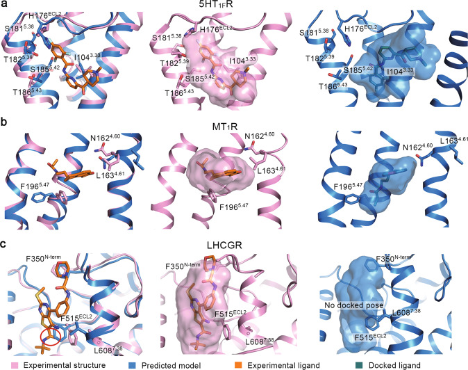
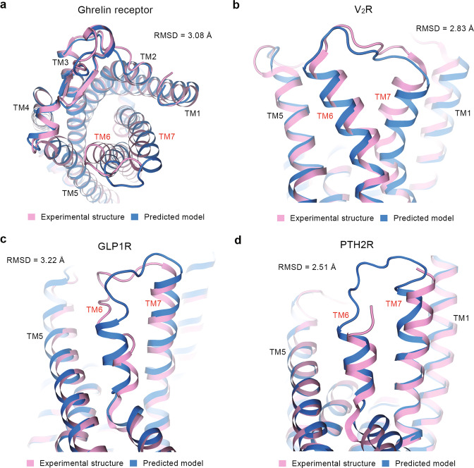
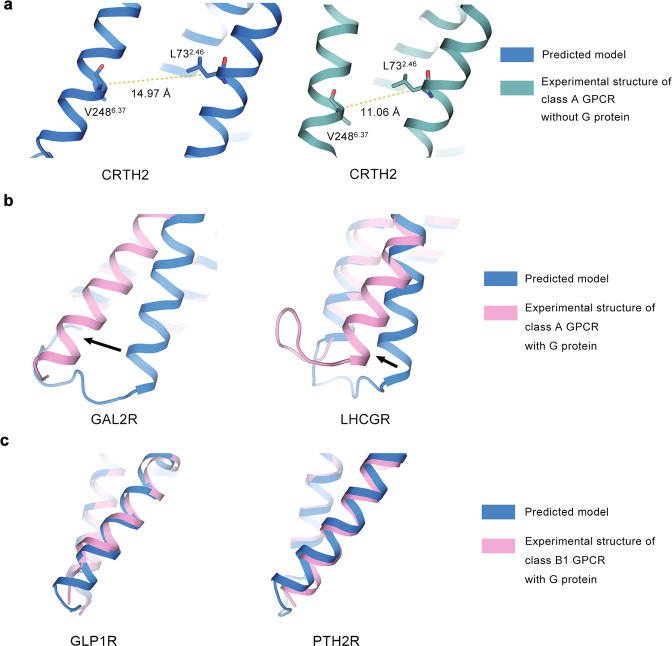
As important drug targets, G protein-coupled receptors (GPCRs) play pivotal roles in a wide range of physiological processes. Extensive efforts of structural biology have been made on the study of GPCRs. However, a large portion of GPCR structures remain unsolved due to structural instability. Recently, AlphaFold2 has been developed to predict structure models of many functionally important proteins including all members of the GPCR family. Herein we evaluated the accuracy of GPCR structure models predicted by AlphaFold2. We revealed that AlphaFold2 could capture the overall backbone features of the receptors. However, the predicted models and experimental structures were different in many aspects including the assembly of the extracellular and transmembrane domains, the shape of the ligand-binding pockets, and the conformation of the transducer-binding interfaces. These differences impeded the use of predicted structure models in the functional study and structure-based drug design of GPCRs, which required reliable high-resolution structural information.

Keywords: AlphaFold2; G protein-coupled receptors; drug design; protein structure prediction; structural biology.


Conflict of interest statement

The authors declare no competing interests.

Figures

Fig. 1
Fig. 1. Comparison between predicted models and experimental structures with large ECD.
Experimental structures and predicted models for glucagon-like peptide 1 receptor (GLP1R) (a), parathyroid hormone 2 receptor (PTH2R) (b), active (c) and inactive (d) luteinizing hormone and choriogonadotropin receptor (LHCGR) in different ways of alignments. Cryo-EM structures of GLP1R (PDB ID: 7KI0), PTH2R (PDB ID: 7F16), LHCGR (PDB IDs: 7FIG and 7FIJ) are selected for calculation. Pink (or cyan) and blue cartoons represent experimental structures and predicted models, respectively. Two conflicts between the predicted model of GLP1R and the peptide ligand from the experimental structure are highlighted in red circles.
Fig. 2
Fig. 2. Evaluation for the orthosteric ligand-binding sites.
Ligands binding to the orthosteric sites in experimental structures and predicted models of 5-hydroxytryptamine receptor 1F receptor (5HT1FR) (a), melatonin receptor 1A (MT1R) (b), and luteinizing hormone and choriogonadotropin receptor (LHCGR) (c). Cryo-EM structures of 5HT1FR (PDB ID: 7EXD), MT1R (PDB ID: 7DB6), LHCGR (PDB ID: 7FIH) are selected for analysis. Key residues and ligands are shown as sticks. Pink, blue, orange, and green represent experimental structures, predicted models, ligands from experimental complex structures, and docked ligands, respectively. The ligand-binding pockets are shown as surfaces.
Fig. 3
Fig. 3. Comparison of TMD conformations at extracellular side.
TM6 and TM7 helices in experimental structures and predicted models of ghrelin receptor (a), vasopressin V2 receptor (V2R) (b), glucagon-like peptide 1 receptor (GLP1R) (c), and parathyroid hormone 2 receptor (PTH2R) (d). Cryo-EM structures of ghrelin receptor (PDB ID: 7F9Y), V2R (PDB ID: 7DW9), GLP1R (PDB ID: 7KI1) and PTH2R (PDB ID: 7F16) are selected for analysis. Pink and blue cartoons represent experimental structures and predicted models, respectively. TM6–TM7 heavy-atom RMSD values are shown in each plot.
Fig. 4
Fig. 4. Comparison of TM6 conformations at intracellular side.
TM6 helices of predicted models and experimental structures for a class A GPCR without G protein (a), a class A GPCR with G protein (b), and a class B1 GPCR with G protein (c). Structures without G protein, structures with G protein, and predicted models are shown in green, pink, and blue cartoons, respectively. PDB IDs of experimental structures are labeled in each plot. Dashed lines indicate the TM6 tilt distance (TD). Arrows indicate the orientational differences of TM6 between predicted models and experimental structures. All structures are aligned with respect to TMD.

Similar articles

Cited by

References

    1. Venter JC, Adams MD, Myers EW, Li PW, Mural RJ, Sutton GG, et al. The sequence of the human genome. Science. 2001;291:1304–51. doi: 10.1126/science.1058040. - DOI - PubMed
    1. Kang Y, Zhou XE, Gao X, He Y, Liu W, Ishchenko A, et al. Crystal structure of rhodopsin bound to arrestin by femtosecond X-ray laser. Nature. 2015;523:561–7. doi: 10.1038/nature14656. - DOI - PMC - PubMed
    1. Xu P, Huang S, Zhang H, Mao C, Zhou XE, Cheng X, et al. Structural insights into the lipid and ligand regulation of serotonin receptors. Nature. 2021;592:469–73. doi: 10.1038/s41586-021-03376-8. - DOI - PubMed
    1. Duan J, Xu P, Cheng X, Mao C, Croll T, He X, et al. Structures of full-length glycoprotein hormone receptor signalling complexes. Nature. 2021;598:688–92. doi: 10.1038/s41586-021-03924-2. - DOI - PubMed
    1. Hauser AS, Attwood MM, Rask-Andersen M, Schiöth HB, Gloriam DE. Trends in GPCR drug discovery: new agents, targets and indications. Nat Rev Drug Discov. 2017;16:829–42. doi: 10.1038/nrd.2017.178. - DOI - PMC - PubMed