. 2022 Jul 1;41(1):215.
doi: 10.1186/s13046-022-02415-0.

Aberrant miR-874-3p/leptin/EGFR/c-Myc signaling contributes to nasopharyngeal carcinoma pathogenesis

Affiliations

Aberrant miR-874-3p/leptin/EGFR/c-Myc signaling contributes to nasopharyngeal carcinoma pathogenesis

Sheng-Dean Luo et al. J Exp Clin Cancer Res. .

Abstract

Background: Leptin is important in physiological and pathological functions in various cancers, however, the significance and mechanisms of leptin in nasopharyngeal carcinoma remain ambiguous.

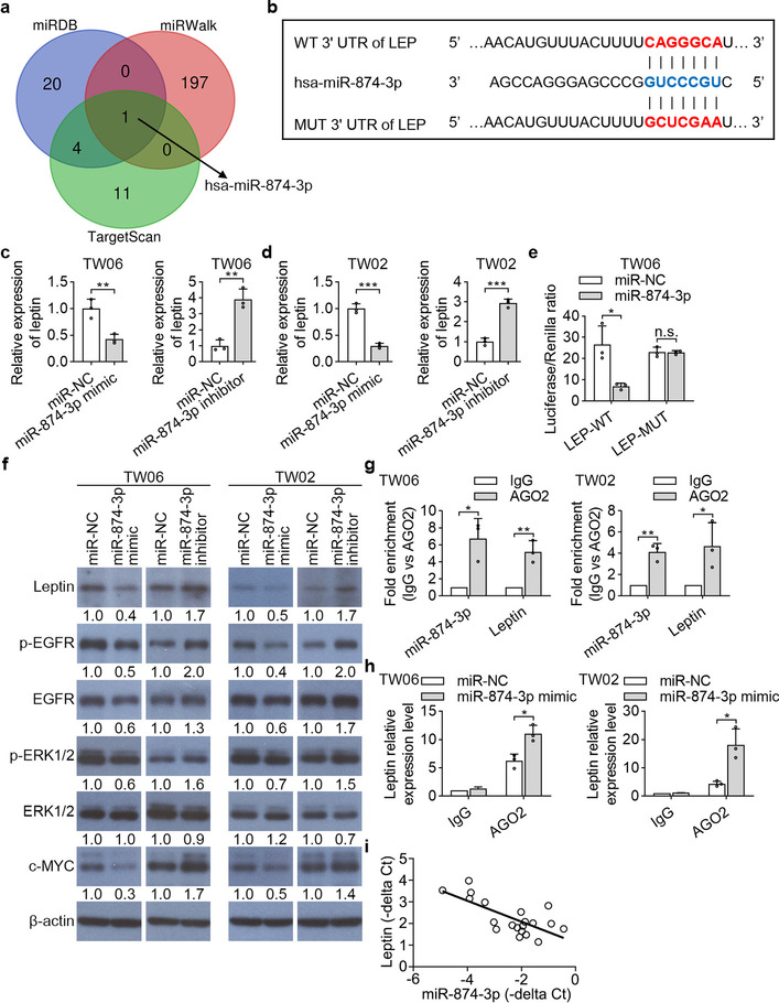
Methods: Leptin expression was analyzed by QPCR, immunohistochemistry, Western blotting, and TCGA database. The impact of gain- or loss-of-function of leptin were determined by MTT, colony formation, wound healing, and Transwell assays in NPC cells, and by a xenograft tumor model. Leptin-modulated glucose consumption and lactate production were assessed by ELISA. Furthermore, leptin-regulated signaling pathways were examined by QPCR and Western blotting assays. The immunoprecipitation assay was conducted to determine interaction between leptin and EGFR. In addition, miR-874-3p-regulated leptin expression was evaluated using bioinformatics, QPCR, luciferase assay, AGO2-RIP assay, and Western blotting.

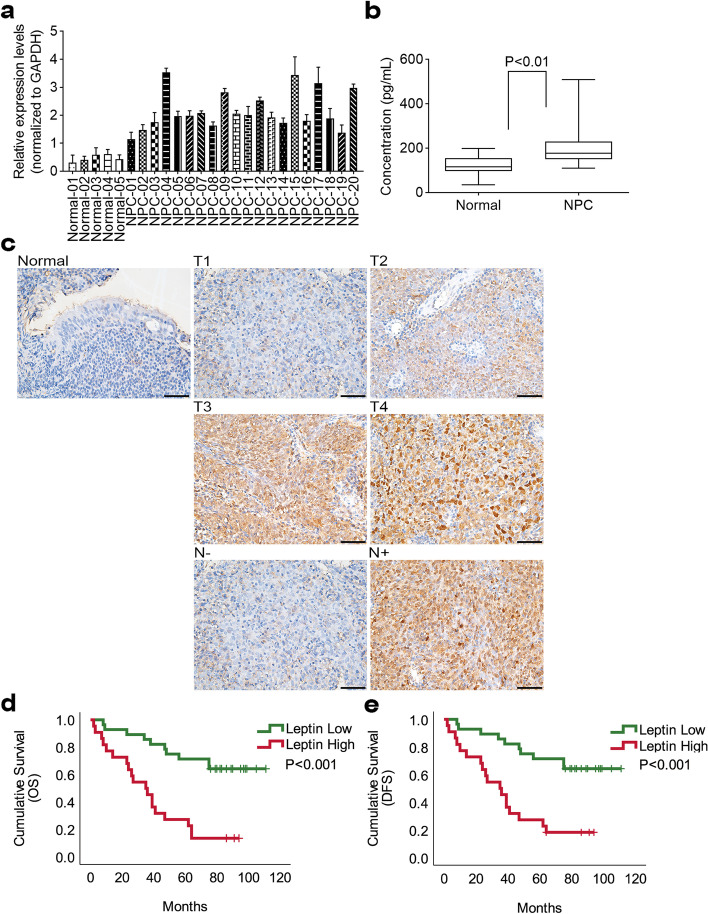
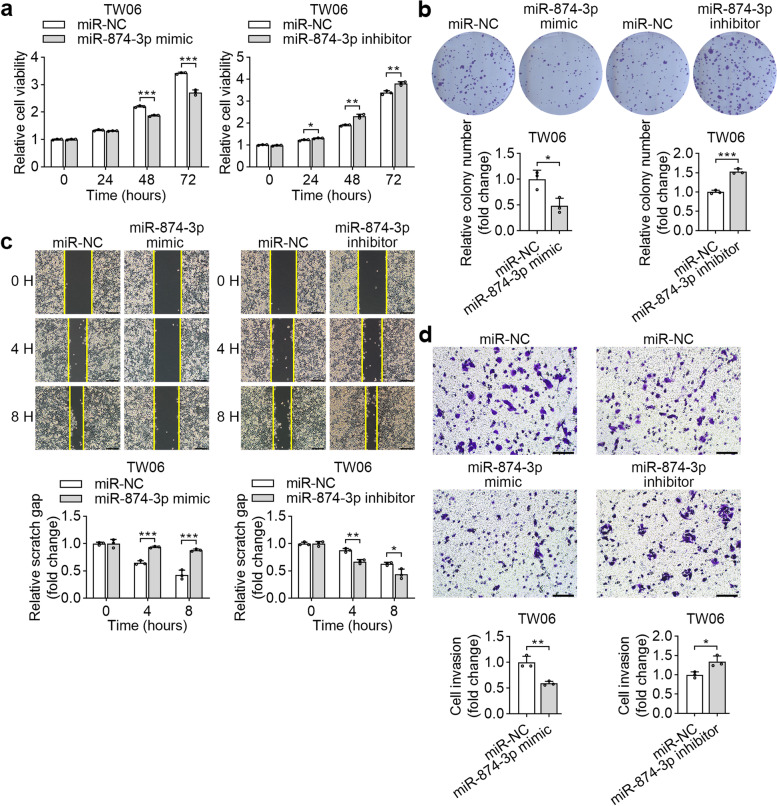
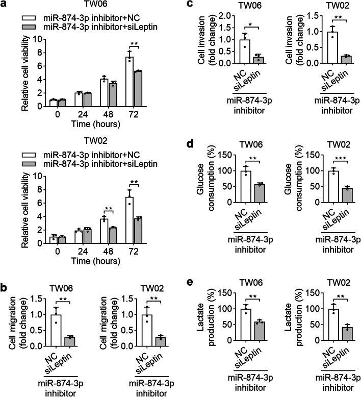
Results: In this study, we found that leptin was highly expressed in the sera and tumor tissues of patients with NPC, and elevated leptin expression was associated with advanced clinical features and poor prognosis. Functional assays demonstrated that leptin remarkably promoted NPC cell growth, motility, and glycolysis in vitro and in vivo. Mechanistically, leptin associated with EGFR, resulting in enhanced cell growth through the regulation of cell-cycle related markers, glycolysis-related genes, and EGFR/AKT/c-Myc signaling. Moreover, leptin potentiated the invasive capacity of NPC cells by promoting EMT. We further explored that miR-874-3p influenced leptin-mediated NPC progression. Overexpression of miR-874-3p prevented cell growth, motility, glucose consumption, and lactate production in NPC cells, whereas miR-874-3p inhibition had the opposite effects. AGO-RIP assays confirmed that Argonaute 2 (AGO2), a protein associated with miR-874-3p, regulated leptin expression in NPC cells. The rescue assays indicated that inhibition of leptin suppressed the effects of miR-874-3p inhibitor. In clinical specimens, miR-874-3p was negatively correlated with leptin.

Conclusions: Leptin may serve as a novel prognostic factor and potential therapeutic target for patients with NPC. In addition, a newly discovered regulatory axis of leptin/EGFR/AKT/c-Myc can provide a novel therapeutic strategy for NPC.

Keywords: EGFR; Leptin; NPC.


Conflict of interest statement

The authors declare no conflicts of interest.

Figures

Fig. 1
Fig. 1
Leptin is overexpression in nasopharyngeal carcinoma. a The mRNA expression level of leptin was determined in 5 benign nasopharyngeal inflammatory tissues and 20 NPC tissues. b Serum level of leptin in NPC patients and healthy controls were detected by ELISA. c Leptin expression was determined by immunohistochemistry in 50-paired paraffin-embedding NPC tissues and adjacent non-tumor tissues, representative images of pathological tumor stage (T) and lymph node stage (N). d and e Kaplan–Meier survival analysis and log-rank tests indicate that high expression level of leptin was associated with poor OS and DFS in NPC patients
Fig. 2
Fig. 2
Leptin expression modulates the cell growth of NPC. a The mRNA and protein expression levels of leptin were assessed in TW06 cells by QPCR and Western blotting. b Cell growth ability was detected by MST assay over four consecutive days. Relative cell growth was normalized to day 0. c The colony formation assays evaluated the effect of leptin on cell proliferation. The representative images and fold change of foci formation were shown. d Leptin expression levels were detected by QPCR and Western blotting after transfection with either a negative control or leptin siRNA. e and f The effect of siLeptin-TW06 on cell proliferation was determined by MTS and colony formation assays. g-h shLeptin-TW06 or sh-NC-TW06 cells were injected into the right flank of null mice for 8 weeks. The tumor volumes were measured every week. The tumor weight in each group was calculated. i Representative images of Ki67 staining were conducted to demonstrate the proliferative cells. *P < 0.05, **P < 0.01, ***P < 0.001
Fig. 3
Fig. 3
Leptin facilitates cell migration and invasion in NPC through EMT. a and c Wound healing assays were conducted to detect change in migratory ability in gain- or loss-of-function of leptin in TW06 cells. b and d TW06 cells with leptin overexpression or silence were subjected to Transwell assays. The representative images and the fold change of cell invasion were presented. e Expressions of EMT markers were detected by Western blotting in gain- or loss-of-function of leptin in TW06 cells. *P < 0.05, **P < 0.01, ***P < 0.001
Fig. 4
Fig. 4
Leptin potentiates glycolysis in NPC cells. a and b The glucose consumption and lactate production were measured in NPC cells with gain- or loss-of-function of leptin. c The association of leptin and glycolytic molecules in GEPIA database was analyzed. d The transcriptional profiles of glycolytic molecules were examined in leptin-overexpressed and leptin-depleted NPC cell lines by QPCR. *P < 0.05, **P < 0.01, ***P < 0.001
Fig. 5
Fig. 5
Leptin triggers cell cycle-related protein expression and EGFR/MAPK/c-Myc pathway in NPC. a Western blot indicated the expressions of cyclin D1, cyclin E, CDK4, p21 and p27 in leptin-overexpression and leptin-depleted TW06 cells. b Western blot analysis was performed to detect the protein levels of p-EGFR, EGFR, p-ERK1/2, ERK1/2, p-AKT, AKT, p-mTOR, mTOR and c-Myc in leptin-overexpression and leptin-depleted TW06 cells. c The IHC was performed to determine the protein expressions of leptin, p-EGFR, EGFR, p-ERK, ERK and c-Myc in the tumor tissues from shleptin-bearing xenograft model. d The EGFR mRNA levels were determined in gain-of-function of leptin in NPC cell lines. e TW06 cell lysates were subjected to EGFR immunoprecipitation with an anti-EGFR antibody or a control antibody, or subjected to leptin immunoprecipitation with an anti-leptin antibody, followed by Western blot of immunoprecipitation with an anti-leptin antibody or with an anti-EGFR antibody, respectively. f The transcriptional profiles of glycolytic molecules were examined by QPCR in leptin-overexpressed TW06 cell lines with c-Myc knockdown. n.s. no significant; *P < 0.05, **P < 0.01
Fig. 6
Fig. 6
Leptin is targeted by miR-874-3p. a Venn diagram of putative miRNA targeting to leptin, and target prediction software miRDB, miRWalk and TargetScan were used for this study. b Predicted binding sites of miR-874-3p within the 3’UTR of leptin mRNA. c and d TW06 and TW02 cells were transfected with miR-874-3p mimics or miR-874-3p inhibitor for 48 h. The leptin mRNA expression levels were examined by QPCR. e Luciferase reporter assays were performed to determine the effect of miR-874-3p on the activity of leptin 3’UTR. f Western blot analysis of leptin and leptin downstream molecules in NPC cells transfected with miR-874-3p mimic, or miR-874-3p inhibitor, and corresponding negative control (NC). g The RIP assay was performed using AGO2 or IgG antibodies in NPC cells and the enrichment of miR-874-3p and leptin were detected by QPCR. h The RIP assays were also performed using AGO2 or IgG to estimate the enrichment of leptin in NPC cells transfected with miR-874-3p mimics and negative control. i The mRNA level of leptin was negatively correlated with miR-874-3p in NPC samples. *P < 0.05, **P < 0.01, ***P < 0.001
Fig. 7
Fig. 7
miR-874-3p suppresses the malignant properties of TW06 cells. a and b MTS assays and colony formation assays were performed to assess the cell proliferation of TW06 cells after transfecting miR-874-3p mimics or miR-874-3p inhibitor and their corresponding negative control. The representative images and fold change of foci formation were shown. c and d The migratory and invasive abilities of TW06 cells transfected with miR-874-3p mimics or miR-874-3p inhibitor and their corresponding negative control were assessed by wound healing and Transwell assays. *P < 0.05, **P < 0.01, ***P < 0.001
Fig. 8
Fig. 8
Leptin mediates the effects of miR-874-3p on NPC. NPC cells were cotransfected with sileptin or the siNC along with the miR-874-3p inhibitor to determine the abilities of cell growth (a), migration (b), invasion (c). The effects of miR-874-3p inhibitor on modulating glucose consumption (d) and lactate production (e) were also assessed by assay kits. *P < 0.05, **P < 0.01, ***P < 0.001
Fig. 9
Fig. 9
A proposed model for an unbalanced miR-874-3p/leptin/EGFR/c-Myc axis regulatory circuit in promoting NPC progression

Similar articles

Cited by

References

    1. Luo SD, Chen WC, Wu CN, Yang YH, Li SH, Fang FM, Huang TL, Wang YM, Chiu TJ, Wu SC: Low-dose aspirin use significantly improves the survival of late-stage NPC: a propensity score-matched cohort study in taiwan. Cancers (Basel). 2020;12(6):1551. - PMC - PubMed
    1. Mahdavifar N, Towhidi F, Makhsosi BR, Pakzad R, Moini A, Ahmadi A, Lotfi S, Salehiniya H. Incidence and mortality of nasopharynx cancer and its relationship with human development index in the world in 2012. World J Oncol. 2016;7:109–118. doi: 10.14740/wjon980w. - DOI - PMC - PubMed
    1. Lo EJ, Bell D, Woo J, Li G, Hanna EY, El-Naggar AK, Sturgis EM. Human papillomavirus & WHO type I nasopharyngeal carcinoma. Laryngoscope. 2010;120(Suppl 4):S185. doi: 10.1002/lary.21649. - DOI - PubMed
    1. Niedobitek G. Epstein-Barr virus infection in the pathogenesis of nasopharyngeal carcinoma. Mol Pathol. 2000;53:248–254. doi: 10.1136/mp.53.5.248. - DOI - PMC - PubMed
    1. Tsao SW, Tsang CM, Lo KW: Epstein-Barr virus infection and nasopharyngeal carcinoma. Philos Trans R Soc Lond B Biol Sci. 2017;372(1732):20160270. - PMC - PubMed

MeSH terms