Clinical Trial
. 2022 Jul 1;19(1):67.
doi: 10.1186/s12984-022-01045-z.

Myoelectric interface training enables targeted reduction in abnormal muscle co-activation

Affiliations
Clinical Trial

Myoelectric interface training enables targeted reduction in abnormal muscle co-activation

Gang Seo et al. J Neuroeng Rehabil. .

Abstract

Background: Abnormal patterns of muscle co-activation contribute to impaired movement after stroke. Previously, we developed a myoelectric computer interface (MyoCI) training paradigm to improve stroke-induced arm motor impairment by reducing the abnormal co-activation of arm muscle pairs. However, it is unclear to what extent the paradigm induced changes in the overall intermuscular coordination in the arm, as opposed to changing just the muscles trained with the MyoCI. This study examined the intermuscular coordination patterns of thirty-two stroke survivors who participated in 6 weeks of MyoCI training.

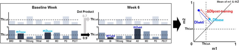
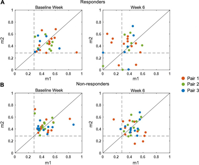
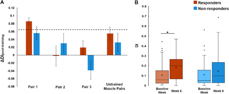
Methods: We used non-negative matrix factorization to identify the arm muscle synergies (coordinated patterns of muscle activity) during a reaching task before and after the training. We examined the extent to which synergies changed as the training reduced motor impairment. In addition, we introduced a new synergy analysis metric, disparity index (DI), to capture the changes in the individual muscle weights within a synergy.

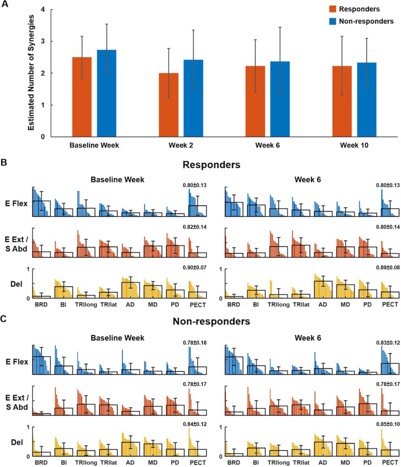
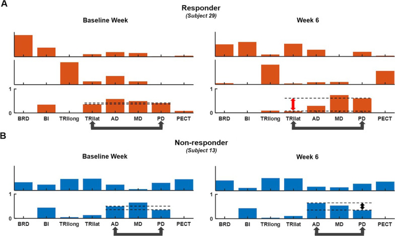
Results: There was no consistent pattern of change in the number of synergies across the subjects after the training. The composition of muscle synergies, calculated using a traditional synergy similarity metric, also did not change after the training. However, the disparity of muscle weights within synergies increased after the training in the participants who responded to MyoCI training-that is, the specific muscles that the MyoCI was targeting became less correlated within a synergy. This trend was not observed in participants who did not respond to the training.

Conclusions: These findings suggest that MyoCI training reduced arm impairment by decoupling only the muscles trained while leaving other muscles relatively unaffected. This suggests that, even after injury, the nervous system is capable of motor learning on a highly fractionated level. It also suggests that MyoCI training can do what it was designed to do-enable stroke survivors to reduce abnormal co-activation in targeted muscles. Trial registration This study was registered at ClinicalTrials.gov (NCT03579992, Registered 09 July 2018-Retrospectively registered, https://clinicaltrials.gov/ct2/show/NCT03579992?term=NCT03579992&draw=2&rank=1 ).

Keywords: Intermuscular coordination; Muscle synergy; Myoelectric computer interface; Stroke motor rehabilitation.


Conflict of interest statement

The authors declare that there are no potential conflicts of interest with respect to the research, authorship, and/or publication of this article.

Figures

Fig. 1
Fig. 1
A reaching task that participants performed to assess the training effects using EMG data. During the motor assessment, participants performed reaches (only away-from-body reaches) three times to each of six targets (approximately 4″ in diameter and spaced 1ft apart), placed at the waist and shoulder height, in front of, and lateral to the impaired arm. The participants were asked to bring the arm back to the rest position at the side (elbow extended maximally, shoulder adducted, not flexed nor rotated with the thumb facing anteriorly) after each reach. The arrows indicate three directions of reach at the waist and shoulder height
Fig. 2
Fig. 2
Calculating the disparity index (DI). From the synergy set of the baseline week, the synergy with the highest co-activation (lowest difference) between the targeted muscle pair (e.g., BI and AD in the second synergy (dark gray); m1base and m2base, their muscle weights) is identified. Both m1base and m2base are higher than ThCoA, the threshold of muscle co-activation. The synergy at week 6 (the second synergy of week 6) corresponding to the synergy of the baseline week is selected by calculating the scalar product with the baseline synergy 2. DI is calculated (right) from m1 and m2 at both time points, as the distance from the mean of m1 and m2 (Eq. 3). Finally, the change in the muscle weight disparity after training (ΔDIpost-training in red) is obtained by subtracting DIbase from DIwk6
Fig. 3
Fig. 3
The number and composition of muscle synergies in responders and non-responders. A Estimation of the number of synergies identified across weeks including pre-training (Baseline week), interim (week 2), post-training (week 6), and retention (week 10) in the responder and non-responder groups. The number of synergies did not differ between the two groups nor across weeks in each group. B, C, The mean and SD of muscle weights, superimposed on the distribution of the muscle weights of responders (B, n = 15) and non-responders (C, n = 17), per each of three synergies identified in pre- and post-training (Baseline week and week 6, respectively). The muscle weights per muscle were displayed in descending order. Eight muscles included: brachioradialis (BRD); biceps brachii (BI); triceps (long (TRIlong) and lateral (TRIlat) heads); anterior, middle, and posterior deltoids (AD, MD, and PD); and pectoralis clavicular fiber (PECT). The synergies were characterized as (1) E Flex: elbow flexors, (2) E Ext/S Abd: elbow extensors, and 3) Del: three heads of the deltoid. The r-value next to each muscle synergy indicates the scalar products between the norm synergy (the mean across the subjects) and its corresponding synergy of each participant (mean ± SD)
Fig. 4
Fig. 4
Exemplary synergies identified pre- and post-training (Baseline week and week 6) of a responder (A Subject 29) and a non-responder (B Subject 13). Arrows below the abbreviation of muscle names indicate the first trained muscle pair for each participant. Dashed lines show the difference between the weights of the trained muscle pair
Fig. 5
Fig. 5
The two-dimensional mapping of activation weights of the trained muscle pairs whose initial co-activation level exceeded ThCoA (dashed lines). m1 and m2 indicate the activation weights of a pair of trained muscles, muscles 1 and 2. Red, green, and blue indicate the muscle pairs trained for the first, second, and third two weeks in the MyoCI training, respectively. The disparity index (DI) measured the average of the distance between each dot and the solid diagonal line (representing co-activation since the weights were equal) for responders (A) and non-responders (B). The increase in DI indicated a decrease in co-activation
Fig. 6
Fig. 6
Changes in the disparity of activation weight of the trained muscle pairs. A ΔDIpost-training (mean ± SD) of each of three trained muscle pairs and all possible pair-wise combinations of untrained muscles in responder and non-responder groups when 5–100% of the ThCoA was applied. The dashed line indicates the average 95th percentile of the bootstrapped ΔDIpre-training. B DI of all of the first trained muscle pairs, computed pre- and post-training in the responder and non-responder groups, respectively, when no weight threshold was applied (*p = 0.011)

Similar articles

Cited by

References

    1. Dewald JP, Pope PS, Given JD, Buchanan TS, Rymer WZ. Abnormal muscle coactivation patterns during isometric torque generation at the elbow and shoulder in hemiparetic subjects. Brain. 1995;118(Pt 2):495–510. doi: 10.1093/brain/118.2.495. - DOI - PubMed
    1. Twitchell TE. The restoration of motor function following hemiplegia in man. Brain. 1951;74(4):443–480. doi: 10.1093/brain/74.4.443. - DOI - PubMed
    1. Beer RF, Dewald JPA, Dawson ML, Rymer WZ. Target-dependent differences between free and constrained arm movements in chronic hemiparesis. Exp Brain Res. 2004;156(4):458–470. doi: 10.1007/s00221-003-1807-8. - DOI - PubMed
    1. Ellis MD, Holubar BG, Acosta AM, Beer RF, Dewald JPA. Modifiability of abnormal isometric elbow and shoulder joint torque coupling after stroke. Muscle Nerve. 2005;32(2):170–178. doi: 10.1002/mus.20343. - DOI - PMC - PubMed
    1. Tresch MC, Cheung VC, d'Avella A. Matrix factorization algorithms for the identification of muscle synergies: evaluation on simulated and experimental data sets. J Neurophysiol. 2006;95(4):2199–2212. doi: 10.1152/jn.00222.2005. - DOI - PubMed

Publication types

Associated data