. 2022 Aug;9(8):1147-1162.
doi: 10.1002/acn3.51611. Epub 2022 Jul 1.

Guanabenz ameliorates disease in vanishing white matter mice in contrast to sephin1

Affiliations

Guanabenz ameliorates disease in vanishing white matter mice in contrast to sephin1

Diede Witkamp et al. Ann Clin Transl Neurol. 2022 Aug.

Abstract

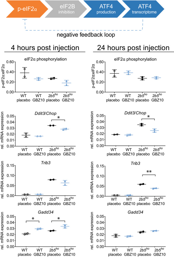
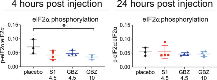
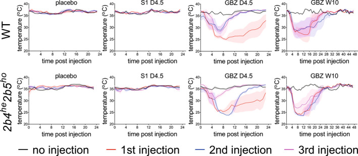
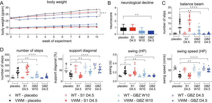
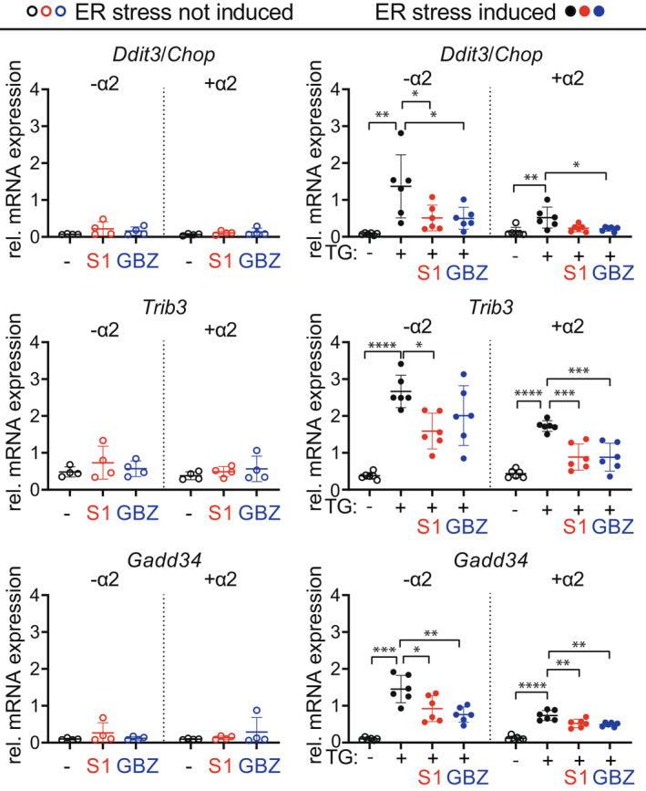
Objective: Vanishing white matter (VWM) is a leukodystrophy, characterized by stress-sensitive neurological deterioration and premature death. It is currently without curative treatment. It is caused by bi-allelic pathogenic variants in the genes encoding eukaryotic initiation factor 2B (eIF2B). eIF2B is essential for the regulation of the integrated stress response (ISR), a physiological response to cellular stress. Preclinical studies on VWM mouse models revealed that deregulated ISR is key in the pathophysiology of VWM and an effective treatment target. Guanabenz, an α2-adrenergic agonist, attenuates the ISR and has beneficial effects on VWM neuropathology. The current study aimed at elucidating guanabenz's disease-modifying potential and mechanism of action in VWM mice. Sephin1, an ISR-modulating guanabenz analog without α2-adrenergic agonistic properties, was included to separate effects on the ISR from α2-adrenergic effects.

Methods: Wild-type and VWM mice were subjected to placebo, guanabenz or sephin1 treatments. Effects on clinical signs, neuropathology, and ISR deregulation were determined. Guanabenz's and sephin1's ISR-modifying effects were tested in cultured cells that expressed or lacked the α2-adrenergic receptor.

Results: Guanabenz improved clinical signs, neuropathological hallmarks, and ISR regulation in VWM mice, but sephin1 did not. Guanabenz's effects on the ISR in VWM mice were not replicated in cell cultures and the contribution of α2-adrenergic effects on the deregulated ISR could therefore not be assessed.

Interpretation: Guanabenz proved itself as a viable treatment option for VWM. The exact mechanism through which guanabenz exerts its ameliorating impact on VWM requires further studies. Sephin1 is not simply a guanabenz replacement without α2-adrenergic effects.


Conflict of interest statement

MSvdK and TEMA have a patent PCT/NL2018/050293 on guanabenz in VWM pending to VUmc. Otherwise, the authors have declared no competing interest exists.

Figures

Figure 1
Figure 1
One i.p. injection with 10 mg/kg GBZ reduces ATF4 transcriptome in WT and 2b5 ho mice. Four‐month‐old male WT (open symbols) and early symptomatic 2b5 ho mice (closed symbols) received one i.p. injection of saline (placebo, black) or 10 mg/kg GBZ (blue). Mice were terminated 4 or 24 h after injection. Brains were taken out and sagittally cut. One half was lysed to obtain protein samples and total RNA samples as described., Western blot and qPCR were performed for indicated ISR markers (Akt was used as reference in qPCR). A simplified overview of the ISR pathway is shown; Ddit3/Chop, Trib3, and Gadd34 are part of the ATF4‐regulated transcriptome. GADD34 is part of the negative feedback loop and dephosphorylates eIF2 when bound to protein phosphatase 1c. Graphs show individual and mean values, ±SD. qPCR and Western blot samples were derived from the same mouse. n = 2 animals per treatment group. Statistically significant differences between placebo‐treated WT and placebo‐treated 2b5 ho mice are not indicated. Treatment effects on protein levels were statistically analyzed with an unpaired t‐test. *p < 0.05, **p < 0.01. [Colour figure can be viewed at wileyonlinelibrary.com]
Figure 2
Figure 2
One i.p. injection with 4.5 mg/kg S1, 4.5 or 10 mg/kg GBZ transiently reduces eIF2α phosphorylation in 2b4 he 2b5 ho mice. Two‐and‐a‐half‐month‐old male early symptomatic 2b4 he 2b5 ho mice received one i.p. injection of 2.25% PEG300. 4.5 or 10 mg/kg GBZ. Mice were terminated 4 or 24 h after injection. Brains were taken out. Cerebella were lysed and samples were subjected to SDS‐PAGE and Western blot to detect Ser51‐phosphorylated eIF2α and total eIF2α. Graphs show individual and mean values, ±SD. Results were statistically analyzed with an unpaired t‐test. *p < 0.05. [Colour figure can be viewed at wileyonlinelibrary.com]
Figure 3
Figure 3
GBZ injection causes temporary hypothermia similarly in WT as in 2b4 he 2b5 ho mice. Temperature loggers were inserted in 2‐month‐old, male WT and pre‐symptomatic 2b4 he 2b5 ho mice. Treatments of daily injection with 2.25% PEG300 (placebo), 4.5 mg/kg S1 (S1 D4.5), 4.5 mg/kg GBZ (GBZ D4.5) or weekly injection with 10 mg/kg GBZ (GBZ W10) were started 1 week after surgery. Injections were placed alternating on left‐ or right‐hand side of the abdominal midline. Body temperature was registered once per hour (h). Temperature at time point 0 was recorded directly before each injection. Graphs show mean body temperature ± SEM. Body temperature is shown for the first three consecutive injections, which follow a daily or weekly interval. Injection of 10 mg/kg GBZ lowered body temperature for more than 24 h, which is the reason for showing a 48‐h time window. Mean baseline temperature for WT and 2b4 he 2b5 ho mice were determined from placebo‐injected animals (WT, 36.6°C, 2b4 he 2b5 ho , 35.4°C) and used for AUC calculations for each injection per animal. AUCs are shown in Figure S1. [Colour figure can be viewed at wileyonlinelibrary.com]
Figure 4
Figure 4
GBZ ameliorates clinical signs in 2b4 he 2b5 ho mice. Eight WT (open symbols) and 16 2b4 he 2b5 ho (VWM) mice (closed symbols) were injected daily with placebo (2.25% PEG300), 4.5 mg/kg S1 (S1 D4.5), 4.5 mg/kg GBZ (GBZ D4.5) or weekly with 10 mg/kg GBZ (GBZ W10) from an age of 6–8 weeks onwards. Injections were placed alternating on left‐ or right‐hand side of the abdominal midline. One mouse displayed signs of epilepsy 2 days after daily injections with 4.5 mg/kg GBZ, was found dead the day after and was omitted from the study. Autopsy did not show signs of infection or other causes for early demise. Graphs show mean phenotypic measures: (A) body weight; (B) neuroscores in VWM mice indicating neurological deterioration; (C) number of slips on balance beam (one S1‐treated VWM mouse was unable to traverse the balance beam and was excluded from analysis); D‐G, gait parameters on the CatWalk in different categories (HP, hind paws): (D) run characterization; E, interlimb coordination; F, temporal; G, kinetic. Neuroscores are 0 in all WT mice (not plotted). Treatment effects by GBZ and S1 were analyzed only for parameters that statistically differed between placebo‐treated WT and placebo‐treated VWM mice. Statistical analyses investigating compound‐related differences in body weight were performed with a repeated measures two‐way ANOVA followed by post hoc Dunnett's correction. Balance beam performance was examined with a Welch's ANOVA and a Dunnett's correction and neuroscore with a Kruskal–Wallis test followed by Dunn's correction. CatWalk data was analyzed with a Kruskal–Wallis with Mann–Whitney U correction. *p < 0.05, **p < 0.01,***p < 0.001, ****p < 0.0001. [Colour figure can be viewed at wileyonlinelibrary.com]
Figure 5
Figure 5
Daily GBZ 4.5 mg/kg reduces Bergmann glia mislocalization in 2b4 he 2b5 ho mice. WT (open symbols) and 2b4 he 2b5 ho (VWM) mice (closed symbols) were injected daily with placebo, 4.5 mg/kg S1 (S1 D4.5), 4.5 mg/kg GBZ (GBZ D4.5) or weekly with 10 mg/kg GBZ (GBZ W10). Sagittally cut brain sections were subjected to immunostaining for S100β (green) and nuclear staining with DAPI (blue). Bergmann glia are double positive for S100β and nuclear DAPI staining. Numbers of mice are n = 2 for WT placebo, WT S1 D4.5, and WT GBZ D4.5, n = 1 for WT GBZ W10, n = 5 for VWM placebo, n = 3 for VWM S1 D4.5 and VWM GBZ D4.5 and n = 4 for VWM GBZ W10. Per mouse 3–6 images were taken. Images show illustrative examples of Bergmann glia localized in the Purkinje layer in WT mice and in the Purkinje and molecular layers in 2b4 he 2b5 ho mice. Bergmann glia were counted in the Purkinje layer (normal location) and in the molecular layer (mislocalized) by a blinded researcher. Percentages of mislocalized Bergmann glia were determined by dividing the number of mislocalized by the total number of Bergmann glia (100%). Graph shows individual and mean percentages of mislocalized Bergmann glia ± SD. The percentage of mislocalized Bergmann glia in placebo‐treated 2b4 he 2b5 ho mice is significantly higher than in WT controls (p < 0.001; nested t‐test; not indicated). Treatment effects on Bergmann glia mislocalization were assessed with a nested one‐way ANOVA followed by Dunnett's correction. *p < 0.05. White bars, 15 μm. [Colour figure can be viewed at wileyonlinelibrary.com]
Figure 6
Figure 6
Daily GBZ 4.5 mg/kg regionally reduces number of immature astrocytes in the corpus callosum in 2b4 he 2b5 ho mice. WT and 2b4 he 2b5 ho mice were injected daily with placebo, 4.5 mg/kg S1 (S1 D4.5), 4.5 mg/kg GBZ (GBZ D4.5) or weekly with 10 mg/kg GBZ (GBZ W10). Sagittally cut brain sections of 3 WT (open symbols) and 5 VWM (closed symbols) mice were stained for nestin (green), GFAP (red), and nuclei (DAPI, blue). Counts of nestin‐GFAP double positive astrocytes (orange) and DAPI‐positive nuclei were determined in two images per rostrum and per splenium of the corpus callosum of each section. (A) Sagittal view of the corpus callosum (shown in white) with the rostrum located anterior and the splenium posterior. (B) Graph shows individual and mean percentages of nestin‐GFAP double positive astrocytes in the corpus callosum ± SD per genotype and treatment. (C and D) Images show representative stainings of VWM brains in indicated sections per indicated treatments. Graphs show percentages of nestin‐GFAP double positive astrocytes in the rostrum (upper panel) or splenium (lower panel) in VWM mice ±SD. Treatment effects on nestin‐GFAP double positive astrocytes were assessed with one‐way ANOVA. Treatment effects on nestin‐GFAP double positive astrocytes in the splenium were corrected with post hoc Dunnett's. C, claustrum; CP, caudoputamen; F, fornix; HC, hippocampus; IC, isocortex; LV, lateral ventricle; S, subiculum *p < 0.05. White bars, 20 μm. [Colour figure can be viewed at wileyonlinelibrary.com]
Figure 7
Figure 7
Daily GBZ 4.5 mg/kg regionally ameliorates myelin pathology the corpus callosum in 2b4 he 2b5 ho mice. WT and 2b4 he 2b5 ho (VWM) mice were injected daily with placebo, 4.5 mg/kg S1 (S1 D4.5), 4.5 mg/kg GBZ (GBZ D4.5) or weekly with 10 mg/kg GBZ (GBZ W10). Sagittally cut brain sections of 2 WT and 2 VWM mice were stained for MOG (brown) and counter stained with H&E (purple, indicating nuclei). Images show representative stainings of indicated sections per indicated treatment. Images are from the same section and represent one mouse per condition. White bars, 50 μm. [Colour figure can be viewed at wileyonlinelibrary.com]
Figure 8
Figure 8
GBZ attenuates ISR parameters in 2b4 he 2b5 ho mouse cerebella. WT (open symbols) and 2b4 he 2b5 ho (VWM, closed symbols) mice were injected daily with placebo, 4.5 mg/kg S1 (S1 D4.5), 4.5 mg/kg GBZ (GBZ D4.5) or weekly with 10 mg/kg GBZ (GBZ W10) from an age of 6–8 weeks onwards for at least 10 weeks. ISR mRNA expression and eIF2α phosphorylation were quantified with qPCR (Hprt as reference) and Western blot in n = 2–3 WT and n = 4–6 2b4 he 2b5 ho VWM cerebella, respectively. RNA and proteins samples were derived from the same cerebellar lysate. Graphs indicate individual data points and means ± SD. Shown ISR markers differ significantly in placebo‐treated WT versus placebo‐treated VWM mice (p < 0.05; not indicated). Treatments effects on ISR markers were statistically analyzed with a one‐way ANOVA followed by post hoc Dunnett's correction. eIF2α phosphorylation findings in WT were statistically analyzed with a one‐way ANOVA followed by post hoc Dunnett's correction (p = 0.0375) and in VWM mice with a Kruskal–Wallis test followed by post hoc Dunn's correction (p = 0.0207). *p < 0.05, **p < 0.01. [Colour figure can be viewed at wileyonlinelibrary.com]
Figure 9
Figure 9
GBZ and S1 attenuates ISR parameters similarly in the absence or presence of the α2‐AR. AtT‐20 cells that express (+α2) or not express (−α2) the α2‐AR subtype 2A were treated with vehicle (−) or thapsigargin (TG: +) for 6 h, in the presence or absence of 50 μmol/L S1 or GBZ to induce ER stress. ISR mRNA marker expression was quantified with qPCR (Akt as reference). Graphs indicate individual data points and means ± SD. Shown ISR markers differ significantly in vehicle‐treated versus TG‐treated cells, as analyzed with an unpaired t‐test or the appropriate non‐parametric alternative. GBZ‐ and S1‐mediated differences were statistically analyzed with one‐way ANOVAs followed by post hoc Dunnett's correction or with a Kruskal–Wallis test followed by post hoc Dunn's correction. *p < 0.05, **p < 0.01, ***p < 0.001, ****p < 0.0001. [Colour figure can be viewed at wileyonlinelibrary.com]

Similar articles

Cited by

References

    1. Hamilton EMC, van der Lei HDW, Vermeulen G, et al. The natural history of vanishing white matter. Ann Neurol. 2018;84:274‐288. - PMC - PubMed
    1. Bugiani M, Boor I, van Kollenburg B, et al. Defective glial maturation in vanishing white matter disease. J Neuropathol Exp Neurol. 2011;70:69‐82. - PMC - PubMed
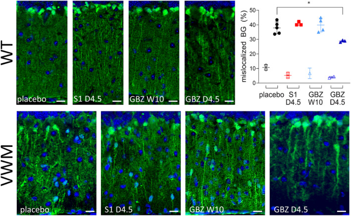
    1. Dooves S, Bugiani M, Wisse LE, Abbink TEM, van der Knaap MS, Heine VM. Bergmann glia translocation: a new disease marker for vanishing white matter identifies therapeutic effects of Guanabenz treatment. Neuropathol Appl Neurobiol. 2018;44:391‐403. - PubMed
    1. Dooves S, Bugiani M, Postma NL, et al. Astrocytes are central in the pathomechanisms of vanishing white matter. J Clin Invest. 2016;126:1512‐1524. - PMC - PubMed
    1. van der Knaap MS, Leegwater PA, Konst AA, et al. Mutations in each of the five subunits of translation initiation factor eIF2B can cause leukoencephalopathy with vanishing white matter. Ann Neurol. 2002;51:264‐270. - PubMed

Publication types

LinkOut - more resources