. 2022 Oct 4;121(19):3651-3662.
doi: 10.1016/j.bpj.2022.06.028. Epub 2022 Jun 30.

Labeling of a mutant estrogen receptor with an Affimer in a breast cancer cell line

Affiliations

Labeling of a mutant estrogen receptor with an Affimer in a breast cancer cell line

Pin Ren et al. Biophys J. .

Abstract

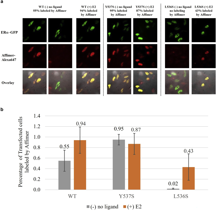
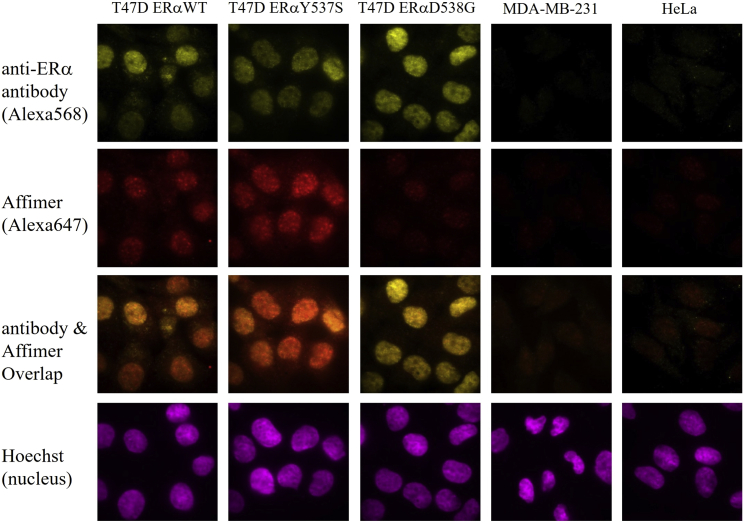
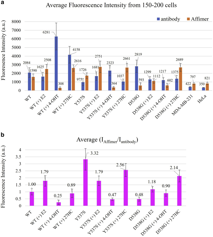
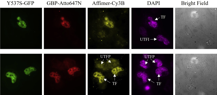
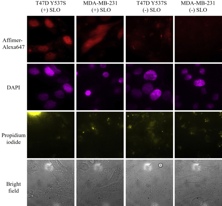
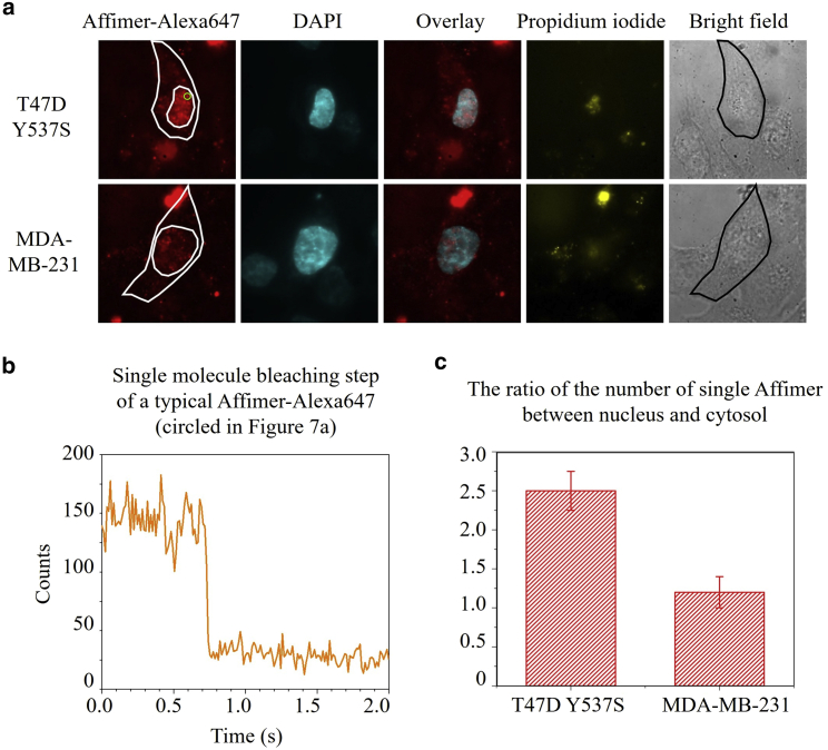
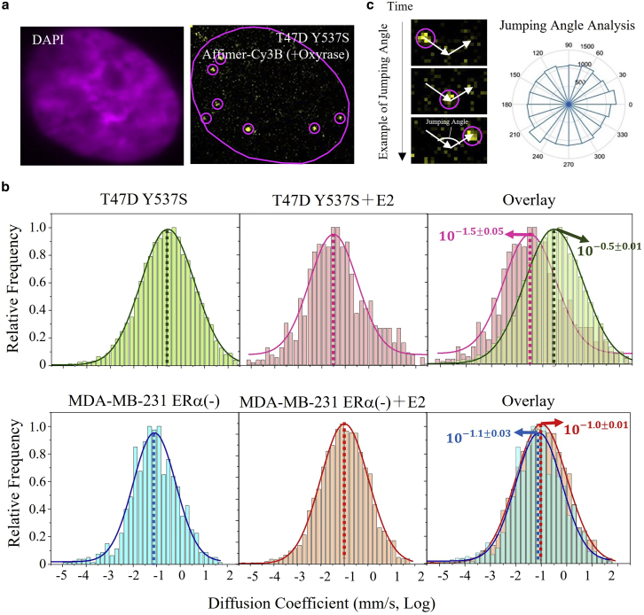
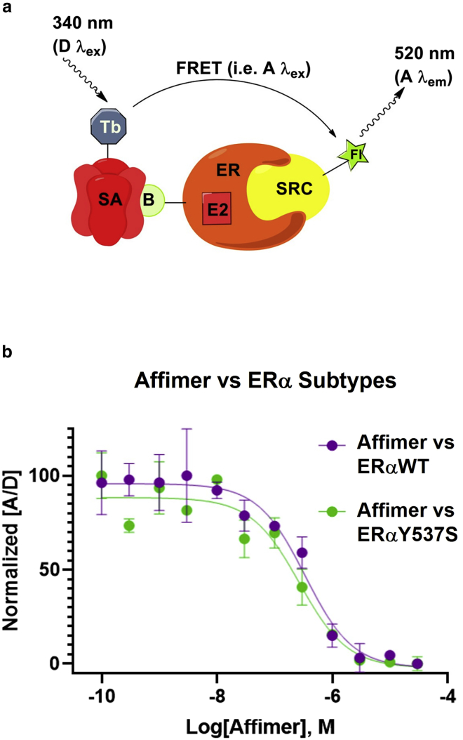
Mutations of the intracellular estrogen receptor alpha (ERα) is implicated in 70% of breast cancers. Therefore, it is of considerable interest to image various mutants (L536S, Y537S, D538G) in living cancer cell lines, particularly as a function of various anticancer drugs. We therefore developed a small (13 kDa) Affimer, which, after fluorescent labeling, is able to efficiently label ERα by traveling through temporary pores in the cell membrane, created by the toxin streptolysin O. The Affimer, selected by a phage display, predominantly labels the Y537S mutant and can tell the difference between L536S and D538G mutants. The vast majority of Affimer-ERαY537S is in the nucleus and is capable of an efficient, unrestricted navigation to its target DNA sequence, as visualized by single-molecule fluorescence. The Affimer can also differentiate the effect of selective estrogen receptor modulators. More generally, this is an example of a small binding reagent-an Affimer protein-that can be inserted into living cells with minimal perturbation and high efficiency, to image an endogenous protein.


Conflict of interest statement

Declaration of interests The authors declare no competing interests.

Figures

Figure 1
Figure 1
a) HeLa cells were individually transfected with ERαWT-GFP, ERαY537S-GFP, or ERαL536S-GFP, and then fixed, permeabilized, and labeled with Affimer-Alexa647, either with no external ligand or with external E2. (b) The addition of E2 to the mutants. Error bars represent the standard deviation. To see this figure in color, go online.
Figure 2
Figure 2
T47D cells with ERαWT, or CRISPR-established ERαY537S or ERαD538G were fixed, permeabilized, and labeled by Affimer-Alexa647 and anti-ERα primary antibody with anti-mouse secondary antibody-Alexa568, and Hoechst (as nucleus marker). MDA-MB-231 (triple-negative breast cancer cell) and HeLa cells (ERα-negative cells) were labeled the same way as the negative controls. To see this figure in color, go online.
Figure 3
Figure 3
a) T47D cells with WT, Y537S, or D538G were treated with 10 nM E2, 1 μM 4-OHT, or 10 μM 27HC for 24 h separately, then fixed, permeabilized, and labeled with Affimer-Alexa647, anti-ERα primary antibody, anti-mouse secondary antibody-Alexa568, and Hoechst (nuclear stain). The absolute fluorescence intensity of Affimer and antibody in the nucleus labeled with Hoechst were recorded and averaged among around 150–200 cells in each sample. MDA-MB-231 and HeLa cells were treated in the same way as negative controls. (b) The average ratio of fluorescence intensity between Affimer and antibody in each single cell (average among 150–200 cells in each sample, normalized to T47D with WT). Error bars represent the standard deviation.To see this figure in color, go online.
Figure 4
Figure 4
In vivo Y537S labeling in live HeLa cells via the SLO technique. External probes, GBP-Atto647N (red) and Affimer-Cy3B (yellow), were delivered into live HeLa cells to label transfected Y537S-GFP (green). A cell-permeable probe, DAPI (magenta), was used as a nucleus marker. Transfected (TF), untransfected and impermeabilized (UTFI), and untransfected and permeabilized (UTFP) cells are shown. To see this figure in color, go online.
Figure 5
Figure 5
In vivo Y537S labeling in T47D Y537S cells and MDA-MB-231 cells via the SLO technique. External probe, Affimer-Alexa647 (red), was delivered into living T47D cells to label endogenous Y537S. DAPI (magenta) was used as nucleus marker. MDA-MB-231 cells were labeled in the same way as the negative control. After recovery, cells were stained with propidium iodide (yellow) to check the integrity of cell membrane. To see this figure in color, go online.
Figure 6
Figure 6
In vivo single Y537S labeling in live T47D and MDA-MB-231 cells via the SLO technique. (a) T47D Y537S cells and MDA-MB-231 cells were labeled by Affimer-Alexa647 and DAPI. Propidium iodide was added to check cell viability after cell recovery. (b) A representative single bleaching step of Affimer-Alexa647 in T47D Y537S cells. (c) Bar graph of the number ratio of single Affimer-Alexa647 between nucleus and cytosol in T47D Y537S and MDA-MB-231 cells. Error bars represent the standard deviation.To see this figure in color, go online.
Figure 7
Figure 7
a) T47D cells with Y537S labeled with Affimer-Cy3B and DAPI by the SLO technique. (b) Diffusion coefficient distribution of single-molecule Affimer fitted by Gaussian function under different conditions. Left, T47D cells with WT, T47D cells with Y537S, and MDA-MB-231 cells without any ligands. Middle, the same cells with 10 nM E2 treatment for 24 h. Right, overlaid graph of no ligand (left) and E2 treatment condition (middle). The mean value of diffusion coefficient from Gaussian fitting in the left and middle graph are indicated by the arrows in the overlaid graphs. (c) Left, example of a jumping angle between two steps on the trajectory of a single molecule circled in Fig. 8a. Right, jumping angle distribution from all detected single-molecule Affimer. Uncertainties in the overlay represent on stabard deviation. To see this figure in color, go online.
Figure 8
Figure 8
a) Schematic of TR-FRET measurement from a terbium-chelate donor excited at 340 nm, which transfers energy to a fluorescein-labeled steroid receptor coactivator (SRC2) peptide bound to ERα. Emission from fluorescein occurs at 520 nm (16). (b) FRET signal recorded in a TR-FRET assay to measure ERα-coactivator interaction in the presence of E2. ERα and coactivator are conjugated with a pair of FRET dyes, terbium donor (D) and fluorescein acceptor (A). When ERαWT and ERαY537S are treated with increasing concentrations of Affimer, the FRET signal is recorded to monitor the interaction between ERα and coactivator. Competition or conformational changes that obscure the SRC2-binding site result in a decrease in TR-FRET signal, as measured by the ratio of acceptor and donor emissions. Error bars represent the standard deviation. To see this figure in color, go online.

Similar articles

Cited by

References

    1. Clusan L., Le Goff P., et al. Pakdel F. A closer look at estrogen receptor mutations in breast cancer and their implications for estrogen and antiestrogen responses. Int. J. Mol. Sci. 2021;22:756. doi: 10.3390/ijms22020756. - DOI - PMC - PubMed
    1. Teng K.W., Ishitsuka Y., et al. Selvin P.R. Labeling proteins inside living cells using external fluorophores for fluorescence microscopy. Elife. 2017;6:e25460. doi: 10.7554/eLife.25460. - DOI - PMC - PubMed
    1. Tai W., Zhao P., Gao X. Cytosolic delivery of proteins by cholesterol tagging. Sci. Adv. 2020;6:eabb0310. doi: 10.1126/sciadv.abb0310. - DOI - PMC - PubMed
    1. Tiede C., Tang A.A.S., et al. McPherson M.J. Adhiron: a stable and versatile peptide display scaffold for molecular recognition applications. Protein Eng. Des. Sel. 2014;27:145–155. doi: 10.1093/protein/gzu007. - DOI - PMC - PubMed
    1. Tiede C., Bedford R., et al. Tomlinson D.C. Affimer proteins are versatile and renewable affinity reagents. Elife. 2017;6:e24903. doi: 10.7554/eLife.24903. - DOI - PMC - PubMed

Publication types

MeSH terms

Substances