

Multifaceted amelioration of cutaneous photoageing by (0.3%) retinol
- PMID: 35778881
- PMCID: PMC9826105
- DOI: 10.1111/ics.12799
Multifaceted amelioration of cutaneous photoageing by (0.3%) retinol
Abstract
Background: Although retinol skin care products improve the appearance of photoaged skin, there is a need for an effective retinol concentration that provides skin benefits without irritation.
Objective: To compare the efficacy of topical 0.1%, 0.3% and 1% retinol in remodelling the cutaneous architecture in an in vivo experimental patch test study, and to determine tolerance of the most effective formulations when used in a daily in-use escalation study.
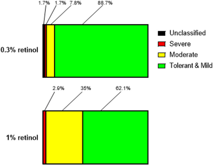
Methods: For the patch test study, retinol products were applied under occlusion, to the extensor forearm of photoaged volunteers (n = 5; age range 66-84 years), and 3 mm skin biopsies obtained after 12 days. Effects of different retinol concentrations, and a vehicle control, on key epidermal and dermal biomarkers of cellular proliferation and dermal remodelling were compared to untreated baseline. Separately, participants (n = 218) recorded their tolerance to 0.3% or 1% retinol over a six-week, approved regimen, which gradually increased the facial applications to once nightly.
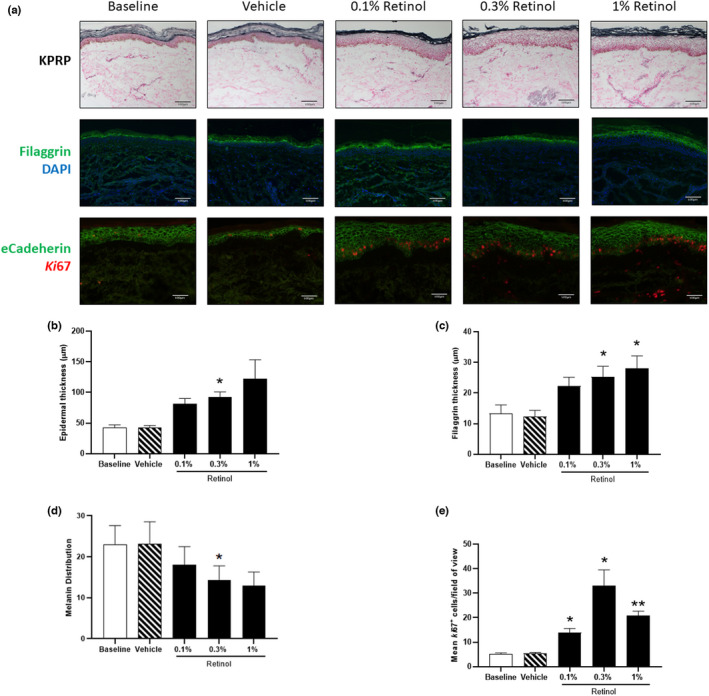
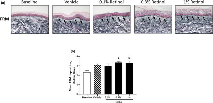
Results: Retinol treatment induced a stepwise increase in epidermal thickness and induced the expression of stratum corneum proteins, filaggrin and KPRP. 0.3% retinol and 1% retinol were comparably effective at inducing keratinocyte proliferation in the epidermis, whilst reducing e-cadherin expression. Fibrillin-rich microfibril deposition was increased following treatment with 0.3% and 1% retinol (p < 0.01); other dermal components remained unaltered (e.g., fibronectin, collagen fibrils, elastin), and no evidence of local inflammation was detected. The in-use study found that 0.3% retinol was better tolerated than 1% retinol, with fewer and milder adverse events reported (χ2 (1) = 23.97; p < 0.001).
Conclusions: This study suggests that 1% and 0.3% retinol concentrations were similarly effective at remodelling photodamaged skin in an in vivo model of long-term use. Use of 0.3% retinol in the escalation study was associated with fewer adverse reactions when applied daily. Hence, 0.3% retinol may be better tolerated than 1% retinol, thereby allowing longer-term topical application.
Contexte: Même si les produits de soins pour la peau à base de rétinol améliorent l'apparence de la peau photovieillie, il est nécessaire d'obtenir une concentration efficace de rétinol procurant des bénéfices cutanés sans irritation.
Objectif: Comparer l'efficacité du rétinol à 0.1%, 0.3% et 1% en application locale dans le remodelage de l'architecture cutanée dans une étude d'irritation cutanée in vivo expérimental, et déterminer la tolérance des formulations les plus efficaces lorsqu'elles sont utilisées dans une étude à doses progressives quotidiennes en cours d'utilisation. MÉTHODES: Pour l'étude d'irritation cutanée, des produits à base de rétinol ont été appliqués sous occlusion, sur le muscle extenseur de l'avant-bras de volontaires présentant des signes de photovieillissement (n = 5; tranche d'âge: 66 à 84 ans), et des biopsies cutanées de 3 mm ont été obtenues après 12 jours. Les effets des différentes concentrations de rétinol, et d'un véhicule témoin sur les principaux biomarqueurs épidermiques et dermiques de la prolifération cellulaire et du remodelage dermique ont été comparés à ceux observés à une région non traitée. Séparément, les participants (n = 218) ont enregistré leur tolérance au rétinol à 0.3% ou 1% au cours d'un schéma posologique approuvé de six semaines, qui a progressivement augmenté les applications faciales à une fois par nuit. RÉSULTATS: Le traitement par rétinol a induit une augmentation progressive de l'épaisseur épidermique, et a induit l'expression des protéines de la couche cornée, la filaggrine et le KPRP. Le rétinol à 0.3% et le rétinol à 1% étaient aussi efficaces pour induire la prolifération des kératinocytes dans l'épiderme, tout en réduisant l'expression de la cadhérine E. Le dépôt de microfibrilles riches en fibrilline a augmenté après un traitement par rétinol à 0.3% et 1% (p < 0.001).
Conclusions: Cette étude suggère que les concentrations de rétinol de 1% et 0.3% étaient aussi efficaces pour remodeler la peau photolésée dans un modèle in vivo lors d'une utilisation à long terme. L'utilisation de rétinol à 0.3% dans l'étude à doses progressives a été associée à moins d'effets indésirables lorsqu'il est appliqué quotidiennement. Par conséquent, le rétinol à 0.3% peut être mieux toléré que le rétinol à 1%, permettant ainsi une application topique à plus long terme.
Keywords: formulation; photodamage; skin barrier; skin physiology/structure.
© 2022 The Authors. International Journal of Cosmetic Science published by John Wiley & Sons Ltd on behalf of Society of Cosmetic Scientists and Societe Francaise de Cosmetologie.
Conflict of interest statement
None.
Figures
Similar articles
-
Cosmetic retinoid use in photoaged skin: A review of the compounds, their use and mechanisms of action.Int J Cosmet Sci. 2025 Feb;47(1):45-57. doi: 10.1111/ics.13013. Epub 2024 Aug 11. Int J Cosmet Sci. 2025. PMID: 39128883 Free PMC article. Review.
-
Retinoids in the treatment of photoageing: A histological study of topical retinoid efficacy in black skin.J Eur Acad Dermatol Venereol. 2024 Aug;38(8):1618-1627. doi: 10.1111/jdv.20043. Epub 2024 Apr 29. J Eur Acad Dermatol Venereol. 2024. PMID: 38682699
-
Histological evaluation of a topically applied retinol-vitamin C combination.Skin Pharmacol Physiol. 2005 Mar-Apr;18(2):81-7. doi: 10.1159/000083708. Skin Pharmacol Physiol. 2005. PMID: 15767769
-
Treatment of facial photodamage using a novel retinol formulation.J Drugs Dermatol. 2013 May;12(5):533-40. J Drugs Dermatol. 2013. PMID: 23652947 Clinical Trial.
-
Amended final report on the safety assessment of glyceryl dilaurate, glyceryl diarachidate, glyceryl dibehenate, glyceryl dierucate, glyceryl dihydroxystearate, glyceryl diisopalmitate, glyceryl diisostearate, glyceryl dilinoleate, glyceryl dimyristate, glyceryl dioleate, glyceryl diricinoleate, glyceryl dipalmitate, glyceryl dipalmitoleate, glyceryl distearate, glyceryl palmitate lactate, glyceryl stearate citrate, glyceryl stearate lactate, and glyceryl stearate succinate.Int J Toxicol. 2007;26 Suppl 3:1-30. doi: 10.1080/10915810701663143. Int J Toxicol. 2007. PMID: 18273450 Review.
Cited by
-
Malnutrition and Allergies: Tipping the Immune Balance towards Health.J Clin Med. 2024 Aug 11;13(16):4713. doi: 10.3390/jcm13164713. J Clin Med. 2024. PMID: 39200855 Free PMC article. Review.
-
Cosmetic retinoid use in photoaged skin: A review of the compounds, their use and mechanisms of action.Int J Cosmet Sci. 2025 Feb;47(1):45-57. doi: 10.1111/ics.13013. Epub 2024 Aug 11. Int J Cosmet Sci. 2025. PMID: 39128883 Free PMC article. Review.
-
Evaluating the Effectiveness and Safety of Home Facial Antiaging Beauty Devices Based on Meridian and Acupoint Theory.J Cosmet Dermatol. 2025 Mar;24(3):e70096. doi: 10.1111/jocd.70096. J Cosmet Dermatol. 2025. PMID: 40099405 Free PMC article. Clinical Trial.
-
Detection of Retinoic Acid in Cosmetics Using Reactive Paper Spray Ionization Mass Spectrometry.Molecules. 2025 Apr 25;30(9):1906. doi: 10.3390/molecules30091906. Molecules. 2025. PMID: 40363713 Free PMC article.
References
-
- Kligman AM. Early destructive effect of sunlight on human skin. JAMA. 1969;210(13):2377–80. - PubMed
-
- Cerimele D, Celleno L, Serri F. Physiological changes in ageing skin. Br J Dermatol. 1990;122(Suppl 35):13–20. - PubMed
-
- Newton VL, Bradley RS, Seroul P, Cherel M, Griffiths CEM, Rawlings AV, et al. Novel approaches to characterize age‐related remodelling of the dermal‐epidermal junction in 2D, 3D and in vivo . Skin Res Technol. 2017;23(2):131–48. - PubMed
-
- Tsuji T. Scanning electron microscope studies of solar elastosis. Br J Dermatol. 1980;103(3):307–12. - PubMed
MeSH terms
Substances
Grants and funding
LinkOut - more resources
Full Text Sources
Medical
