Review
. 2022 Aug;45(8):621-634.
doi: 10.1016/j.tins.2022.05.008. Epub 2022 Jun 30.

Magnetoencephalography with optically pumped magnetometers (OPM-MEG): the next generation of functional neuroimaging

Affiliations
Review

Magnetoencephalography with optically pumped magnetometers (OPM-MEG): the next generation of functional neuroimaging

Matthew J Brookes et al. Trends Neurosci. 2022 Aug.

Abstract

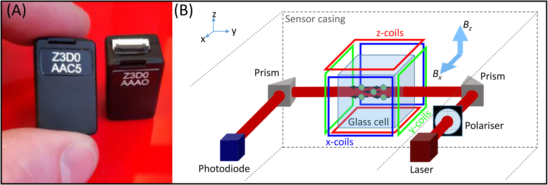
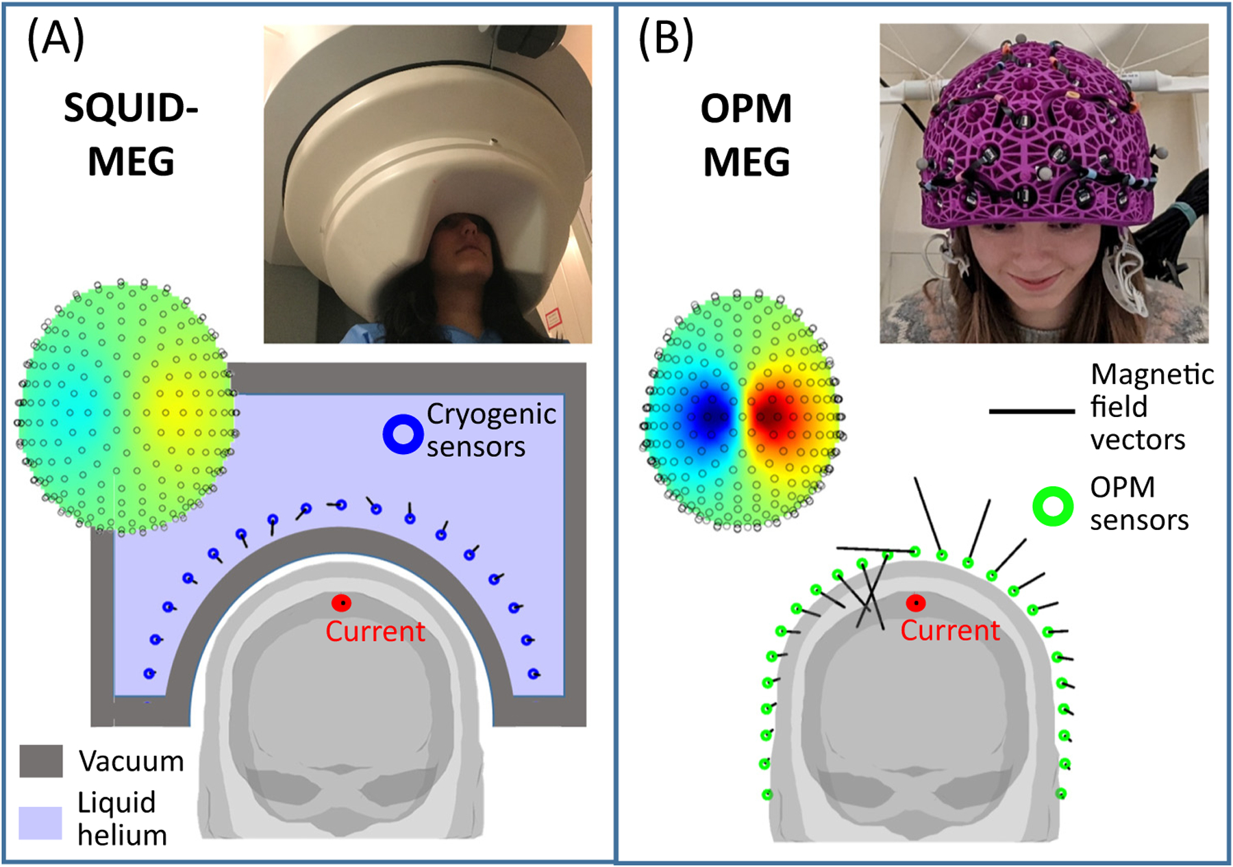
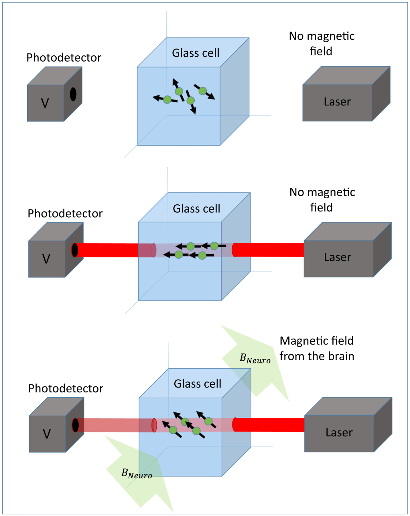
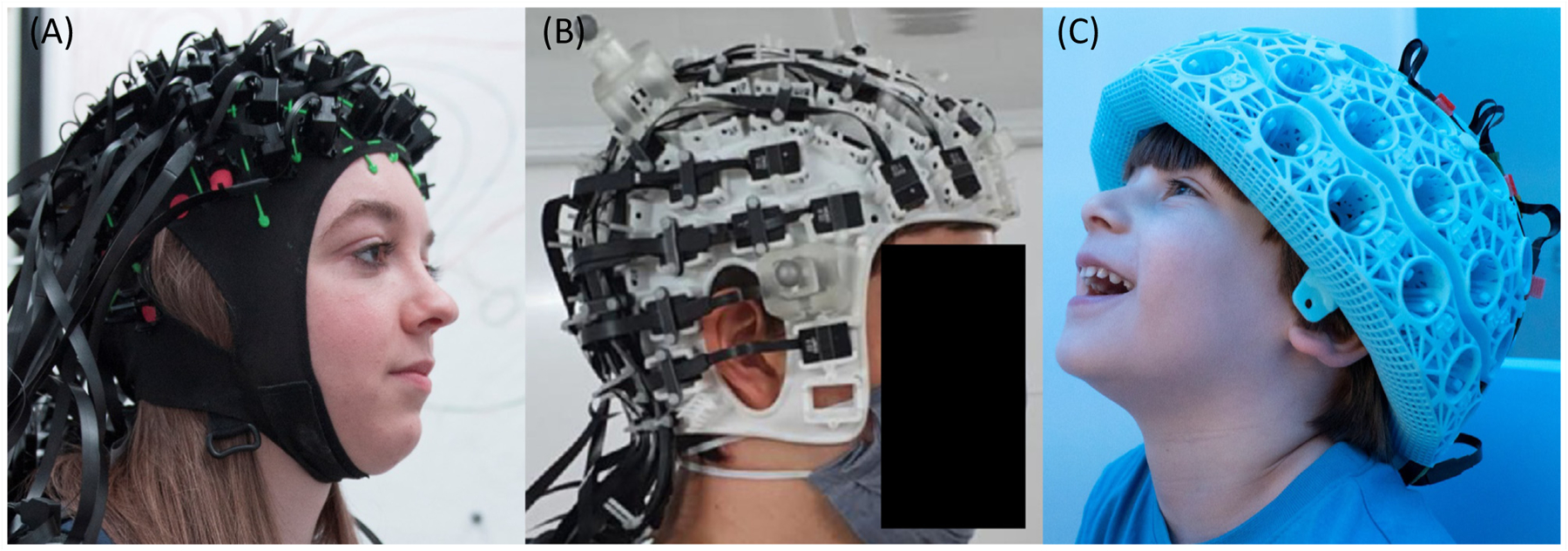
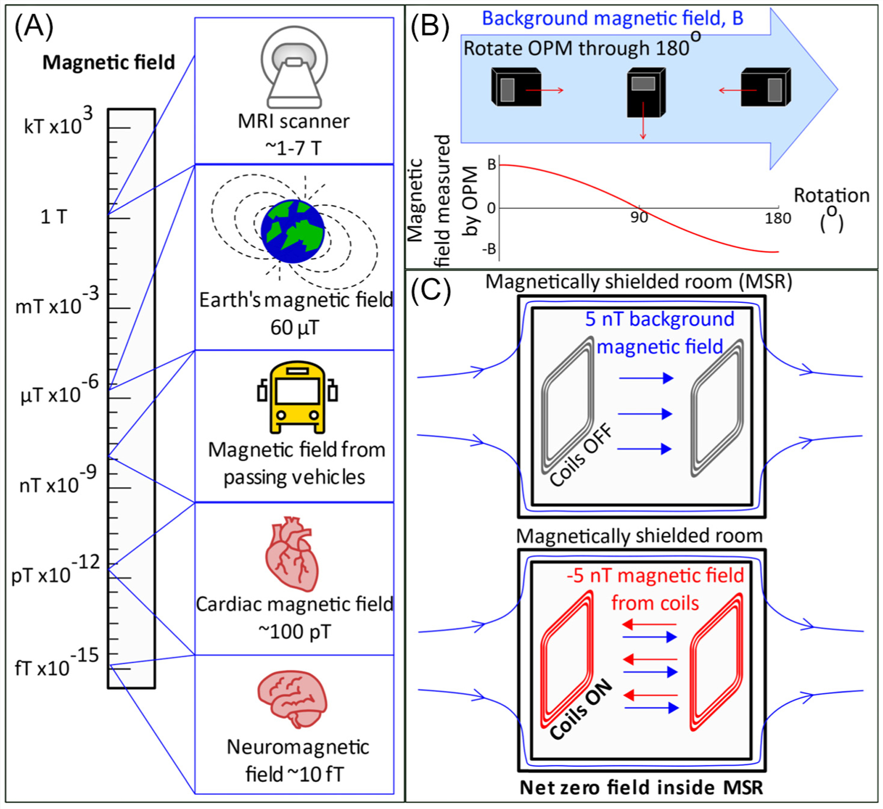
Magnetoencephalography (MEG) measures human brain function via assessment of the magnetic fields generated by electrical activity in neurons. Despite providing high-quality spatiotemporal maps of electrophysiological activity, current MEG instrumentation is limited by cumbersome field sensing technologies, resulting in major barriers to utility. Here, we review a new generation of MEG technology that is beginning to lift many of these barriers. By exploiting quantum sensors, known as optically pumped magnetometers (OPMs), 'OPM-MEG' has the potential to dramatically outperform the current state of the art, promising enhanced data quality (better sensitivity and spatial resolution), adaptability to any head size/shape (from babies to adults), motion robustness (participants can move freely during scanning), and a less complex imaging platform (without reliance on cryogenics). We discuss the current state of this emerging technique and describe its far-reaching implications for neuroscience.

Keywords: OPM-MEG; biomagnetism; electrophysiology; functional brain imaging; neurophysiology; quantum technology.


Conflict of interest statement

Declaration of interests E.B. and M.J.B. are directors of Cerca Magnetics Limited, a spin-out company whose aim is to commercialise aspects of OPM-MEG technology. E.B., M.J.B., R.B., N.H., and R.H. hold founding equity in Cerca Magnetics Limited, and R.B., N.H., and R.H. sit on the scientific advisory board. E.B. is on the scientific advisory board of MyndSpan. The authors are involved in UK patent application numbers 2015427.4, 2106961.2, and 2108360.5, all of which relate to OPM-MEG.

Figures

Figure 1.
Figure 1.. Optically pumped magnetometers (OPMs).
(A) Two OPM sensors, made by QuSpin Inc. (www.quspin.com). Sensors are approximately the size and shape of a (2 × 4) Lego brick. (B) A schematic diagram of the inside of an OPM, showing the component parts. Laser light is directed through a glass cell to interact with atoms in a 87Rb vapour. Coils placed around the cell enable control of the magnetic field within the cell, along all three Cartesian axes. With the laser beam oriented in the y direction, fields oriented in both x and z (Bx and Bz respectively) can be measured.
Figure 2.
Figure 2.. Advantages of optically pumped magnetometer (OPM)-magnetoencephalography (MEG) compared with conventional MEG.
(A) A schematic representation of conventional MEG [superconducting quantum interference device (SQUID)-MEG]. A participant sits with their head in a static helmet (see inset photo, adapted from [16]), containing an array of field sensors (blue circles). Sensors require cryogenic cooling and are consequently bathed in liquid helium. The requirement for thermal insulation (provided by a vacuum, shown in grey) limits sensor proximity to the head, hence the size of the measured magnetic field (represented by the length of the black lines) is limited. For participants with smaller heads (and particularly infants or babies) the sensors would be even further away and, consequently, the signal-to-noise ratio even lower. (B) A schematic representation of OPM-MEG. OPMs do not require cryogenic cooling and so can be mounted flexibly in a lightweight helmet (see inset photo, adapted from [54]) that can be made to fit any head shape. Because sensors are closer to the head compared with conventional MEG, the measured fields (black lines) are larger, increasing sensitivity. In addition, closer proximity allows denser sampling of focal field patterns (examples of which are also shown in the insets), which increases spatial resolution.
Figure I.
Figure I.. Schematic diagram of the operation of an optically pumped magnetometer (OPM).
Laser light is passed through a glass cell containing an Rb vapour onto a photo detector. Interaction of the light with the vapour causes the amount of light passing through the vapour to become a sensitive marker of magnetic field.
Figure I.
Figure I.. Example optically pumped magnetometer (OPM) helmets.
(A) A flexible, ‘EEG-like’ cap (QuSpin Inc.; photo credit: Lisa Gilligan Lee, University of Nottingham). (B) A bespoke helmet, designed to fit an individual’s scalp and specific experimental paradigm (image from [49]; helmet designed by Chalk Studios Ltd.). (C) A generic 3D-printed helmet, which can be used by several participants with approximately similar head size and offers a compromise in terms of cost and performance between individually designed helmets and a one-size-fits-all helmet (Cerca Magnetics Ltd.; photo credit: Lisa Gilligan Lee, University of Nottingham).
Figure I.
Figure I.. Magnetic shielding.
(A) The scale of magnetic fields. (B) The effect of rotating an optically pumped magnetometer (OPM) through 180° in a background field, B. (C) Schematic showing the principle of field nulling.

Similar articles

Cited by

References

    1. Cohen D (1968) Magnetoencephalography: evidence of magnetic fields produced by alpha-rhythm currents. Science 161, 784–786 - PubMed
    1. Bonaiuto JJ et al. (2018) Non-invasive laminar inference with MEG: comparison of methods and source inversion algorithms. NeuroImage 167, 372–383 - PMC - PubMed
    1. Baillet S (2017) Magnetoencephalography for brain electro-physiology and imaging. Nat. Neurosci 20, 327–339 - PubMed
    1. Cohen D (1972) Magnetoencephalography: detection of the brain’s electrical activity with a superconducting magnetometer. Science 5, 664–666 - PubMed
    1. Hamalainen MS et al. (1993) Magnetoencephalography: theory, instrumentation, and applications to non-invasive studies of the working human brain. Rev. Mod. Phys 65, 413–497

Publication types