

A clinical trial on 3D CT scan and polysomnographyc changes after rapid maxillary expansion in children with snoring
- PMID: 35780010
- PMCID: PMC9801059
- DOI: 10.1016/j.bjorl.2022.04.004
A clinical trial on 3D CT scan and polysomnographyc changes after rapid maxillary expansion in children with snoring
Abstract
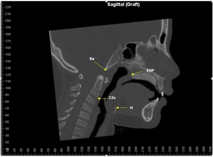
Objective: The present prospective clinical study aimed to investigate the effects of rapid maxillary expansion on the airway, correlating airway volumes obtained on multi-slice computed tomography and polysomnography assessment of oxygen saturation and apnea/hypopnea index.
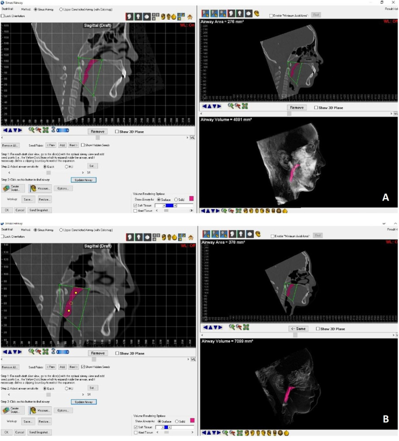
Methods: Twenty-four patients (11 with obstructive sleep apnea and 13 with residual snoring, mean age 10.0 (1.8), were enrolled in the study. Each patient underwent multislice computed tomography and nocturnal polysomnography before rapid maxillary expansion and after removal of maxillary expansion after six months. Airway regions were segmented, and volumes were computed.
Results: The increase in oropharyngeal volume was significant in both groups. Oxygen saturation and apnea/hypopnea index were not statistically significant. No correlation was found between total airway volume, oxygen saturation, and apnea/hypopnea index changes between the time points examined.
Conclusions: This study showed that when rapid maxillary expansion is performed in individuals with sleep-disordered breathing, there were statistically significant differences in oropharyngeal volume between pre- and post-rapid maxillary expansion, but there was no correlation between oxygen saturation values and oropharyngeal volume increase.
Level of evidence: The article is classified as Evidence Level 3 (Three).
Keywords: Craniofacial abnormalities; Imaging, three-dimensional; Palatal expansion technique; Sleep apnea syndromes; Sleep apnea, obstructive.
Copyright © 2022 Associação Brasileira de Otorrinolaringologia e Cirurgia Cérvico-Facial. Published by Elsevier Editora Ltda. All rights reserved.
Figures
References
-
- Carroll J.L. Obstructive sleep-disordered breathing in children: new controversies, new directions. Clin Chest Med. 2003;24:261–282. - PubMed
-
- Brockbank J.C. Update on pathophysiology and treatment of childhood obstructive sleep apnea syndrome. Paediatr Respir Rev. 2017;24:21–23. - PubMed
-
- Li Z., Celestin J., Lockey R.F. Pediatric sleep apnea syndrome: an update. J Allergy Clin Immunol Pract. 2016;4:852–861. - PubMed
Publication types
MeSH terms
LinkOut - more resources
Full Text Sources
Research Materials
