. 2022 Jul 2;19(1):172.
doi: 10.1186/s12974-022-02534-7.

Spermidine reduces neuroinflammation and soluble amyloid beta in an Alzheimer's disease mouse model

Affiliations

Spermidine reduces neuroinflammation and soluble amyloid beta in an Alzheimer's disease mouse model

Kiara Freitag et al. J Neuroinflammation. .

Abstract

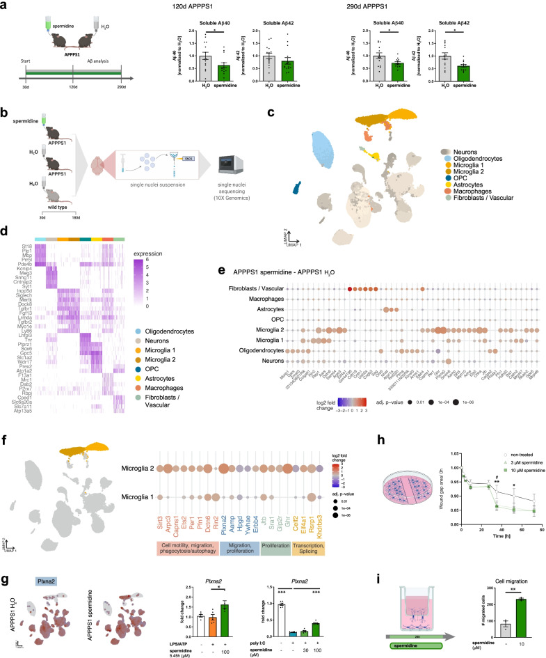
Background: Deposition of amyloid beta (Aβ) and hyperphosphorylated tau along with glial cell-mediated neuroinflammation are prominent pathogenic hallmarks of Alzheimer's disease (AD). In recent years, impairment of autophagy has been identified as another important feature contributing to AD progression. Therefore, the potential of the autophagy activator spermidine, a small body-endogenous polyamine often used as dietary supplement, was assessed on Aβ pathology and glial cell-mediated neuroinflammation.

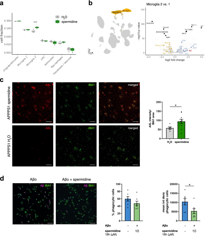
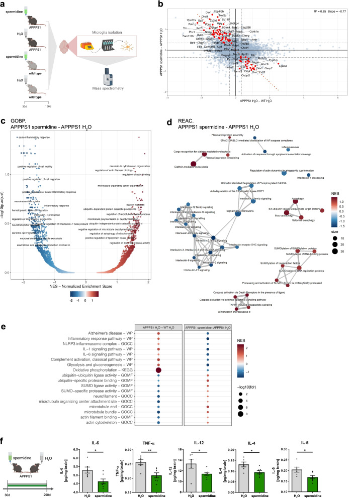
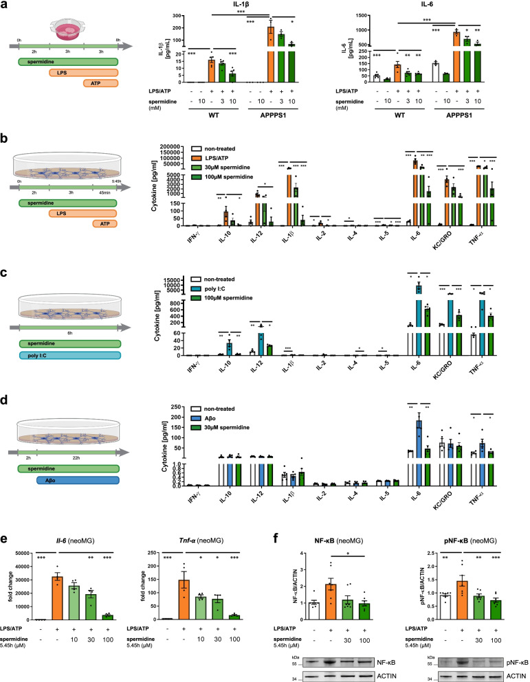
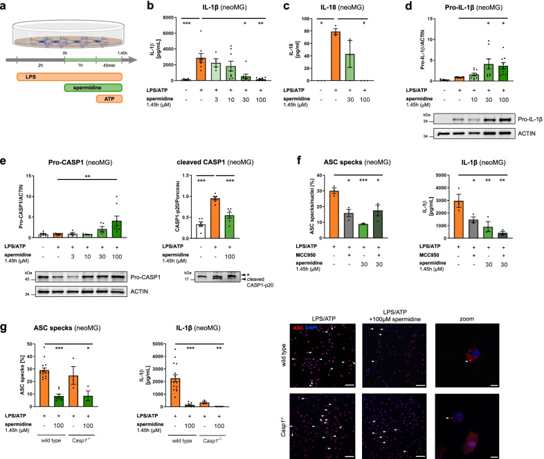
Results: Oral treatment of the amyloid prone AD-like APPPS1 mice with spermidine reduced neurotoxic soluble Aβ and decreased AD-associated neuroinflammation. Mechanistically, single nuclei sequencing revealed AD-associated microglia to be the main target of spermidine. This microglia population was characterized by increased AXL levels and expression of genes implicated in cell migration and phagocytosis. A subsequent proteome analysis of isolated microglia confirmed the anti-inflammatory and cytoskeletal effects of spermidine in APPPS1 mice. In primary microglia and astrocytes, spermidine-induced autophagy subsequently affected TLR3- and TLR4-mediated inflammatory processes, phagocytosis of Aβ and motility. Interestingly, spermidine regulated the neuroinflammatory response of microglia beyond transcriptional control by interfering with the assembly of the inflammasome.

Conclusions: Our data highlight that the autophagy activator spermidine holds the potential to enhance Aβ degradation and to counteract glia-mediated neuroinflammation in AD pathology.

Keywords: Alzheimer’s disease; Astrocytes; Autophagy; Dietary supplement; Liquid chromatography tandem mass spectrometry; Microglia; Neuroinflammation; Phagocytosis; Single nuclei sequencing; Spermidine.


Conflict of interest statement

The authors declare that they have no competing interests.

Figures

Fig. 1
Fig. 1
Spermidine reduced soluble Aβ and induced transcriptomic alterations in microglia of APPPS1 mice. a APPPS1 mice were treated with 3 mM spermidine via their drinking water starting at 30 days (d) until mice reached an age of 120 days or 290 days according to the depicted treatment scheme. Spermidine-treated APPPS1 mice were compared to non-treated controls (H2O). The Aβ40 and Aβ42 content was measured in the TBS (soluble) fraction of brain homogenates of 120-day-old or 290-day-old spermidine-treated mice and water controls (mixed sex) using electrochemiluminescence (MesoScale Discovery panel). Values were normalized to water controls. 120d APPPS1 H2O (n = 14), 120d APPPS1 spermidine (n = 14), 290d APPPS1 H2O (n = 14), 290d APPPS1 spermidine (n = 12); two‐tailed t‐test, Aβ42 in 120d mice: Mann–Whitney U test. b Single nuclei sequencing of hemispheres harvested from 180-day-old male spermidine-treated APPPS1, H2O APPPS1 and H2O control mice was performed of FACS-sorted DAPI-stained nuclei using the 10x Genomics platform (n = 3). c UMAP embedding and clustering of the snRNA-seq data, together with annotation of the major cell types. d Heatmap showing the top 5 marker genes for 300 cells in each of the major cell types. e Dot plot for the top 50 genes in a cell-type-specific differential expression analysis between spermidine-treated APPPS1 and H2O APPPS1 mice. Color scale indicates log2 fold change, dot size indicates adjusted p value. f Same as e, for selected genes differentially expressed in microglia clusters 1 or 2. Associated pathways are color-coded. g Expression of Plxna2 in APPPS1 spermidine and APPPS1 H2O mice. Color scale indicates normalized expression, grey dots represent no data (left panels). For validation, neonatal microglia were treated with the indicated concentrations of spermidine in combination with LPS (1 µg/ml) and ATP (2 mM) or with poly I:C (50 µg/ml) and the gene expression was assessed by RT-qPCR (right panels). Plxna2 expression was normalized to Actin and displayed as fold change compared to non-treated control cells; n = 5–6, one-way ANOVA, Dunnett’s post hoc test. h Neonatal microglia were pre-treated with 3 or 10 µM spermidine for 15 h. The confluent cell layer was scratched and the scratch area was imaged for 72 h at the indicated timepoints. The gap area normalized to timepoint 0 h is displayed; n = 5–6, two-way ANOVA, Dunnett’s post hoc test. i Neonatal microglia were non-treated or treated with 10 µM spermidine and their migration towards 300 µM ATP was quantified after 24 h using a transwell migration assay; two‐tailed t‐test. *p < 0.05, **p < 0.01, ***p < 0.001
Fig. 2
Fig. 2
Spermidine altered AD-associated microglia and their capacity to degrade Aβ. a Cluster abundance in snRNA-seq of male spermidine-treated APPPS1 and H2O APPPS1 mice with p values from mixed-effects binomial model. b Volcano plot of genes differentially expressed in microglia cluster 2 vs. 1. The top 5 up- and down-regulated genes are indicated as well as previously published markers for homeostatic (yellow) and disease-associated (blue) microglia [1]. Significance threshold of adj. p value < 0.01 was used. Axl as a gene of interest is highlighted in red. c Tissue sections of male 180-day-old mice were stained for the microglia cluster 2 marker AXL (red) and IBA1 (green). The AXL intensity normalized to the IBA1 area was determined by ImageJ analysis; n = 6–10, two‐tailed t‐test. d Neonatal microglia were pre-treated for 18 h with 10 µM spermidine and fluorescently labelled oligomeric (Aβo) Aβ (magenta) was added for further 24 h. Microglia cells were stained for IBA1 (green). The percentage of phagocytic cells and the Aβ mean intensity density per phagocytic cell was assessed by confocal microscopy. Representative images are shown; n = 7, two‐tailed t‐test. *p < 0.05, **p < 0.01, ***p < 0.001
Fig. 3
Fig. 3
Spermidine treatment reduced progressive neuroinflammation in APPPS1 mice. a Male APPPS1 mice were treated with 3 mM spermidine via their drinking water starting at 30 days (d) until mice reached an age of 180 days. Microglia were isolated by MACS and the proteome assessed by mass spectrometry. b Scatterplot of protein regulation in Contrast2 (APPPS1 spermidine—APPPS1 H2O, y axis) vs. its regulation in Contrast1 (APPPS1 H2O,—WT H2O, x axis). Contrast 2 shows regulation due to spermidine effect, Contrast 1 shows the regulation of proteins by Alzheimer disease. Proteins that are regulated by spermidine and show significant anti-APPPS1 effect were marked in red. As such we selected proteins that show significant (alpha = 0.04) regulation due to spermidine in APPPS1 mice (Contrast2) and simultaneously, show significant (alpha = 0.04) effect in Contrast5 = (Contrast2 − Contrast1)/2, in the direction, opposite to the effect of the AD-like model. c Volcano plot of GSEA enrichment of GO BP terms. x-axis shows normalized enrichment score of functional term, y-axis represent the − log10 of its false discovery rate. Labelled are only terms that have relation to neurodegeneration and inflammation. As such we selected terms that have in their names following strings: neuro, inflamm, Clathrin, interleukin, Caspase, TNF, ubiquitin, SUMO, Alzheimer, Parkinson, Huntington, lipoprotein, autophagy, cell migration, cell motility, microtubule, actin, actin-, glia, amyloid. Not all labels appear due to strong overlap, especially at high fdr > ~ 0.5 (− log10(fdr) < ~ 0.3). Long term names are truncated to 50 characters. d GSEA enrichment map using top 50 REACTOME terms from list of neurodegeneration and inflammation terms. As such we selected terms that have in their names following strings: neuro, inflamm, Clathrin, interleukin, Caspase, TNF, ubiquitin, SUMO, Alzheimer, Parkinson, Huntington, lipoprotein, autophagy, cell migration, cell motility, microtubule, actin, actin-, glia, amyloid. e Dot plot of selected functional terms related to neuroinflammation and degeneration. f APPPS1 mice were treated with 3 mM spermidine via their drinking water starting at 30 days until mice reached an age of 290 days. The content of the indicated pro-inflammatory cytokines was measured in the TBS (soluble) fraction of brain homogenates of male spermidine-treated mice and water controls using electrochemiluminescence (MesoScale Discovery panel). Values were normalized to water controls. 290d APPPS1 H2O (n = 14), 290d APPPS1 spermidine (n = 12); two‐tailed t‐test. *p < 0.05, **p < 0.01, ***p < 0.01
Fig. 4
Fig. 4
Spermidine exhibits direct anti-inflammatory effects on microglia. a Hemispheres of wild type (WT) and APPPS1 mice were coronally sliced and treated with the indicated spermidine concentration, LPS (10 µg/ml) and ATP (5 mM) as depicted. The IL-1β and IL-6 concentration in the supernatant was determined by ELISA; n = 3–5, two-way ANOVA, Tukey’s post hoc test. b–f Neonatal microglia (neoMG) were either treated with LPS (1 µg/ml) and ATP (2 mM), with poly I:C (50 µg/ml) or with oligomeric Aβ (Aβo, 5 µM) and the indicated spermidine concentrations as depicted in the schemes. b–d Amount of cytokines in the cell supernatant was determined by electrochemiluminescence (MesoScale Discovery panel); n = 4–5. b IFN-γ, IL-10, IL-12, IL-2: Kruskal–Wallis, Dunn’s multiple comparison; IL-1β, IL-4, IL-5, IL-6, KC/GRO, TNF-α: one-way ANOVA, Dunnett’s post hoc test. c INF-γ, IL-2, IL-4: Kruskal–Wallis, Dunn’s multiple comparison; IL-10, IL-12, IL-1β, IL-5, IL-6, KC/GRO, TNF-α: one-way ANOVA, Dunnett´s post hoc test. d IL-10, IL-12, IL-4, KC/GRO: Kruskal–Wallis, Dunn’s multiple comparison; INF-γ, IL-1β, IL-2, IL-5, IL-6, TNF-α: one-way ANOVA, Dunnett’s post hoc test. e The gene expression of Tnf-α and Il-6 was assessed by RT-qPCR after treatment of neonatal microglia as depicted in b. Their expression was normalized to Actin and displayed as fold change compared to non-treated control cells; n = 4. Il-6: one-way ANOVA, Dunnett’s post hoc test; Tnf-α: Kruskal–Wallis, Dunn’s multiple comparison. f Levels of phosphorylated NF-κB (pNF-κB) and NF-κB were determined by western blot in neonatal microglia treated as depicted in b. Representative images are shown and protein levels are displayed as fold changes compared to non-treated controls normalized to ACTIN; n = 7. NF-κB: Kruskal–Wallis, Dunn’s multiple comparison; pNF-κB: one-way ANOVA, Dunnett’s post hoc test. *p < 0.05, **p < 0.01, ***p < 0.001
Fig. 5
Fig. 5
Spermidine regulates neuroinflammation beyond transcription by interfering with inflammasome assembly. Neonatal microglia (neoMG) were treated with LPS (1 µg/ml) and spermidine at indicated concentrations for 1.45 h and ATP (2 mM) as depicted in the scheme (a). b IL-1β concentration in the cell supernatant was determined by ELISA; n = 4–8; Kruskal–Wallis, Dunn´s multiple comparison. c IL-18 concentration in the cell supernatant was determined by ELISA; n = 3; Kruskal–Wallis, Dunn’s multiple comparison. d Pro-IL-1β protein levels were determined by western blot and normalized to ACTIN. Representative images are shown and values are displayed as fold changes compared to LPS/ATP-treated cells; n = 8–9; Kruskal–Wallis, Dunn’s multiple comparison. e Cellular Pro-CASP1 and cleaved CASP1 p20 levels in the supernatant were determined by western blot (* non-specific band). Pro-CASP1 was normalized to ACTIN (n = 4–8) and CASP1 p20 was normalized on whole protein content determined by Ponceau S staining (n = 3). Values are displayed as fold changes compared to LPS/ATP-treated cells; Pro-CASP1: Kruskal–Wallis, Dunn´s multiple comparison; cleaved CASP1: one-way ANOVA, Dunnett’s post hoc test. f Neonatal microglia were stimulated as shown in a and MCC950 was added 15 min before addition of ATP. Cells were stained for ASC to visualize inflammasomes and with DAPI for nuclear staining. The percentage of ASC specks in respect to the number of total cells (DAPI positive cells) was determined (left). The IL-1β concentration in the cell supernatant was assessed by ELISA (right); n = 3; one-way ANOVA, Dunnett’s post hoc test. g Neonatal WT and Casp1−/− microglia were stimulated as shown in a but with 4 mM ATP to increase the number of inflammasomes. Cells were stained for ASC (red) to visualize inflammasomes and with DAPI (blue) for nuclear staining as shown in the representative images (scale bar = 100 µm). Arrowheads highlight ASC specks within microglia. The percentage of ASC specks in respect to the number of total cells (DAPI positive cells) was determined (left). The IL-1β concentration in the cell supernatant was assessed by ELISA (right); WT: n = 8–16; Casp1−/−: n = 3. Kruskal–Wallis, Dunn’s multiple comparison. *p < 0.05, **p < 0.01, ***p < 0.001

References

    1. Keren-Shaul H, Spinrad A, Weiner A, Matcovitch-Natan O, Dvir-Szternfeld R, Ulland TK, et al. A unique microglia type associated with restricting development of Alzheimer’s disease. Cell. 2017;169(7):1276–90.e17. doi: 10.1016/j.cell.2017.05.018. - DOI - PubMed
    1. Heppner FL, Ransohoff RM, Becher B. Immune attack: the role of inflammation in Alzheimer disease. Nat Rev Neurosci. 2015;16(6):358–372. doi: 10.1038/nrn3880. - DOI - PubMed
    1. LaRocca TJ, Cavalier AN, Roberts CM, Lemieux MR, Ramesh P, Garcia MA, et al. Amyloid beta acts synergistically as a pro-inflammatory cytokine. Neurobiol Dis. 2021;159:105493. doi: 10.1016/j.nbd.2021.105493. - DOI - PMC - PubMed
    1. Montoliu-Gaya L, Mulder SD, Herrebout MAC, Baayen JC, Villegas S, Veerhuis R. Aβ-oligomer uptake and the resulting inflammatory response in adult human astrocytes are precluded by an anti-Aβ single chain variable fragment in combination with an apoE mimetic peptide. Mol Cell Neurosci. 2018;89:49–59. doi: 10.1016/j.mcn.2018.03.015. - DOI - PubMed
    1. Vuillermot S, Joodmardi E, Perlmann T, Ove Ögren S, Feldon J, Meyer U. Prenatal immune activation interacts with genetic <em>Nurr1</em> deficiency in the development of attentional impairments. J Neurosci. 2012;32(2):436–451. doi: 10.1523/JNEUROSCI.4831-11.2012. - DOI - PMC - PubMed

MeSH terms