Review
. 2022 Oct;474(10):1043-1051.
doi: 10.1007/s00424-022-02719-2. Epub 2022 Jul 2.

Regulation of mitochondrial temperature in health and disease

Affiliations
Review

Regulation of mitochondrial temperature in health and disease

Zaynab El-Gammal et al. Pflugers Arch. 2022 Oct.

Abstract

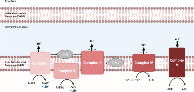
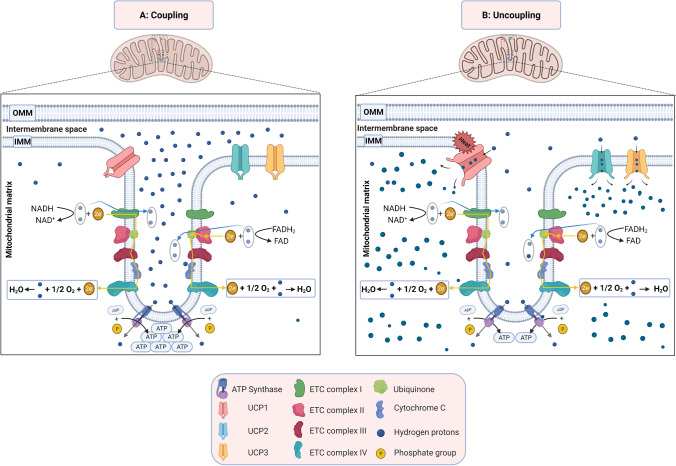
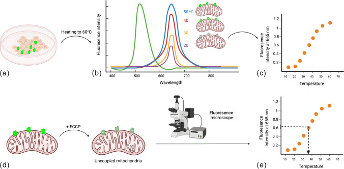
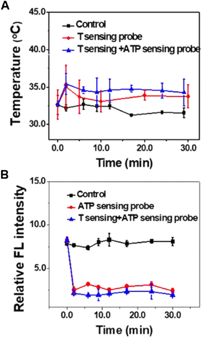
Mitochondrial temperature is produced by various metabolic processes inside the mitochondria, particularly oxidative phosphorylation. It was recently reported that mitochondria could normally operate at high temperatures that can reach 50℃. The aim of this review is to identify mitochondrial temperature differences between normal cells and cancer cells. Herein, we discussed the different types of mitochondrial thermosensors and their advantages and disadvantages. We reviewed the studies assessing the mitochondrial temperature in cancer cells and normal cells. We shed the light on the factors involved in maintaining the mitochondrial temperature of normal cells compared to cancer cells.

Keywords: Heat shock proteins; Mitochondria; Mitochondrial temperature; Oxidative phosphorylation; Uncoupling proteins.


Conflict of interest statement

The authors declare no competing interests.

Figures

Fig. 1
Fig. 1
Electron transport chain transfer protons H+ across a membrane to synthesize ATP. Created with BioRender.com
Fig. 2
Fig. 2
Mitochondrial coupling and uncoupling. A Mitochondrial coupling: proton pumps of the electron transport chain uses redox energy to generate proton motive force. This force will regenerate ATP by ATP synthase. B Mitochondrial uncoupling inhibits the coupling between electron transport and ATP-synthetic reactions. This in turn causes loss of the energy as heat. Created with BioRender.com
Fig. 3
Fig. 3
Measuring mitochondrial temperature by fluorescent thermosensors. a Cells are stained by fluorescent thermosensors and gradually heated. b Fluorescence intensity, measured by means of fluorescent microscopy at different temperatures will be recorded. c A plot of fluorescence intensity versus temperature will be drawn “Calibration plot.” d Uncoupler reagent (such as FCCP) will be added to induce mitochondrial uncoupling. e By means of fluorescence microscope, the fluorescence intensity of the cells will be measured by fluorescence microscopy and plotted (c) to conclude the cell temperature
Fig. 4
Fig. 4
Mitochondrial temperature fluctuation response to FCCP inhibition in HeLa cells. Live HeLa cells were prestained with the T sensing probe (0.5 mg mL−1, 20 min) and the ATP sensing probe (5.0 μM, 20 min). The intensity data was obtained from 25 live HeLa cells. Reprinted with permission from “Qiao, J., Chen, C., Shangguan, D., Mu, X., Wang, S., Jiang, L., & Qi, L. (2018). Simultaneous monitoring of mitochondrial temperature and ATP fluctuation using fluorescent probes in living cells. Analytical chemistry90(21), 12,553–12,558”, Fig. 6A. Copyright 2022 American Chemical Society
Fig. 5
Fig. 5
Visualization of mitochondrial thermal dynamics in HeLa cells response to glucose stimulations. (Left) Upconversion nanoparticles (UCNPs) at (3carboxypropyl) triphenylphosphonium bromide (TPP) images. MitoTracker (red) and UCNPs@TPP (green) from different treatments. (Middle) Mitochondrial temperature dynamics in the presence of 5 mg/mL glucose within 30 min. (Right) Student’s t test of both no glucose and glucose at 10 min (p < 0.0001). Reprinted with permission from “Di, X., Wang, D., Zhou, J., Zhang, L., Stenzel, M. H., Su, Q. P., & Jin, D. (2021). Quantitatively monitoring in situ mitochondrial thermal dynamics by upconversion nanoparticles. Nano letters, 21(4), 1651–1658”, Fig. 4A. Copyright 2022 American Chemical Society

References

    1. Alberts B (2018). Molecular biology of the cell.
    1. Andrews ZB, Diano S, Horvath TL. Mitochondrial uncoupling proteins in the CNS: in support of function and survival. Nat Rev Neurosci. 2005;6(11):829–840. - PubMed
    1. Arai S, Suzuki M, Park S-J, Yoo JS, Wang L, Kang N-Y, Ha H-H, Chang Y-T. Mitochondria-targeted fluorescent thermometer monitors intracellular temperature gradient. Chem Commun. 2015;51(38):8044–8047. - PubMed
    1. Archibald JM. Origin of eukaryotic cells: 40 years on. Symbiosis. 2011;54(2):69–86.
    1. Arulkumaran N, Deutschman CS, Pinsky MR, Zuckerbraun B, Schumacker PT, Gomez H, Gomez A, Murray P, Kellum JA, Workgroup AX. Mitochondrial function in sepsis SHOCK. 2016;45(3):271–281. - PMC - PubMed

Publication types

Substances

LinkOut - more resources