. 2022 Sep:134:18-24.
doi: 10.1016/j.pediatrneurol.2022.06.007. Epub 2022 Jun 16.

Cerebrovascular Complications of COVID-19 Disease in Children: A Single-Center Case Series

Affiliations

Cerebrovascular Complications of COVID-19 Disease in Children: A Single-Center Case Series

Klára Španělová et al. Pediatr Neurol. 2022 Sep.

Abstract

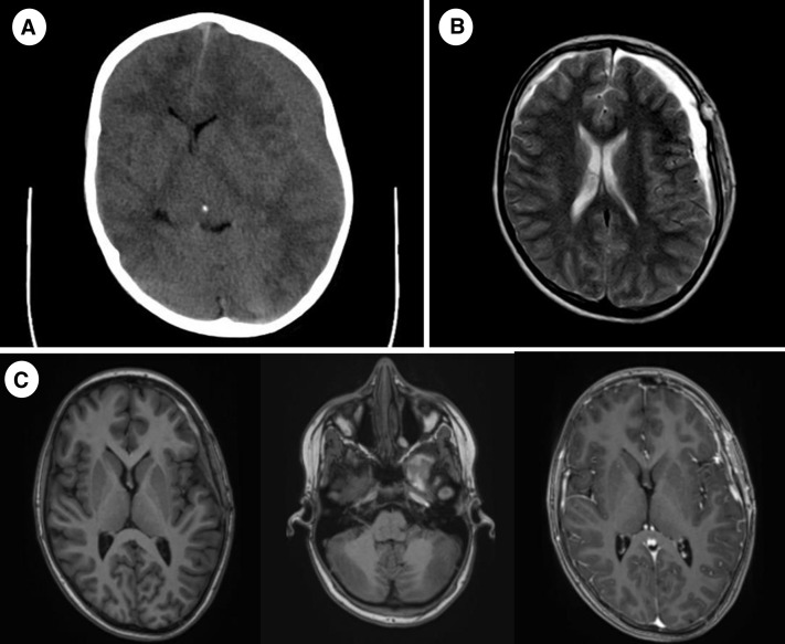
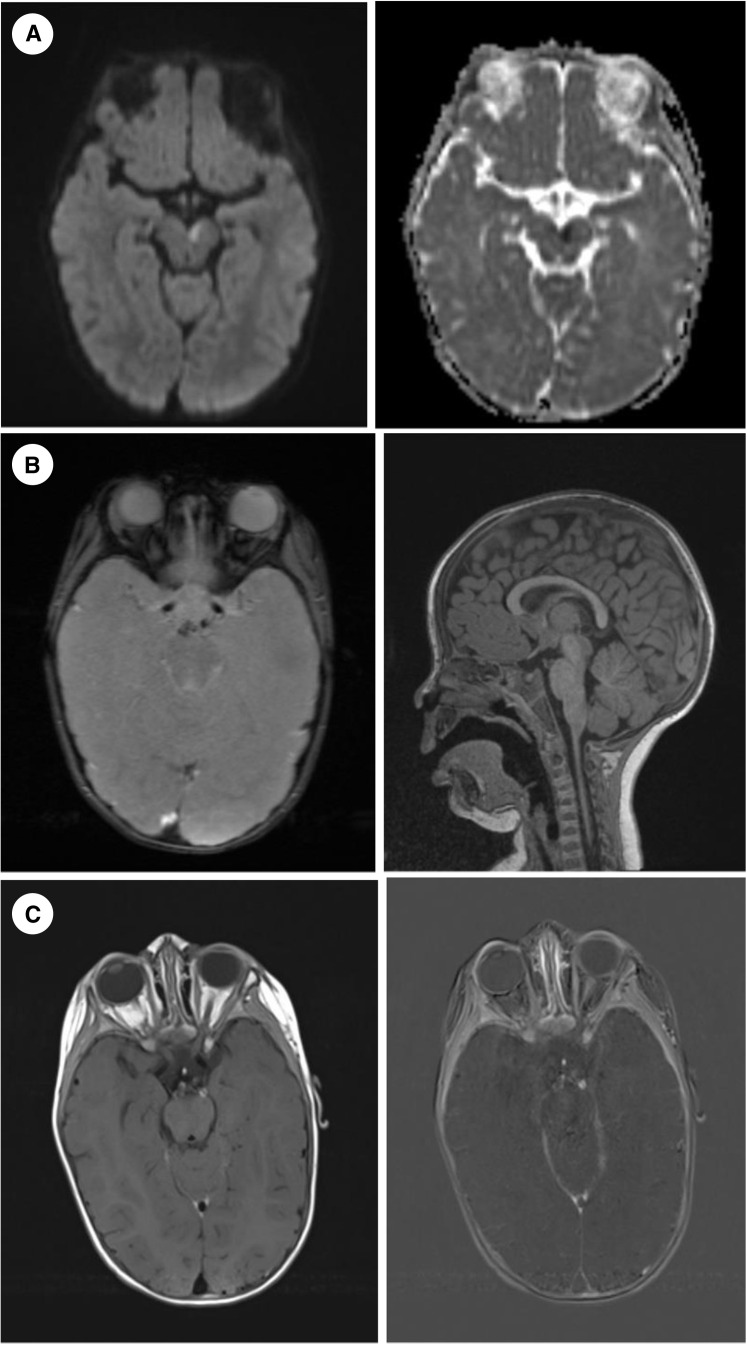
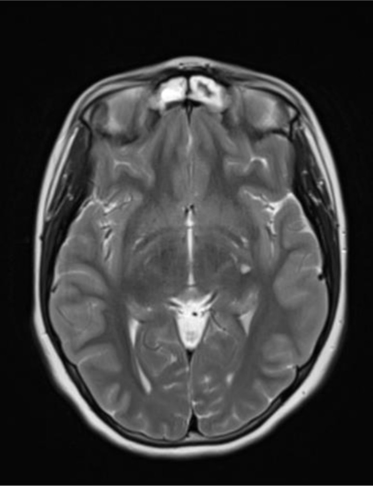
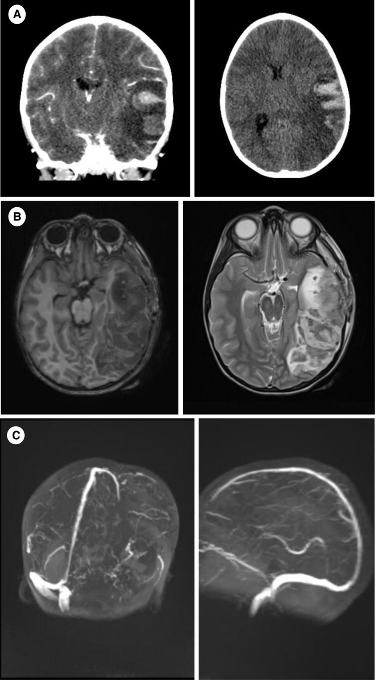
This work presents a case series of four children diagnosed with severe cerebrovascular disease in association with recent severe acute respiratory syndrome coronavirus 2 (SARS-CoV-2) infection, yet no patient from the group met typical diagnostic criteria for multisystem inflammatory syndrome in children. Our aim was to highlight the possible vascular involvement and coagulopathies associated with SARS-CoV-2 infection in the pediatric population. Further data are needed to better understand the pathophysiological basis of this condition in children and to ensure its optimal management.

Keywords: Brain vasculopathy; COVID-19; Children; SARS-CoV-2; Stroke.


Figures

FIGURE 1
FIGURE 1
(A) Noncontrast brain CT, at onset (axial plane): Left-sided subdural hematoma. (B) Brain MRI, day 6 after surgery, T2 tse (axial plane): Bilateral subdural hematoma. (C) Brain MRI, 1-month follow-up, T1 mprage precontrast and postcontrast (axial plane): Left temporal arachnoideal cyst with older hemorrhagic component, bilateral residual subdural hematoma and pachymeningeal contrast enhancement more pronounced on the left side. CT, computed tomography; MRI, magnetic resonance imaging.
FIGURE 2
FIGURE 2
(A) Brain MRI, at onset, DWI and ADC maps (axial plane): Area of cytotoxic edema of left mesencephalon. (B) Brain MRI, at onset, GRE (axial plane) and T1 mprage (sagittal plane): Discrete subarachnoideal hemorrhage in the prepontine cistern. (C) Brain MRI, at onset, T1 tse blood suppression postcontrast and subtracted (axial plane): Left P1 of ACP wall enhancement. ACP, arteria cerebri posterior; ADC, apparent diffusion coefficient; DWI, diffusion weighted imaging; GRE, multiecho gradient recalled echo; MRI, magnetic resonance imaging.
FIGURE 3
FIGURE 3
Brain MRI, 1 week after onset, T2 tse (axial plane): Lacunar lesion in the left basal nuclei. MRI, magnetic resonance imaging.
FIGURE 4
FIGURE 4
(A) Brain CT, at onset (coronary and axial plane): Large hypodense ischemic area of left temporal, occipital and parietal lobe with profound hemorrhagic component and moderate midline shift. (B) Brain MRI, day 1 follow-up, T1 tse and T2 tse (axial plane): Extensive hemorrhagic venous infarction of left temporal, occipital and parietal lobe. (C) Brain MRI, day 1 follow-up, TOF 3D venography (3D rec): Left-sided cerebral venous sinus thrombosis propagating into the left internal jugular vein. CT, computed tomography; MRI, magnetic resonance imaging; TOF, time-of-flight.

Comment in

References

    1. Hanafi R., Roger P.A., Kuchcinski G., et al. COVID-19 neurologic complications with CNS vasculitis-like pattern. AJNR Am J Neuroradiol. 2020;41:1384–1387. - PMC - PubMed
    1. Kirschenbaum D., Imbach L.L., Rushing E.J., et al. Intracerebral endotheliitis and microbleeds are neuropathological features of COVID-19. Neuropathol Appl Neurobiol. 2021;47:454–459. - PMC - PubMed
    1. Vaschetto R., Cena T., Sainaghi P.P., et al. Cerebral nervous system vasculitis in a Covid-19 patient with pneumonia. J Clin Neurosci. 2020;79:71–73. - PMC - PubMed
    1. Stevens S.M., Woller S.C., Kreuziger L.B., et al. Antithrombotic therapy for VTE disease: second update of the CHEST guideline and expert panel report. Chest. 2021;160:e545–e608. - PubMed
    1. Gutierrez Amezcua J.M., Jain R., Kleinman G., et al. COVID-19-induced neurovascular injury: a case series with emphasis on pathophysiological mechanisms. SN Compr Clin Med. 2020;2:2109–2125. - PMC - PubMed

Supplementary concepts