. 2022;14(4):366-379.
doi: 10.1159/000518277. Epub 2022 Apr 22.

SP1 Promotes HDAC4 Expression and Inhibits HMGB1 Expression to Reduce Intestinal Barrier Dysfunction, Oxidative Stress, and Inflammatory Response after Sepsis

Affiliations

SP1 Promotes HDAC4 Expression and Inhibits HMGB1 Expression to Reduce Intestinal Barrier Dysfunction, Oxidative Stress, and Inflammatory Response after Sepsis

Zhen-Mi Liu et al. J Innate Immun. 2022.

Abstract

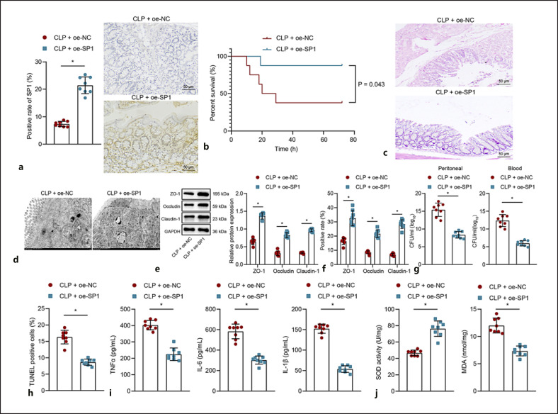
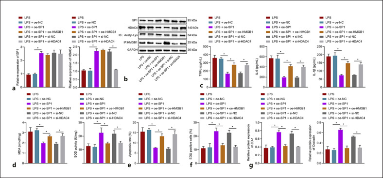
As a serious and elusive syndrome caused by infection, sepsis causes a high rate of mortality around the world. Our investigation aims at exploring the role and possible mechanism of specificity protein-1 (SP1) in the development of sepsis. A mouse model of sepsis was established by cecal ligation perforation, and a cellular model was stimulated by lipopolysaccharide (LPS), followed by determination of the SP1 expression. It was determined that SP1 was poorly expressed in the intestinal tissues of septic mice and LPS-treated cells. Next, we examined the interactions among SP1, histone deacetylase 4 (HDAC4), and high mobility group box 1 (HMGB1) and found that SP1 bound to the HDAC4 promoter to upregulate its expression, thereby promoting the deacetylation of HMGB1. Meanwhile, gain- or loss-of-function approaches were applied to evaluate the intestinal barrier dysfunction, oxidative stress, and inflammatory response. Overexpression of SP1 or underexpression of HMGB1 was observed to reduce intestinal barrier dysfunction, oxidative stress, and inflammatory injury. Collectively, these experimental data provide evidence reporting that SP1 could promote the HDAC4-mediated HMGB1 deacetylation to reduce intestinal barrier dysfunction, oxidative stress, and inflammatory response induced by sepsis, providing a novel therapeutic target for sepsis prevention and treatment.

Keywords: High mobility group box 1; Histone deacetylase 4; Inflammatory response; Intestinal barrier dysfunction; Oxidative stress; Sepsis; Specificity protein-1.


Conflict of interest statement

The authors have no conflicts of interest relevant to this article.

Figures

Fig. 1
Fig. 1
Characterization of the CLP-induced mouse sepsis model and significance of SP1 in sepsis. a Venn diagram of the predicted genes related to sepsis by the CTD, GeneCards, and Phenolyzer databases. b A bar chart of regulatory factors of the obtained 88 candidate targets predicted by the Metascape database. c Survival rate of Sham-operated and septic mice. d HE staining analysis of intestinal tissues of Sham-operated and septic mice. e Pathological changes of intestinal tissues of Sham-operated and septic mice under a TEM. f Western blot analysis of ZO-1 and occludin proteins in intestinal tissues of Sham-operated and septic mice. g Immunohistochemistry staining analysis of SP1 protein in intestinal tissues of Sham-operated and septic mice. n = 8 for Sham-operated and septic mice. *p < 0.05 between groups. The experiment was repeated 3 times independently. HE, hematoxylin-eosin; TEM, transmission electron microscope; CLP, cecal ligation perforation; SP1, specificity protein-1; GAPDH, glyceraldehyde-3-phosphate dehydrogenase.
Fig. 2
Fig. 2
Upregulation of SP1 improves intestinal barrier dysfunction and suppresses oxidative stress and epithelial cell apoptosis in septic mice. a Immunohistochemistry staining analysis of SP1 protein in intestinal tissues of septic mice treated with oe-SP1. b Survival rate of septic mice treated with oe-SP1. c HE staining analysis of intestinal tissues of septic mice treated with oe-SP1. d Pathological changes of intestinal tissues of septic mice treated with oe-SP1 under a TEM. e Western blot analysis of ZO-1, occludin, and claudin-1 proteins in intestinal tissues of septic mice treated with oe-SP1. f Immunohistochemistry staining analysis of ZO-1, occludin, and claudin-1 proteins in intestinal tissues of septic mice treated with oe-SP1. g Colony-forming unit in the PLF and serum of septic mice treated with oe-SP1. h Apoptosis of intestinal epithelial cells measured by TUNEL assay. i Serum levels of TNF-α, IL-6, and IL-1β in septic mice treated with oe-SP1 measured by ELISA. j MDA content and SOD activity in intestinal tissues of septic mice treated with oe-SP1. n = 8 for mice following each treatment. *p < 0.05 between groups. The experiment was repeated 3 times independently. HE, hematoxylin-eosin; TEM, transmission electron microscope; TUNEL, terminal deoxynucleotidyl transferase-mediated dUTP-biotin nick end labeling; SP1, specificity protein-1; PLF, peritoneal lavage fluid; ELISA, enzyme-linked immunosorbent assay; MDA, malondialdehyde; SOD, superoxide dismutase; CLP, cecal ligation perforation; oe-NC, overexpression-negative control; GAPDH, glyceraldehyde-3-phosphate dehydrogenase.
Fig. 3
Fig. 3
Upregulation of SP1 suppresses LPS-induced inflammatory response, oxidative stress, and apoptosis of intestinal epithelial cells. a SP1 mRNA expression determined by RT-qPCR in LPS-induced cells transfected with oe-SP1. b Levels of TNF-α, IL-6, and IL-1β measured by ELISA in the culture medium of LPS-induced cells transfected with oe-SP1. c MDA content and SOD activity in LPS-induced cells transfected with oe-SP1. d Flow cytometric analysis of the apoptosis rate of LPS-induced cells transfected with oe-SP1. e Proliferation of LPS-induced cells transfected with oe-SP1 measured by EdU assay. f Western blot analysis of ZO-1 and occludin proteins in LPS-induced cells transfected with oe-SP1. *p < 0.05 between groups. The experiment was repeated 3 times independently. RT-qPCR, reverse transcription-quantitative polymerase chain reaction; SP1, specificity protein-1; LPS, lipopolysaccharide; ELISA, enzyme-linked immunosorbent assay; MDA, malondialdehyde; SOD, superoxide dismutase; GAPDH, glyceraldehyde-3-phosphate dehydrogenase; oe-NC, overexpression-negative control.
Fig. 4
Fig. 4
SP1 increases HDAC4 transcription and expression, causing inhibition of HMGB1 acetylation level and its translocation into nuclei. a PPI network of the SP1-related genes predicted by the GeneCards database. b Venn diagram of the downstream target genes of SP1 analyzed by the TRRUST database and SP1-related genes. c Binding sites of SP1 in the HDAC4 predicted by the ChIPBase database. d SP1 enrichment in the HDAC4 promoter analyzed by ChIP. e Binding of SP1 to HDAC4F confirmed by dual-luciferase reporter assay in 293T cells. f HDAC4 mRNA expression determined by RT-qPCR in MODE-K cells transfected with oe-SP1. g Western blot analysis of HDAC4 protein in MODE-K cells transfected with oe-SP1. h Venn diagram of the HDAC4-related genes analyzed by the GeneCards and BioGRID databases and sepsis-related genes. i The interaction between HDAC4 and other genes analyzed by MEM dataset; each square represents a sample. j Immunohistochemistry staining analysis of HDAC4 and HMGB1 proteins in intestinal tissues of septic mice. k Immunoprecipitation analysis of HMGB1 protein and acetylation level in oe-HDAC4-transfected MODE-K cells. l HMGB1 translocation into nuclei analyzed by fractionation of nuclear/cytoplasmic RNA in SP1-overexpressing MODE-K cells. m HMGB1 translocation into nuclei analyzed by fractionation of nuclear/cytoplasmic RNA in HDAC4-overexpressing MODE-K cells. n Immunofluorescence analysis of HMGB1 protein in HDAC4-overexpressing MODE-K cells. *p < 0.05 between groups. The experiment was repeated 3 times independently. RT-qPCR, reverse transcription-quantitative polymerase chain reaction; ChIP, chromatin immunoprecipitation; PPI, protein-protein interaction; MEM, Multi-Experiment Matrix; SP1, specificity protein-1; HDAC4, histone deacetylase 4; HMGB1, high mobility group box 1; oe-NC, overexpression-negative control; GAPDH, glyceraldehyde-3-phosphate dehydrogenase; CLP, cecal ligation perforation; LPS, lipopolysaccharide.
Fig. 5
Fig. 5
SP1 advances HDAC4-mediated HMGB1 deacetylation and prevents inflammatory response, oxidative stress, and apoptosis of intestinal epithelial cells. LPS-induced MODE-K cells were transfected with oe-SP1, oe-SP1 + oe-HMGB1, or oe-SP1 + si-HDAC4. a SP1 and HDAC4 mRNA expressions determined by RT-qPCR in MODE-K cells. b HDAC4 protein expression and acetylation level determined by Western blot analysis and immunoprecipitation assay in MODE-K cells. c TNF-α, IL-6, and IL-1β expressions determined by ELISA in the supernatant of MODE-K cells. d MDA content and SOD activity in MODE-K cells. e Flow cytometric analysis of the apoptosis rate of MODE-K cells. f Proliferation of MODE-K cells measured by EdU assay. g Western blot analysis of ZO-1 and occludin proteins in MODE-K cells. *p < 0.05 between groups. The experiment was repeated 3 times independently. RT-qPCR, reverse transcription-quantitative polymerase chain reaction; SP1, specificity protein-1; HDAC4, histone deacetylase 4; HMGB1, high mobility group box 1; LPS, lipopolysaccharide; oe-NC, overexpression-negative control; GAPDH, glyceraldehyde-3-phosphate dehydrogenase; MDA, malondialdehyde.
Fig. 6
Fig. 6
HMGB1 knockdown delays the intestinal barrier injury, oxidative stress, and inflammatory response in septic mice. a Immunohistochemistry staining analysis of HMGB1 protein in intestinal tissues of HMGB1−/− septic mice. b Survival rate of HMGB1−/− septic mice. c HE staining analysis of intestinal tissues of HMGB1−/− septic mice. d Pathological changes of intestinal tissues of HMGB1−/− septic mice under a TEM. e Western blot analysis of ZO-1 and occludin proteins in intestinal tissues of HMGB1−/− septic mice. f Colony-forming unit in the PLF and serum of HMGB1−/− septic mice. g Apoptosis of intestinal epithelial cells in intestinal tissues of HMGB1−/− septic mice measured by TUNEL assay. h Serum levels of TNF-α, IL-6, and IL-1β in HMGB1−/− septic mice measured by ELISA. i MDA content and SOD activity in intestinal tissues of HMGB1−/− septic mice. n = 8 for mice following each treatment. *p < 0.05 between groups. The experiment was repeated 3 times independently. HE, hematoxylin-eosin; TEM, transmission electron microscope; TUNEL, Terminal deoxynucleotidyl transferase-mediated dUTP-biotin nick end labeling; HMGB1, high mobility group box 1; PLF, peritoneal lavage fluid; ELISA, enzyme-linked immunosorbent assay; MDA, malondialdehyde; SOD, superoxide dismutase; CLP, cecal ligation perforation.
Fig. 7
Fig. 7
The mechanism diagram of SP1/HDAC4/HMGB1 axis in sepsis. SP1 stimulated the HDAC4 transcription and decreased HMGB1 level and its translocation into nuclei, consequently preventing intestinal barrier dysfunction, oxidative stress, and inflammatory response, therefore delaying the progression of sepsis. HMGB1, high mobility group box 1; SP1, specificity protein-1; HDAC4, histone deacetylase 4.

Similar articles

Cited by

References

    1. Taeb AM, Hooper MH, Marik PE. Sepsis: current definition, pathophysiology, diagnosis, and management. Nutr Clin Pract. 2017;32((3)):296–308. - PubMed
    1. Yoseph BP, Klingensmith NJ, Liang Z, Breed ER, Burd EM, Mittal R, et al. Mechanisms of intestinal barrier dysfunction in sepsis. Shock. 2016;46((1)):52–9. - PMC - PubMed
    1. Xie S, Yang T, Wang Z, Li M, Ding L, Hu X, et al. Astragaloside IV attenuates sepsis-induced intestinal barrier dysfunction via suppressing RhoA/NLRP3 inflammasome signaling. Int Immunopharmacol. 2020;78:106066. - PubMed
    1. Berretta M, Quagliariello V, Maurea N, Di Francia R, Sharifi S, Facchini G, et al. Multiple effects of ascorbic acid against chronic diseases: updated evidence from preclinical and clinical studies. Antioxidants. 2020;9((12)):1182. - PMC - PubMed
    1. Kumar S, Tripathy S, Jyoti A, Singh SG. Recent advances in biosensors for diagnosis and detection of sepsis: a comprehensive review. Biosens Bioelectron. 2019;124–125:205–15. - PubMed

Publication types