. 2022 Aug;298(8):102218.
doi: 10.1016/j.jbc.2022.102218. Epub 2022 Jun 30.

Neurotoxic and cytotoxic peptides underlie the painful stings of the tree nettle Urtica ferox

Affiliations

Neurotoxic and cytotoxic peptides underlie the painful stings of the tree nettle Urtica ferox

Jing Xie et al. J Biol Chem. 2022 Aug.

Abstract

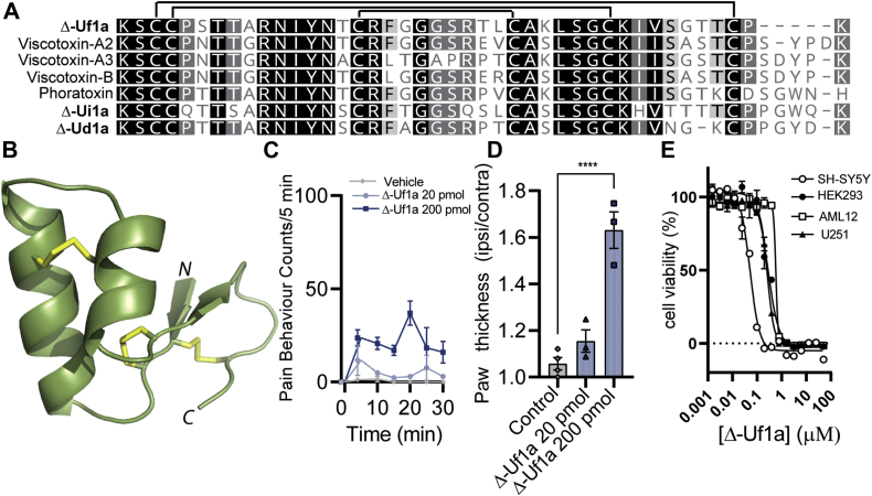
The stinging hairs of plants from the family Urticaceae inject compounds that inflict pain to deter herbivores. The sting of the New Zealand tree nettle (Urtica ferox) is among the most painful of these and can cause systemic symptoms that can even be life-threatening; however, the molecular species effecting this response have not been elucidated. Here we reveal that two classes of peptide toxin are responsible for the symptoms of U. ferox stings: Δ-Uf1a is a cytotoxic thionin that causes pain via disruption of cell membranes, while β/δ-Uf2a defines a new class of neurotoxin that causes pain and systemic symptoms via modulation of voltage-gated sodium (NaV) channels. We demonstrate using whole-cell patch-clamp electrophysiology experiments that β/δ-Uf2a is a potent modulator of human NaV1.5 (EC50: 55 nM), NaV1.6 (EC50: 0.86 nM), and NaV1.7 (EC50: 208 nM), where it shifts the activation threshold to more negative potentials and slows fast inactivation. We further found that both toxin classes are widespread among members of the Urticeae tribe within Urticaceae, suggesting that they are likely to be pain-causing agents underlying the stings of other Urtica species. Comparative analysis of nettles of Urtica, and the recently described pain-causing peptides from nettles of another genus, Dendrocnide, indicates that members of tribe Urticeae have developed a diverse arsenal of pain-causing peptides.

Keywords: NMR structure; gating modifier toxin; neuropharmacology; sodium channel; trichome.


Conflict of interest statement

Conflict of interest The authors declare that they have no conflicts of interest with the contents of this article.

Figures

Figure 1
Figure 1
Pain-causing toxins in Urtica ferox trichome extracts.A, U. ferox is densely covered in white stinging hairs. The trichomes shown in the inset are approximately 8 mm long. B, fractionation of the crude stinging hair extract via RP-HPLC identified four sequentially eluting fractions (11–14) with pain-causing activity when injected intraplantar into mice (n = 1). C, further subfractionation of fraction 12 yielded six subfractions (12.1–12.6), which elicited varying degrees of nocifensive behaviors (n = 1). D, HR-MALDI-MS spectrum of the principal ions present in F12.2 (left) and F12.6 (right).
Figure 2
Figure 2
Activation of mouse DRG neurons by U. ferox pain-causing toxins.A and B, Ca2+ responses in mouse DRG cells of F12.2 (A) and F12.6 (B). Traces from all cells in a single representative experiment are shown in the left panel. The dark gray trace represents the average response. Pseudocolor images showing cells before (buffer) and after toxin application are shown to the right (The scale bar represents 100 μm). C, distribution of F12.6 responding and nonresponding cells by cell size. D, percentage of DRG neurons activated by F12.6 (440 nM) in the absence or presence of TTX (1 μM). Data are expressed as mean ± SEM.
Figure 3
Figure 3
Δ-Uf1a is an algogenic and cytotoxic plant defensin.A, alignment of the mature peptide sequences of Δ-Uf1a, viscotoxins (Uniprot P32880, P01538, P08943), phoratoxin (Uniprot P01539), Δ-Ui1a (from an Urtica incisa transcriptome, NCBI short read archive [SRA] SRR10567092) and Δ-Ud1a (Urtica dioica transcriptome, SRA ERR2040431). B, 3D structure of synthetic Δ-Uf1a determined by NMR spectroscopy (PDB ID 7S7P). Disulfide bonds are shown as yellow sticks. C, spontaneous pain behaviors in mice following shallow intraplantar injection of Δ-Uf1a (20 or 200 pmol). D, paw swelling caused by Δ-Uf1a when injected intraplantar into mice. E, Δ-Uf1a is a potent cytotoxin. Cell viability was assessed via MTT assay after 24 h. Data are expressed as means ± SEM.
Figure 4
Figure 4
β/δ-Uf2a defines a new class of disulfide-rich plant peptides.A, alignment of the mature primary sequences of β/δ-Uf2a and putative paralogues and orthologues identified in transcriptome data and confirmed on cDNA or genomic DNA levels in U. ferox (Uf2a and Uf2b), U. incisa (Ui2a), Dendrocnide moroides (Dm2a) and D. excelsa (De2a). Numbering is based on β/δ-Uf2a and % sequence identity relative to β/δ-Uf2a is indicated in the boxed section. B, spontaneous pain behaviors in mice following shallow intraplantar injection of β/δ-Uf2a (0.2–200 pmol). All data are expressed as means ± SEM.
Figure 5
Figure 5
β/δ-Uf2a contributes to the symptoms of U. ferox stings via modulation of voltage-gated sodium channels.A, D, G and J, representative NaV1.5, NaV1.6, NaV1.7, and NaV1.8 current responses to a step depolarization from −90 to −20 mV (+10 mV for NaV1.8) in the absence or presence of β/δ-Uf2a. B, E and H, effects of β/δ-Uf2a on sustained current (I40 ms/Ipeak) and kinetics of fast inactivation (τ) of NaV1.5 (B), NaV1.6 (E), and NaV1.7 (H), where response is I40-ms (Current amplitude at 40 ms) after β/δ-Uf2a treatment/peak current (Ipeak) of cell (before toxin addition). Statistical significance compared with buffer control was determined via paired t test, ∗∗p < 0.01, ∗∗∗p <0.001, ∗∗∗∗p <0.0001. C, F and I, superimposed conductance–voltage (GV) and steady-state fast inactivation curves, before (open symbols) and after addition of toxin (filled symbols) for NaV1.5 (C, 1 μM β/δ-Uf2a), NaV1.6 (F, 300 nM β/δ-Uf2a), and NaV1.7 (C, 1 μM β/δ-Uf2a). K, concentration–response relationship of β/δ-Uf2a modulation of human NaV1.5, NaV1.6, and NaV1.7, where response is I10-ms (Current amplitude at 10 ms) after β/δ-Uf2a treatment/peak current (Ipeak) of cell (before toxin addition). All data are expressed as mean ± SEM.
Figure 6
Figure 6
The extracellular loops of domain IV on NaV1.7 mediate the effects of β/δ-Uf2a.A, schematic representation of NaV1.7 depicting the extracellular loops on DII and DIV that were replaced with NaV1.8 sequences. B, the effect of β/δ-Uf2a (1 μM) on the τ of fast inactivation at NaV1.7 where the DII S1-S2, DII S3-S4, DIV S1-S2, and DIV S3-S4 extracellular loops were replaced by NaV1.8 (n = 5–10). Insertion of NaV1.8 DIV S1-S2 and NaV1.8 DIV S3-S4 loops significantly reduced the activity of β/δ-Uf2a. The loop substitutions had no effect on the τ of fast inactivation in the absence of toxin (Fig. S5). Statistical significance was determined using one-way ANOVA with Dunnett’s multiple comparisons test compared with wildtype NaV1.7 with buffer or β/δ-Uf2a as indicated, ∗∗∗p <0.001, ∗∗∗∗p <0.0001. Data are presented as mean ± SEM. C, representative current traces from the NaV1.7/NaV1.8 loop mutant channels before and after addition of β/δ-Uf2a (1 μM). Currents were elicited by a 50-ms pulse to −20 mV (+10 mV for DII S3-S4) from a holding potential of −90 mV. D, superimposed conductance–voltage (GV) curves for NaV1.7 where the DIV S3-S4 extracellular loop has been replaced with the corresponding sequence from NaV1.8, before (white) and after addition of 1 μM β/δ-Uf2a (cyan). V0.5: buffer control −29.1 ± 1.1 mV; β/δ-Uf2a −31.9 ± 1.4 mV, p = 0.0539, paired t test, n = 4.

References

    1. Parthasarathy A., Borrego E.J., Savka M.A., Dobson R.C.J., Hudson A.O. Amino acid-derived defense metabolites from plants: a potential source to facilitate novel antimicrobial development. J. Biol. Chem. 2021;296:100438. - PMC - PubMed
    1. War A.R., Paulraj M.G., Ahmad T., Buhroo A.A., Hussain B., Ignacimuthu S., et al. Mechanisms of plant defense against insect herbivores. Plant Signal. Behav. 2012;7:1306–1320. - PMC - PubMed
    1. Pilgrim R.L.C. Some properties of the sting of the New-Zealand nettle, Urtica-ferox. Proc. R. Soc. B. 1959;151:48–56.
    1. Ensikat H.J., Wessely H., Engeser M., Weigend M. Distribution, ecology, chemistry and toxicology of plant stinging hairs. Toxins. 2021;13:141. - PMC - PubMed
    1. Kittow N.C. A case of canine poisoning with New Zealand Tree Nettle (Ongaonga, Urtica ferox) N. Z. Vet. J. 2013;61:60–62. - PubMed

Publication types