. 2022 May;57(3):267-281.
doi: 10.5152/TurkArchPediatr.2022.22085.

Imaging of Central Nervous System Involvement in Pediatric Hematologic Disorders

Affiliations

Imaging of Central Nervous System Involvement in Pediatric Hematologic Disorders

Sevinç Kalın et al. Turk Arch Pediatr. 2022 May.

Abstract

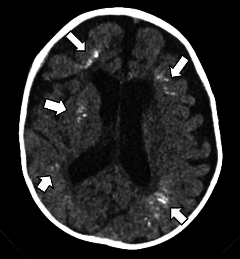
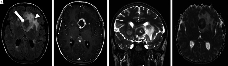
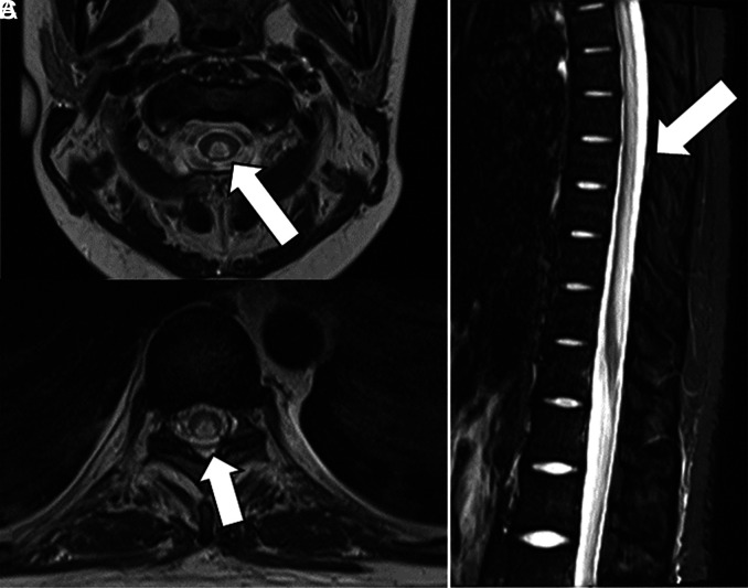
Hematologic disorders and malignancies are commonly encountered in children. The central nervous system is affected by many benign and malignant hematological diseases. Neurologic findings may occur due to central involvement of hematological disease or neurotoxic effects of treatment during the disease. At all these stages, radiological imaging and evaluation have a critical role. In this review article, we aim to describe the imaging findings of central nervous system involvement in pediatric hematologic diseases. This review was prepared based on the latest literature available in the PubMed database in the English language from inception to March 2022. The radiological images of the patients in our archive were obtained from the PACS (Picture Archiving and Communication Systems) to set an example for the diseases described in the text.


Figures

Figure 1.
Figure 1.
A 13-year-old male diagnosed with Factor XIII deficiency has an acute hematoma in the right temporal lobe is seen on axial CT (a) and SWI (b) images. In the same patient, there is an encephalomalastic area secondary to the previous hemorrhage accompanied by calcifications in the right frontal lobe (c, d).
Figure 2.
Figure 2.
A 17-year-old male with protein C deficiency. 2D time of flight magnetic resonance venography image shows nonocclusive thrombus within the dominant right sigmoid sinus.
Figure 3.
Figure 3.
In the axial FLAIR-W images of the patient with Moyamoya syndrome, a continuous chronic ischemic encephalomalasic area is observed in the left temporooccipital region (a). Perimesencephalic collateral vascular structures are selected in axial post-contrast T1W images (b). In the diagnostic angiography examination of the patient, collateral vascular structures that create a puff of smoke appearance are observed (c).
Figure 4.
Figure 4.
Signal changes consistent with ischemia are observed in the bilateral basal ganglia and ventromedial of the thalamus in FLAIR W (a) diffusion-weighted images (b) and ADC mapping (c) in a patient diagnosed with sickle cell anemia who applied to the emergency department with acute neurological findings. An area of acute ischemic tissue is observed in the posterior parietal at the vertex level in the non-contrast CT examination performed in the emergency department at a different time (d).
Figure 5.
Figure 5.
Axial head computed tomography in bone window shows characteristic expansion of the calvarial bone marrow in sickle cell disease.
Figure 6.
Figure 6.
15-year-old with thalassemia. Coronal T2-weighted (T2W) image show marked T2 hypointensity within the pituitary gland due to iron accumulation secondary to chronic blood transfusions. There is marked expansion and T2 hypointensity of the calvarial and skull base bone marrow. A 14-year-old girl with a diagnosis of thalassemia, hypointensity is observed in the vetebral bone marrow (b) and liver (c) in T2W images secondary to coronal iron deposition. The same patient has a nodular lesion consistent with extramedullary hematopoiesis in the spleen (c).
Figure 7.
Figure 7.
An 11 year-old female diagnosed with LCH, presented with diabetes insipidus. There is lack of visualization of the normal neurohypophysis (a, thin arrow) in pre-contrast sagittal T1W images. The suprasellar mass demonstrates heterogenous, intense enhancement (thick arrow). Incidental note is made of a pineal cyst (arrow head).
Figure 8.
Figure 8.
A 2-year-old male diagnosed with hemaphagocytic lymphohistiocytosis, hyperintense areas forming confluence are observed in the subcortical and peiventricular white matter in the left posterior parietal in axial FLAIR-W images (a). In diffusion-weighted studies, there are signal changes consistent with periventricular and subcortical millimetric ischemic diffusion limitations (b). In post-contrast axial T1-weighted images, contrast enhancements in the periventricular white matter at the centrum semiovale level are remarkable (c).
Figure 9.
Figure 9.
A 4-year-old male diagnosed with ALL, in the axial plane T2W, diffusion, and post-contrast T1W images, respectively, there is an extraaxially posterior fossa solid mass (arrow) with subperiosteal component (arrowhead). There is a cytotoxic edema effect by pressing the right cerebellar hemispheres and brain stem with diffusion restriction (b) and heterogeneously contrasting (c). A 4-year-old patient with a diagnosis of ALL has a solid mass lesion in the spinal canal extradurally at L5-S2 levels in sagittal T2W (d), pre (e) and post-contrast T1W images (f), developed during the follow-up. Hypointencity of vertebrae is also seen on T1W images secondary to diffuse leukemic bone marrow infiltration.
Figure 10.
Figure 10.
Bilateral periventricular nodular diffusion restriction is observed at centrum semiovale and corona radiata levels in diffusion-weighted images (a,b) and ADC maps (d,e) in a 12-year-old male who received intrathecal treatment 5 days ago.
Figure 11.
Figure 11.
In a 7-year-old male with ALL, a cortical-subcortical hemorrhagic infarct in the right frontal lobe secondary to superior sagittal sinus thrombosis is observed in the images of T2W (a), SWI (b), and MR venography (c).
Figure 12.
Figure 12.
In a 4-month-old male with ALL, unenhanced axial CT scan shows multiple dystrophic calcifications at bilateral periventricular white matter secondary to mineralized microangiopathy.
Figure 13.
Figure 13.
A 4-year-old male diagnosed with primary CNS lymphoma, coronal T2-weighted (T2W) images show a hypointense solid mass involving the basal ganglia and extending into the ventricle in the right frontoparietal white matter (a). Also, right hemispheric subdural effusion and midline shift are seen in pre-contrast T1-weighted (T1W) images (b). There is diffuse heterogeneous enhancement in post-contrast T1W images and the mass extends to the opposite hemisphere through the corpus callosum (c). In diffusion-weighted images (d) and ADC mapping (e), there is diffusion restriction secondary to high cellularity in the mass. 8-year-old male with spinal Burkitt lymphoma, In sagittal T2W images (f), there is a hypointense mass lesion in the spinal canal. In pre- and post-contrast axial images, we see a diffusely enhancing solid mass located extra-axial space in the spinal canal that compresses the spinal cord (g, h).
Figure 14.
Figure 14.
A 15-year-old patient with a diagnosis of diffuse B-cell lymphoma lost the normal hyperintense signal in the neurohypophysis on pre-contrast T1-weighted (T1W) images. Nodular mass thickening was observed in the pituitary stalk on post-contrast T1W images. After the treatment, the thickening of the stalk regressed and the dimensions of the pituitary gland decreased. Normal brightness in the neurohypophysis has not returned.
Figure 15.
Figure 15.
A 15-year-old male with orophayrngeal B-cell lymphoma and central nervous system involvement. In axial FLAIR-W images (a), there is a periventricular mass (arrow) that compresses the 3rd and lateral ventricle, accompanied by a peripheral large edema (arrowhead). Multiple lesions with ring-like enhancements are seen in post-contrast images (b). Midline shift is better seen in coronal T2W images (c) and there is diffusion restriction in ADC mapping (d).
Figure 16.
Figure 16.
A 16-year-old patient with a diagnosis of thalassemia who underwent bone marrow transplantation, has hyperintense vasogenic edema consistent with PRES in the posterior parietal areas, right temporal lobe, and frontal subcortical areas in FLAIR-W images.
Figure 17.
Figure 17.
In an 8-year-old patient diagnosed with ALL, hyperintensity in the vertebrae on T1 W (a) and T2W (b), fat-free images and hypointensity secondary to fat suppression on coronal STIR (c) images consistent with fatty bone marrow conversion secondary to radiotherapy are observed. There is also a loss of height secondary to a compression fracture in vetebrae.
Figure 18.
Figure 18.
A 1-year-old girl diagnosed with ALL has hypointensity in sagittal T1W (a) and hyperintensity in STIR (b) images, consistent with bone marrow reconstruction.
Figure 19.
Figure 19.
A 15-year-old girl with a diagnosis of primary vertebral diffuse b-cell lymphoma has a solid mass lesion extending to the pre-paravertebral areas, which causes compression by destructing the L4 vertebra on coronal CT (a), T2W (b), and enhanced T1W (c) images.
Figure 20.
Figure 20.
A 17-year-old male was diagnosed with lymphoma. Heterogeneous patchy bone marrow infiltration is observed on sagittal CT (a), pre- (b), and post-contrast (c) T1W and T2W (d) images, respectively.
Figure 21.
Figure 21.
A 14-year-old with T cell lymphoma. Nelarabine neurotoxicity is characterized by nonenhancing, symmetrical T2 prolongation involving the central and posterior spinal cord throughout axial cervical (a), thoracic (b), and sagittal images.

Similar articles

References

    1. Filatova I, Stratchko LL, Kanekar S. Central nervous system complications of hemorrhagic and coagulation disorders. Hematol Oncol Clin North Am. 2016;30(4):757 777. 10.1016/j.hoc.2016.03.003) - DOI - PubMed
    1. McDonald J, Stevenson DA. Hereditary hemorrhagic telangiectasia. GeneReviews® [internet]; 2021. https://www.ncbi.nlm.nih.gov/books/NBK1351/. Accessed 2022 Apr 8.
    1. Jaskolka J, Wu L, Chan RP, Faughnan ME. Imaging of hereditary hemorrhagic telangiectasia. AJR Am J Roentgenol. 2004;183(2):307 314. 10.2214/ajr.183.2.1830307) - DOI - PubMed
    1. Mathew T, Sinha S, Taly AB, Arunodaya GR, Srikanth SG. Neurological manifestations of Ehlers-Danlos syndrome. Neurol India. 2005;53(3):339-341. 10.4103/0028-3886.16938) - DOI - PubMed
    1. North KN, Whiteman DAH, Pepin MG, Byers PH. Cerebrovascular complications in Ehlers-Danlos syndrome type IV. Ann Neurol. 1995;38(6):960 964. 10.1002/ana.410380620) - DOI - PubMed

LinkOut - more resources