. 2022 Jul;10(7):e004325.
doi: 10.1136/jitc-2021-004325.

Novel intravesical bacterial immunotherapy induces rejection of BCG-unresponsive established bladder tumors

Affiliations

Novel intravesical bacterial immunotherapy induces rejection of BCG-unresponsive established bladder tumors

Eduardo Moreo et al. J Immunother Cancer. 2022 Jul.

Abstract

Background: Intravesical BCG is the gold-standard therapy for non-muscle invasive bladder cancer (NMIBC); however, it still fails in a significant proportion of patients, so improved treatment options are urgently needed.

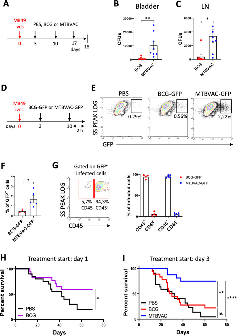
Methods: Here, we compared BCG antitumoral efficacy with another live attenuated mycobacteria, MTBVAC, in an orthotopic mouse model of bladder cancer (BC). We aimed to identify both bacterial and host immunological factors to understand the antitumoral mechanisms behind effective bacterial immunotherapy for BC.

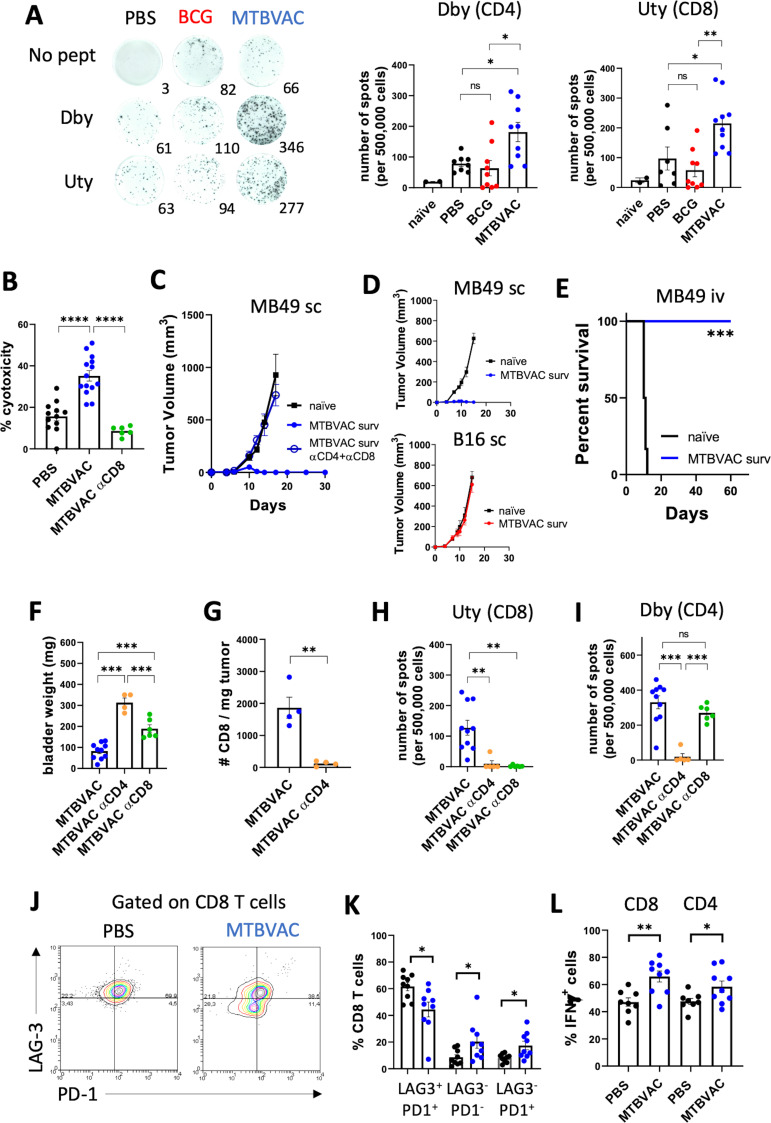
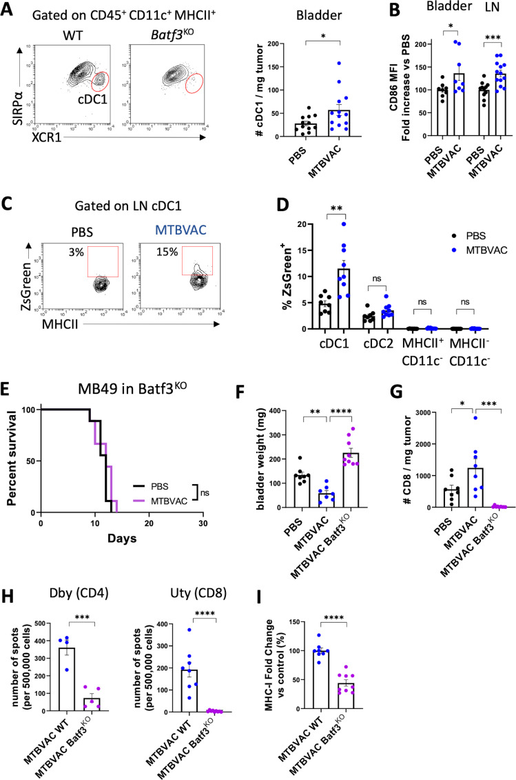
Results: We found that the expression of the BCG-absent proteins ESAT6/CFP10 by MTBVAC was determinant in mediating bladder colonization by the bacteria, which correlated with augmented antitumoral efficacy. We further analyzed the mechanism of action of bacterial immunotherapy and found that it critically relied on the adaptive cytotoxic response. MTBVAC enhanced both tumor antigen-specific CD4+ and CD8+ T-cell responses, in a process dependent on stimulation of type 1 conventional dendritic cells. Importantly, improved intravesical bacterial immunotherapy using MBTVAC induced eradication of fully established bladder tumors, both as a monotherapy and specially in combination with the immune checkpoint inhibitor antiprogrammed cell death ligand 1 (anti PD-L1).

Conclusion: These results contribute to the understanding of the mechanisms behind successful bacterial immunotherapy against BC and characterize a novel therapeutic approach for BCG-unresponsive NMIBC cases.

Keywords: Adaptive Immunity; Dendritic Cells; Immunogenicity, Vaccine; Immunotherapy; Urinary Bladder Neoplasms.


Conflict of interest statement

Competing interests: SU, EP, ER, CM and NA are coinventors of the patent 'Compositions for use as a prophylactic agent to those at risk of infection of tuberculosis, or as secondary agents for treating infected tuberculosis patients' held by the University of Zaragoza and Biofabri. CM is inventor of the patent 'Tuberculosis vaccine' held by the University of Zaragoza. There are no other conflicts of interest.

Figures

Figure 1
Figure 1
Superior efficacy of MTBVAC against bladder cancer correlates with better colonization capacity. (A) Schedule of intravesical treatments of mice bearing orthotopic MB49 bladder tumors with PBS, BCG Tice or MTBVAC. (B, C) Mice were treated as in (A), and at day 18, bladder tumors and draining LNs were extracted and homogenized and total CFUs were enumerated (n=8 mice per group, pooled from two independent experiments). (D) Mice were treated as in (A) with GFP-expressing bacteria and euthanized 2 hours after the second instillation, at day 10. Bladder tumors were extracted; single-cell suspensions were generated; and infected GFP+ cells were identified by flow cytometry. (E) Representative density plots of GFP+ cells. (F) Percentage of GFP+ infected cells (n=4–5 mice per group, pooled from two independent experiments). (G) Representative density plots and distribution of GFP+ infected cells in CD45 and CD45+ subsets in bladder tumors. (H) Survival of mice bearing MB49 bladder tumors treated with intravesical PBS or BCG at days 1, 8 and 15 post tumor implantation (n=20 PBS, n=18 BCG, pooled from two independent experiments). (I) Survival of mice bearing MB49 bladder tumors treated as in (A) (n=22 PBS, n=18 BCG, n=16 MTBVAC, pooled from two independent experiments). Graphs represent mean±SEM. *P<0.05, **P<0.01, ****P<0.0001, unpaired t-test (B, C, F), or log-rank test (H, I). CFU, colony-forming unit; LN, lymph node.
Figure 2
Figure 2
ESAT6 and CFP10 expression on MTBVAC determines improved antitumoral efficacy. (A, B) Tumor-bearing mice were treated with intravesical BCG TICE, MTBVAC or MTBVACΔE6C10 at days 3, 10, and 17, and at day 18, bladders and dLN were extracted and homogenized, and total CFUs were enumerated (n=4 mice per group, from one experiment). (C) Tumor-free mice were intravesically instilled with the indicated bacteria, and 24 hours later bladders were extracted and total CFUs were enumerated (n=6 mice MTBVACΔE6C10 group, n=12 BCG and MTBVAC groups, pooled from two independent experiments). (D) Tumor-free mice were intravesically instilled at days 0, 7, and 14, and at day 15, dLNs were extracted and total CFUs were enumerated (n=6 mice MTBVACΔE6C10 group, n=12 BCG and MTBVAC groups, pooled from two independent experiments). (E) Mice were treated as in (C) with intravesical BCG Pasteur or RD1-recomplemented BCG Pasteur, and total CFUs were enumerated in the bladder 24 hours later (n=5 mice per group, from one experiment). (F–H) Mice were treated as in (D), and at day 15, bladders were extracted; single-ell suspensions were generated; and CD45+, CD4+, and CD8+ cells were identified by flow cytometry (n=6 mice MTBVACΔE6C10 group, n=16 BCG and MTBVAC groups, n=10 PBS group, pooled from three independent experiments). (H) Correlation between CD4+ and CD8+ T cells infiltrated in the bladder and bacterial CFUs in the draining LN, analyzed by linear regression. (I) Survival curve of mice implanted with orthotopic MB49 bladder tumors and treated intravesically with PBS, MTBVAC or MTBVACΔE6C10 at days 3, 10, and 17 (n=5 PBS, n=13 MTBVAC, n=22 MTBVACΔE6C10, pooled from two independent experiments). Graphs represent mean±SEM. *P<0.05, **P<0.01, ***P<0.001, one-way analysis of variance with Bonferroni post-test (A–D, F, G), unpaired t-test (E), or log-rank test (I). CFU, colony-forming unit; dLN, draining lymph node; LN, lymph node.
Figure 3
Figure 3
MTBVAC efficacy relies on the adaptive immune system. (A–C) survival curves of Rag1KO (A), PerfKO (B) and IFN-γKO (C) mice implanted orthotopically with MB49 tumors and treated with either PBS or MTBVAC at days 3, 10, and 17 (n=5–6 mice per group, from one experiment). (D) Survival of mice implanted orthotopically with MB49-B2mKO or MB49-WT cells and treated with either PBS or MTBVAC at days 3, 10, and 17 (n=5 mice per group, from one experiment). (E–I) bladder tumors were induced by intravesical instillation of GFP-expressing MB49 cells and mice were treated with either PBS or MTBVAC at days 3 and 10 and euthanized at day 12. (F) Representative density plots of single-cell suspensions from the MB49 and MB49-GFP tumor-bearing bladders. (G) Bladder weights of mice treated as in (E) (n=4 no tumor, n=9 PBS, n=16 MTBVAC, from three independent experiments). (H) Flow cytometry analysis of MHC-I expression on GFP+ CD45 tumor cells in vivo (n=4–5 mice per group, representative of two independent experiments). (I) Mice were treated as in (E) but were given αCD4 or αCD8 depleting antibodies the day before intravesical treatments (on days 2 and 9). The graph shows tumor cell MHC-I expression fold change versus the control MTBVAC group (n=4–6 mice per group, pooled from two independent experiments). Graphs represent mean±SEM. *P<0.05, **P<0.01, ***P<0.001, ****P<0.0001 one-way analysis of variance with Bonferroni post-test (G–I) or log-rank test (A–D). FMO, fluorescence minus one; IFN-γ, interferon gamma; ns, not significant; WT, wild type.
Figure 4
Figure 4
Intravesical MTBVAC augments CD4+ and CD8+ T-cell tumor antigen-specific immunity. MB49 bladder tumors were implanted orthotopically and mice were treated with PBS, BCG or MTBVAC at days 3 and 10. (A) At day 12 post tumor implantation, IFN-γ- producing splenocytes were assessed by ELISPOT following stimulation with 10 µg/mL of the Dby or Uty peptides. Numbers below the representative images indicate the numbers of spots (n=8–9 mice per group, pooled from two independent experiments). (B) In vitro cytotoxic activity of splenocytes against MB49 cells (n=12 mice PBS, n=14 MTBVAC, n=6 MTBVAC αCD8, pooled from three independent experiments). (C) MTBVAC-treated survivors were subcutaneously rechallenged with MB49 cells in the flank, and tumor growth was measured over time. One group of surviving mice was given CD4 and CD8 depleting antibodies 3 days before the rechallenge and 3 days after. Naïve mice were used as controls (n=8–10 mice per group, from two independent experiments). (D) Mice were subcutaneously rechallenged with MB49 cells in one flank and B16F10 in the contralateral flank, and tumor growth was measured over time. Naïve mice were used as controls (n=5–6 mice per group, from one experiment). (E) MTBVAC-treated surviving mice were intravenously rechallenged with MB49 cells. Naïve mice were used as controls (n=6–7 mice per group, from one experiment). (F–I) MB49 tumor-bearing mice treated with MTBVAC were given CD4 or CD8 depleting antibodies and euthanized at day 12. (F) Bladder weights at day 12 post tumor cell inoculation (n=10 mice MTBVAC, n=4 MTBVAC+αCD4, n=6 MTBVAC+αCD8, pooled from two independent experiments). (G) Number of tumor-infiltrating CD8+ T cells per milligram of tumor, identified by flow cytometry in bladder single cell suspensions (n=4 mice per group, one experiment). (H, I) IFN-γ -producing splenocytes after stimulation with the Uty (H) or Dby (I) peptides measured by ELISPOT assay (n=10 mice MTBVAC, n=4 MTBVAC+αCD4, n=6 MTBVAC+αCD8, pooled from two independent experiments). (J–L) Tumor-infiltrating T cells were analyzed by flow cytometry at day 12 in PBS or MTBVAC-treated mice (n=8–9 mice per group, pooled from two independent experiments). (J) Representative density plots of PD-1 and LAG-3 expression by CD8+ T cells. (K) Quantification of CD8+ cells expressing PD-1 and LAG-3. (L) Quantification of tumor-infiltrating CD4+ and CD8+ T cells expressing IFN-γ after ex vivo restimulation with PMA/ionomycin. Graphs represent mean±SEM. *P<0.05, **P<0.01, ***P<0.001, one-way analysis of variance with Bonferroni post-test (A, B, F, H, I) or unpaired T test (G,K,L). IFN-γ, interferon gamma; ns, not significant; PD-1, programmed cell death 1; PMA, phorbol-12-myristate 13-acetate.
Figure 5
Figure 5
MTBVAC efficacy depends on type 1 conventional DCs. (A–C) MB49 cells were implanted intravesically and mice were treated with PBS or MTBVAC at days 3 and 10. At day 12, DCs were analyzed by flow cytometry in bladder and dLN single-cell suspensions. (A) Representative plot identifying XCR1+ cDC1s. Number of cDC1s per milligram of bladder tumor (n=12–13 mice per group, pooled from three independent experiments). (B) Normalized MFI of CD86 in bladder tumor-infiltrating and LN cDC1s (n=8 mice for bladder, pooled from two independent experiments and n=12–14 for LNs, pooled from three independent experiments). (C) Representative density plot of dLN cDC1s of mice inoculated with ZsGreen-expressing MB49 cells. (D) percentage of ZsGreen+ cells in distinct dLN cell compartments (n=8–9 mice per group, from two independent experiments). (E) Survival curve of Batf3KO mice implanted orthotopically with MB49 cells and treated with PBS or MTBVAC at days 3, 10, and 17 (n=9 mice per group, two independent experiments). (F–I) WT or Batf3KO mice were implanted with MB49 bladder tumors and received PBS or MTBVAC intravesically at days 3 and 10 (n=8–9 mice per group, from two independent experiments). (F) Bladder weights at day 12. (G) Number of infiltrating CD8+ T cells per milligram of tumor analyzed by flow cytometry in bladder single-cell suspensions. (H) IFN-γ-producing splenocytes stimulated with the Dby or Uty peptides. (I) Tumor cell MHC-I expression was measured by flow cytometry. Graph shows MHC-I expression fold change versus the control MTBVAC group (n=8–9 mice per group, from two independent experiments). Graphs represent mean±SEM. *P<0.05, **P<0.01, ***P<0.001, one-way analysis of variance with Bonferroni post-test (G, H), unpaired t-test (A, C, E, I, J), or log-rank test (F). DC, dendritic cell; dLN, draining lymph node; IFN-γ, interferon gamma; LN, lymph node; MFI, mean fluorescence of intensity; ns, not significant; WT, wild type.
Figure 6
Figure 6
Improved bacterial immunotherapy rejects established bladder tumors. (A) Representative histology (H&E staining) of bladders 6 days after MB49 tumor cell inoculation. Tumor area is outlined in yellow. Letters denote the bladder lumen (L), lamina propia (LM), tumor (T) and muscle layer (M). (B) Representative histograms and quantification of PD-L1 MFI on the indicated cell type of mice bearing bladder tumors, treated intravesically on days 3 and 10 and analyzed on day 12. Graph shows PD-L1 expression fold change versus the control PBS of each mouse strain (n=5–9 mice per group, pooled from two independent experiments). (C) Schedule of intravesical treatments of mice bearing orthotopic MB49 bladder tumors with PBS or MTBVAC and intraperitoneal treatments with αPD-L1. (D) Survival of mice treated as in (C), (n=9–15 mice per group, pooled from three independent experiments). *P<0.05, **P<0.01, ***P<0.001, ****P<0.0001, log-rank test in (D), one-way analysis of variance with Bonferroni post-test in (B). IFN-γ, interferon gamma; MFI, mean fluorescence of intensity; ns, not significant; PD-L1, programmed cell death ligand 1; WT, wild type.

References

    1. Antoni S, Ferlay J, Soerjomataram I, et al. . Bladder cancer incidence and mortality: a global overview and recent trends. Eur Urol 2017;71:96–108. 10.1016/j.eururo.2016.06.010 - DOI - PubMed
    1. Morales A, Eidinger D, Bruce AW. Intracavitary Bacillus Calmette-Guerin in the treatment of superficial bladder tumors. J Urol 1976;116:180–2. 10.1016/S0022-5347(17)58737-6 - DOI - PubMed
    1. Gandhi NM, Morales A, Lamm DL. Bacillus Calmette-Guérin immunotherapy for genitourinary cancer. BJU Int 2013;112:288–97. 10.1111/j.1464-410X.2012.11754.x - DOI - PubMed
    1. Decaestecker K, Oosterlinck W. Managing the adverse events of intravesical Bacillus Calmette–Guérin therapy. Res Reports Urol 2015;7:157–63. 10.2147/RRU.S63448 - DOI - PMC - PubMed
    1. Sylvester RJ. Bacillus Calmette-Guérin treatment of non-muscle invasive bladder cancer. Int J Urol 2011;18:113–20. 10.1111/j.1442-2042.2010.02678.x - DOI - PubMed

Publication types