

Clinical grade ACE2 as a universal agent to block SARS-CoV-2 variants
- PMID: 35781796
- PMCID: PMC9350269
- DOI: 10.15252/emmm.202115230
Clinical grade ACE2 as a universal agent to block SARS-CoV-2 variants
Abstract
The recent emergence of multiple SARS-CoV-2 variants has caused considerable concern due to both reduced vaccine efficacy and escape from neutralizing antibody therapeutics. It is, therefore, paramount to develop therapeutic strategies that inhibit all known and future SARS-CoV-2 variants. Here, we report that all SARS-CoV-2 variants analyzed, including variants of concern (VOC) Alpha, Beta, Gamma, Delta, and Omicron, exhibit enhanced binding affinity to clinical grade and phase 2 tested recombinant human soluble ACE2 (APN01). Importantly, soluble ACE2 neutralized infection of VeroE6 cells and human lung epithelial cells by all current VOC strains with markedly enhanced potency when compared to reference SARS-CoV-2 isolates. Effective inhibition of infections with SARS-CoV-2 variants was validated and confirmed in two independent laboratories. These data show that SARS-CoV-2 variants that have emerged around the world, including current VOC and several variants of interest, can be inhibited by soluble ACE2, providing proof of principle of a pan-SARS-CoV-2 therapeutic.
Keywords: COVID-19; clinical trial; treatment; vaccine.
©2022 The Authors. Published under the terms of the CC BY 4.0 license.
Figures
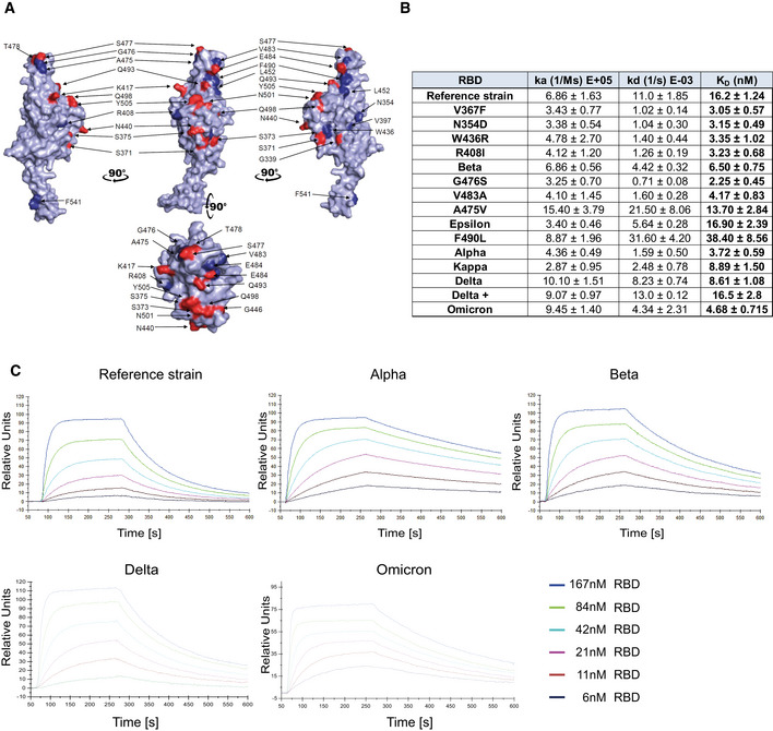
- A
PyMOL rendered visualization of the SARS‐CoV‐2 RBD. Rendering depicts the SARS‐CoV‐2 RBD from the Wuhan reference isolate with mutation sites observed in SARS‐CoV‐2 variants shown in red. Indicated in blue are positions mutated in the various strains of SARS‐CoV‐2 used in experiments in this study.
- B
Surface Plasmon Resonance analysis to derive kinetic constants (ka, kd) and affinity values (KD) of SARS‐CoV‐2 RBD/APN01 interaction. The constants represent mean values and standard deviations obtained from sensorgram fittings performed in quadruplicate. The table lists both the tested variants and the introduced amino acid substitution as well as the designation of the respective Variant of Concern (VOC) or Variant of Interest (VOI) mutations tested in this study. Reference strain RBD sequence corresponds to the Wuhan SARS‐CoV‐2 isolate.
- C
Representative SPR sensorgram images for the SARS‐CoV‐2 RBD/APN01 interaction of current VOCs.
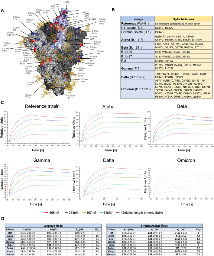
- A
PyMOL rendering of the trimeric full‐length SARS‐CoV‐2 Spike protein. One RBD is shown in violet. Indicated in blue are positions mutated in the various strains of SARS‐CoV‐2 used in experiments in this study. Mutations described for the Omicron VOC are depicted in red. Shown in yellow are the glycan modifications of the spike protein (Capraz et al, 2021).
- B
Table lists the SARS‐CoV‐2 strains and their respective mutations within the Spike protein that were used in this study.
- C
Representative sensorgram images for the SPR analysis conducted with full‐length trimeric spike proteins in pre‐fusion state with APN01. Reference strain corresponds to original Wuhan viral isolate spike sequence. Indicated are VOC Alpha, Beta, Gamma, and Delta, and Omicron.
- D
Tables listing ka, kd, as well as KD values for the interaction of APN01 and full‐length trimeric spike proteins. The constants represent mean values and standard deviations obtained from sensorgram fittings performed in quadruplicate. Values are derived from calculations based upon the Langmuir (left table) or Bivalent Analyte sensorgram fitting (right table).
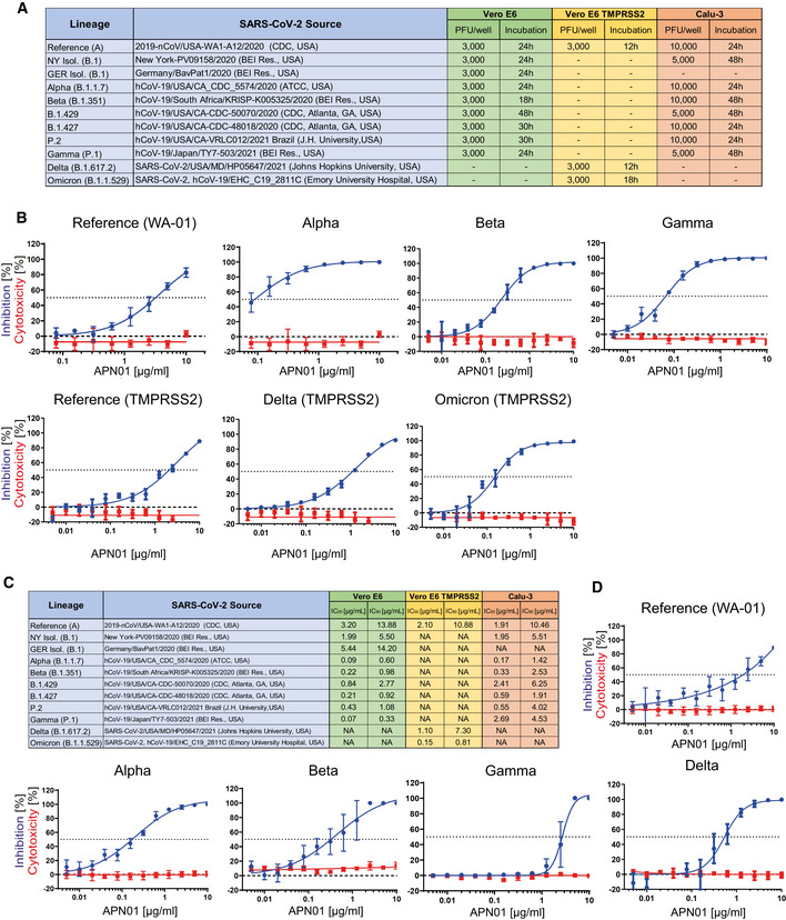
- A
Table depicts source of the tested viral isolates, as well plaque‐forming units (PFU) and the infection time used in these experiments for both VeroE6/VeroE6‐TMPRSS2 and Calu‐3 cells.
- B
Panels depict both neutralization of the indicated SARS‐CoV‐2 isolates (blue line) and cytotoxicity of APN01 (red line) in VeroE6 or VeroE6‐TMPRSS2 cells. Analysis was done in quadruplicate with mean and standard deviations shown. Y‐axis depicts the percentage of neutralization and cytotoxicity, respectively.
- C
Table depicts IC50 and IC90 values for APN01‐mediated neutralization of viral infection in VeroE6/VeroE6‐TMPRSS2 and Calu‐3 cells.
- D
Same experimental setup as in (a) but conducted with the epithelial lung cancer cell line Calu‐3. Analysis was done in quadruplicate with mean and standard deviations shown. Y‐axis depicts the percentage of neutralization and cytotoxicity, respectively.
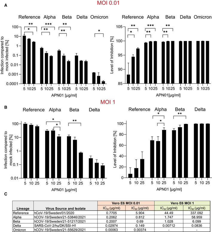
- A, B
Diagrams depict the level of infection with the indicated SARS‐CoV‐2 isolates at MOI 0.01 (A) and MOI 1 (B) of VeroE6 cells after pre‐incubation with increasing concentrations of APN01 as compared to infections in the absence of APN01 pre‐incubation. MOIs as depicted reflect multiplicities of infection before the pre‐incubation with APN01 that was followed by the viral infection of cells. Shown are means of biological replicates (n = 3) analyses with standard deviations. Statistical significance is indicated by asterisks (*P‐value < 0.05; **P‐value < 0.01, ***P‐value < 0.001 as calculated with one‐way ANOVA).
- C
List and source of strains used at the Karolinska Institutet and IC50 and IC90 values obtained for the indicated MOIs. See Materials and Methods section for a detailed list of viral mutations for the strains used.
Update of
-
Clinical grade ACE2 as a universal agent to block SARS-CoV-2 variants.bioRxiv [Preprint]. 2021 Sep 10:2021.09.10.459744. doi: 10.1101/2021.09.10.459744. bioRxiv. 2021. Update in: EMBO Mol Med. 2022 Aug 8;14(8):e15230. doi: 10.15252/emmm.202115230. PMID: 34545368 Free PMC article. Updated. Preprint.
References
Publication types
MeSH terms
Substances
Supplementary concepts
Grants and funding
LinkOut - more resources
Full Text Sources
Medical
Miscellaneous
