

Globe dislocation and optic nerve avulsion following all-terrain vehicle accidents
- PMID: 35782169
- PMCID: PMC9243039
- DOI: 10.1016/j.ajoc.2022.101621
Globe dislocation and optic nerve avulsion following all-terrain vehicle accidents
Abstract
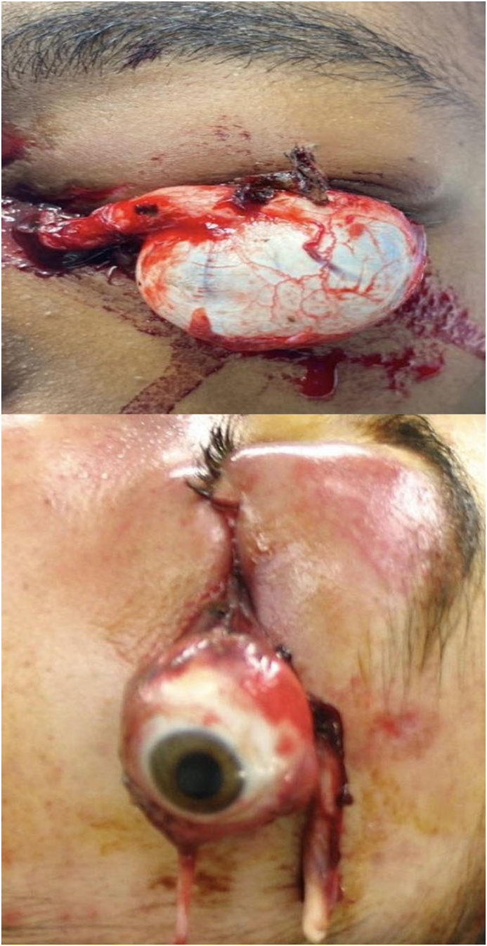
Purpose: Open-air motor vehicles present unique trauma risks to the eyes and face. We describe two patients who suffered a crash while riding an all-terrain vehicle (ATV), leading to globe dislocation with optic nerve avulsion in order to raise awareness about the risks associated with ATV accidents.
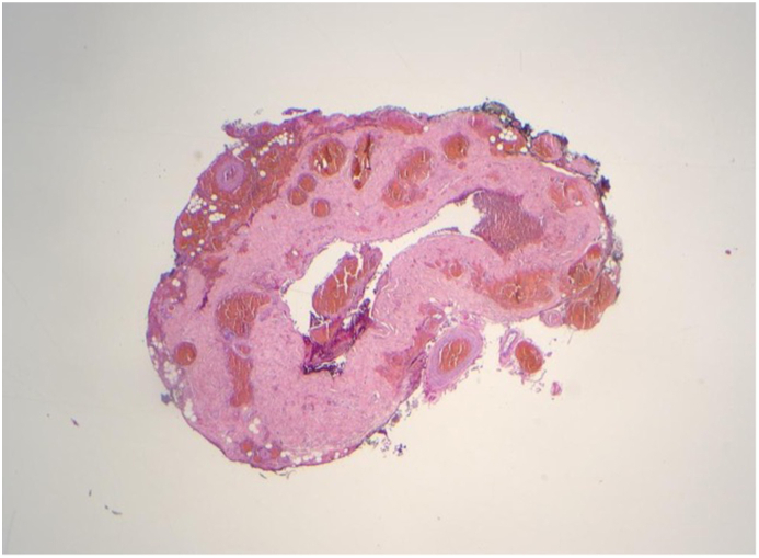
Observations: In both cases, the injury was caused by high-speed trauma to the orbit involving a tree branch. One patient sustained a life threatening arrythmia requiring a short stay in the intensive care unit, and both patients required emergent surgical management and eventual socket reconstruction.
Conclusions and importance: These cases highlight the need for greater advocacy on behalf of rider safety. The authors encourage ophthalmologists to counsel patients who use ATVs to wear helmets, seatbelts, and protective eyewear to prevent these types of injuries in the future.
Keywords: All terrain vehicles; Globe dislocation; Optic nerve avulsion.
© 2022 The Authors.
Conflict of interest statement
The authors declare that there are no conflicts of interest, either financially or personally, regarding the publication of this paper.
Figures


Similar articles
-
Etiologies of pediatric craniofacial injuries: a comparison of injuries involving all-terrain vehicles and golf carts.Int J Pediatr Otorhinolaryngol. 2013 Mar;77(3):414-7. doi: 10.1016/j.ijporl.2012.11.043. Epub 2013 Jan 6. Int J Pediatr Otorhinolaryngol. 2013. PMID: 23299042
-
On-road all-terrain vehicle (ATV) fatalities in the United States.J Safety Res. 2014 Sep;50:117-23. doi: 10.1016/j.jsr.2014.05.001. Epub 2014 May 20. J Safety Res. 2014. PMID: 25142368
-
All-terrain vehicles (ATVs) on the road: a serious traffic safety and public health concern.Traffic Inj Prev. 2013;14(1):78-85. doi: 10.1080/15389588.2012.675110. Traffic Inj Prev. 2013. PMID: 23259522
-
Pediatric and adolescent injury in all-terrain vehicles.Res Sports Med. 2018;26(sup1):38-56. doi: 10.1080/15438627.2018.1438279. Res Sports Med. 2018. PMID: 30431365 Review.
-
Agricultural All-Terrain Vehicle Safety: Hazard Control Methods Using the Haddon Matrix.J Agromedicine. 2021 Oct;26(4):420-435. doi: 10.1080/1059924X.2020.1837705. Epub 2020 Nov 10. J Agromedicine. 2021. PMID: 33169657 Review.
Cited by
-
Traumatic globe avulsion secondary to a penetrating orbital injury from a bicycle handlebar: a case report.J Trauma Inj. 2025 Jun;38(2):147-151. doi: 10.20408/jti.2024.0070. Epub 2025 Apr 1. J Trauma Inj. 2025. PMID: 40174900 Free PMC article.
-
Traumatic Globe Luxation and Optic Nerve Avulsion: A Case Report and Literature Review.Cureus. 2024 Jan 29;16(1):e53150. doi: 10.7759/cureus.53150. eCollection 2024 Jan. Cureus. 2024. PMID: 38420068 Free PMC article.
References
-
- Mayercik V.A., Eller A.W., Stefko S.T. Ocular injuries in all-terrain-vehicle accidents. Injury. 2012 Sep;43(9):1462–1465. - PubMed
-
- Roberts S.P., Schaumberg D.A., Thompson P. Traumatic avulsion of the optic nerve. Optom Vis Sci. 1992 Sep;69(9):721–727. - PubMed
-
- Hughes B. Indirect injury to the optic nerves and chiasma. Bull Hopkins Hosp. 1962;111:98–126. - PubMed
Publication types
LinkOut - more resources
Full Text Sources
Research Materials
