Review
. 2022 Jul;12(7):3947-3986.
doi: 10.21037/qims-2022-04.

The value of magnetic resonance imaging and computed tomography in the study of spinal disorders

Affiliations
Review

The value of magnetic resonance imaging and computed tomography in the study of spinal disorders

Fernando Ruiz Santiago et al. Quant Imaging Med Surg. 2022 Jul.

Abstract

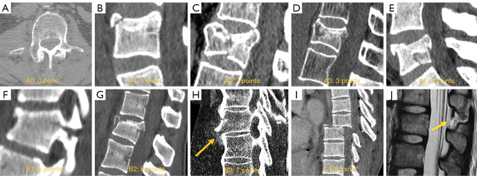
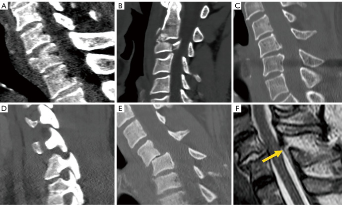
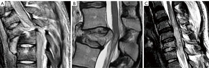
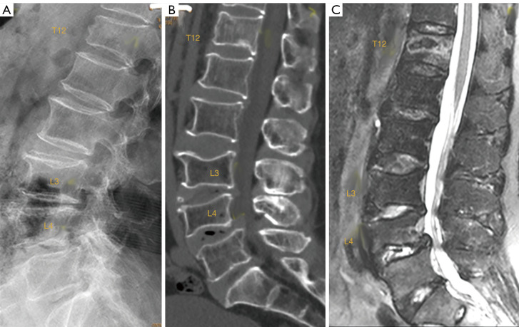
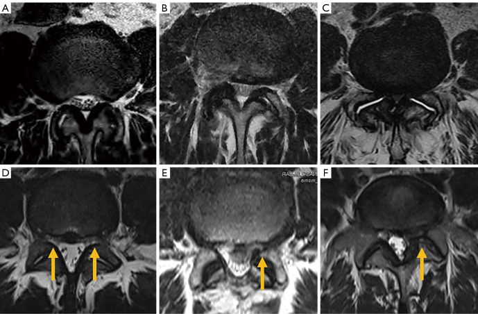
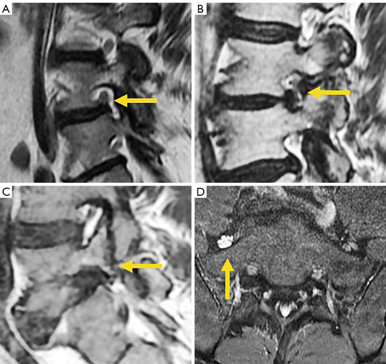
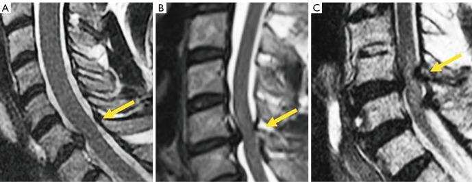
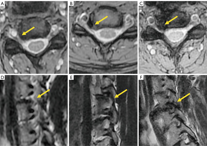
Computed tomography (CT) and magnetic resonance imaging (MRI) have replaced conventional radiography in the study of many spinal conditions, it is essential to know when these techniques are indicated instead of or as complementary tests to radiography, which findings can be expected in different clinical settings, and their significance in the diagnosis of different spinal conditions. Proper use of CT and MRI in spinal disorders may facilitate diagnosis and management of spinal conditions. An adequate clinical approach, a good understanding of the pathological manifestations demonstrated by these imaging techniques and a comprehensive report based on a universally accepted nomenclature represent the indispensable tools to improve the diagnostic approach and the decision-making process in patients with spinal pain. Several guidelines are available to assist clinicians in ordering appropriate imaging techniques to achieve an accurate diagnosis and to ensure appropriate medical care that meets the efficacy and safety needs of patients. This article reviews the clinical indications of CT and MRI in different pathologic conditions affecting the spine, including congenital, traumatic, degenerative, inflammatory, infectious and tumor disorders, as well as their main imaging features. It is intended to be a pictorial guide to clinicians involved in the diagnosis and treatment of spinal disorders.

Keywords: Spine; back pain; computed tomography (CT); magnetic resonance imaging (MRI); osteoporotic fracture; spinal diseases; spinal disorder.


Conflict of interest statement

Conflicts of Interest: All authors have completed the ICMJE uniform disclosure form (available at https://qims.amegroups.com/article/view/10.21037/qims-2022-04/coif). YXJW serves as the Editor-in-Chief of Quantitative Imaging in Medicine and Surgery. The other authors have no conflicts of interest to declare.

Figures

Figure 1
Figure 1
Congenital deformities. (A) Scoliosis secondary to a semi-segmented hemivertebra. (B) Klippel-Feil syndrome with formation and segmentation abnormalities. (C) Scoliosis secondary to asymmetric butterfly vertebra [from reference (10)]. (D) Kyphosis secondary to anterior failure of vertebral formation.
Figure 2
Figure 2
Spinal dysraphism. (A) Myelomeningocele in open dysraphism in intrauterine fetus showing the protruded placode (arrow). (B) Posterior closed dysraphism with lipomyelomeningocele. The lipoma/placode interface (arrow) is outside the spinal canal. (C) Lipomyelocele The lipoma/placode interface (arrow) is within the spinal canal. (D) Dermal sinus (arrow). (E) Tethered cord with filum terminale lipoma (*) and filar thickening (arrow). (F) Split cord/diastematomyelia.
Figure 3
Figure 3
Castellvi types of transitional vertebra in three cases. Type II on CT depicting the neo articulation (arrow) (A) and MRI (B) showing the subchondral edema (arrow). (C) Type III on 3D CT. (D) Type IV on coronal CT. CT, computed tomography; MRI, magnetic resonance imaging.
Figure 4
Figure 4
Transitional vertebral. (A) Iliolumbar ligament at L5 (arrows). (B) Splitting pattern of L4 (arrow) compared to L5 nerve root.
Figure 5
Figure 5
AO-Spine thoracolumbar fracture classification score. (A) A0. Apophyseal fracture. (B) A1. Compression fracture without involvement of the posterior wall. (C) A2. Split fracture without involvement of the posterior wall. (D) A3. Burst fracture with involvement of one endplate. (E) A4. Burst fracture with involvement of both endplates. (F) B1. Trans osseous fracture. (G) B2. Posterior tension band injury fracture. (H) B3. Anterior tension band injury fracture (arrow). (I) Fracture dislocation injury type C. (J) MRI showing flavum (arrow), interspinous and supraspinous ligamentum tear. AO, Arbeitsgemeinschaft für Osteosynthesefragen; MRI, magnetic resonance imaging.
Figure 6
Figure 6
AO-Spine subaxial fracture classification score. (A) Compression fracture; (B) burst fracture; (C) tension band injury fracture; (D) locked facets; (E) fracture dislocation; (F) sagittal T2 weighted images showing ligamentum flavum and supraspinous ligaments tear (arrow). AO, Arbeitsgemeinschaft für Osteosynthesefragen.
Figure 7
Figure 7
Traumatic spinal cord injury. (A) Type I, hemorrhagic lesion; (B) Type II, edematous injury; (C) Type III, mixed injury.
Figure 8
Figure 8
Elderly patient with spine trauma. (A) Radiograph; (B) sagittally reconstructed CT; (C) sagittal T2 weighted fat suppressed MRI. A T12 traumatic fracture, L3 chronic osteoporotic upper endplate fracture, and L4 chronic osteoporotic deformity (i.e., fracture) are detected. For T12, on radiograph, attention should be paid to the anterior cortex fracture and vertebral height loss, while on MRI apparent abnormal high signal is noted. Conversely, L3 and L4 show deformity, but no abnormal high signal is noted on MRI. CT, computed tomography; MRI, magnetic resonance imaging.
Figure 9
Figure 9
Subtle osteoporotic fracture after low energy trauma (arrow). (A) Radiograph; (B) radiograph magnified view for T12; (C) sagittally reconstructed CT; (D) T1 weighted MRI. On radiograph T12 vertebral anterior cortex buckling is noted. There is no apparent height loss of the vertebral body. CT confirms T12 fracture and vertebral anterior cortex break. MRI also confirms T12 fracture. CT, computed tomography; MRI, magnetic resonance imaging.
Figure 10
Figure 10
Osteoporotic fractures. Sagittal T1 (A) and STIR (B) showing the band like edema pattern (arrows). (C) Burst fracture with intravertebral vacuum cleft (arrow) and retropulsion of the posteroinferior margin (*). (D) Sagittal STIR with intravertebral liquid cleft (arrow) and retropulsion of posterosuperior margin (*). STIR, short tau inversion recovery.
Figure 11
Figure 11
Pathologic fracture in several cases of metastatic lung cancer. Sagittal CT (A) and sagittal T2 weighted images (B) showing convex posterior border. Sagittal T1 weighted image without (C) and with (D) contrast in pathologic fracture. In phase (E) and out of phase (F) showing lack of fat in this metastatic vertebra, with a signal intensity out phase/phase ratio of 0.95. CT, computed tomography.
Figure 12
Figure 12
Osteoarthritis of the posterior elements. (A) Axial CT of facet joint osteoarthritis (arrows). (B) Sagittal STIR showing edema in cervical facet joint osteoarthritis (arrow). (C) Sagittal CT showing Baastrup’s disease (arrow). (D) Interspinous bursitis (arrow) demonstrated during injection with dye and steroids of the facets joint. CT, computed tomography; STIR, short tau inversion recovery.
Figure 13
Figure 13
Annular tears on T2 weighted images. Sagittal (A) and axial (B) of transverse tears (arrow). (C) Concentric and radial (D) tears (arrows).
Figure 14
Figure 14
Disc displacement. (A) Diffuse bulge; (B) central protrusion; (C) right paracentral disc extrusion; (D) foraminal extrusion; (E) cranial migration of the disc extrusion; (F) sequestered fragment (arrow).
Figure 15
Figure 15
Modic changes. Sagittal T1 weighted image (A), T2 weighted image (B) and STIR sequence image (C) of Modic type I changes. Sclerosis (arrow) detected at S1 on CT (D) is barely visible on MRI. Sagittal T1 weighted image (E), T2 weighted image (F) and STIR image sequence (G) of Modic type II changes. Sclerosis (arrows) conspicuous on CT (H) is barely suspected on MRI. STIR, short tau inversion recovery; CT, computed tomography; MRI, magnetic resonance imaging.
Figure 16
Figure 16
DISH. (A) Sagittal STIR showing subligamentous edema (arrows). (B) Sagittal reformatted CT showing bridging osteophytes. (C) Sagittal CT of low energy cervical fracture in a patient with DISH (arrow). DISH, diffuse idiopathic skeletal hyperostosis; STIR, short tau inversion recovery; CT, computed tomography.
Figure 17
Figure 17
Lumbar canal stenosis. Axial T2 images of mild (A), moderate (B) and severe (C) central canal stenosis. (D) Mild bilateral lateral recess stenosis (arrows). Moderate (E) and severe (F) left foraminal stenosis (arrows).
Figure 18
Figure 18
Lumbar foraminal stenosis. Sagittal T2 weighted images of mild (A), moderate (B) and severe (C) foraminal stenosis (arrow). (D) Axial intermediate weighted fat saturated image showing increased signal of L5 nerve root (arrow) in severe foraminal stenosis.
Figure 19
Figure 19
Cervical canal stenosis. Sagittal T2 weighted of mild (A), moderate (B) and severe (C) central canal stenosis (arrow).
Figure 20
Figure 20
Cervical foraminal stenosis. Axial T2* weighted images of mild (A), moderate (B) and severe (C) foraminal stenosis (arrow). Sagittal T2 weighted images of mild (D), moderate (E) and severe (F) foraminal stenosis (arrow).
Figure 21
Figure 21
MRI of spondylolysis. (A) Sagittal STIR image of grade 1 spondylolysis showing edema without pars interarticularis defect (arrow). (B) Grade 2 spondylolysis, partial defect with edema (arrow). (C) Grade 2A spondylolysis, partial defect without edema (arrow). (D) Grade 3 spondylolysis, complete defect with edema (arrow). (E) Grade 4 spondylolysis, chronic defect without edema (arrow). (F) Bilateral pedicle spondylolysis (arrow). MRI, magnetic resonance imaging; STIR, short tau inversion recovery.
Figure 22
Figure 22
MRI Indirect signs of spondylolysis and spondylolisthesis. (A) Epidural fat interposition between the thecal sac and the L5 spinous process in spondylolysis without spondylolisthesis. (B) Widening of the central canal (arrow) and foraminal stenosis (arrow) (C) in spondylolysis with spondylolisthesis. MRI, magnetic resonance imaging.
Figure 23
Figure 23
Alignment abnormalities in three patients. (A) Sagittal T2 weighted image of a patient with degenerative spondylolisthesis with narrowing of the central canal and anterior shifting of the spinous process (arrow). (B) Sagittal CT with retrolisthesis of L5. (C) CT with MIP reconstruction in Hangman’s fracture. (D) Axial T2 of a hyperintense vertebral artery (arrow) secondary to arterial dissection in Hangman’s fracture. CT, computed tomography; MIP, maximum intensity projection.
Figure 24
Figure 24
Alterations in spinal curvature. (A) Sagittal T2 weighted image of a case with Scheuermann’s disease. (B) Coronal T2 weighted image of a case with neuromuscular scoliosis in Rett syndrome. (C) Coronal T2 weighted image of a patient with scoliosis secondary to osteoid osteoma (arrow).
Figure 25
Figure 25
Sacroiliitis in four patients. (A) Axial STIR in active inflammatory spondyloarthropathy. (B) CT showing structural changes at the sacroiliac joints. (C) T2 weighted image of a patient with chronic sacroiliitis with fused joints. (D) Axial STIR sequence image of a patient with post-partum subchondral edema at both iliac wings. STIR, short tau inversion recovery; CT, computed tomography.
Figure 26
Figure 26
Spinal involvement in axial spondyloarthropathy. Sagittal STIR images showing Romanus lesions (arrows) (A), costovertebral joints inflammation (arrows) (B), and Andersson lesions (arrows) (C). STIR, short tau inversion recovery.
Figure 27
Figure 27
Infectious pathology of the spine. (A) Sagittal STIR image of a case of pseudomonas discitis. (B) Sagittal T1 weighted image of a case of brucella osteomyelitis (arrow). (C) Sagittal T2 weighted image of a case of tuberculous prevertebral subligamentous abscess (arrow). (D) Axial CT of a case of chronic bilateral psoas abscess showing muscle enlargement with fluid collection and small calcifications (arrows). STIR, short tau inversion recovery; CT, computed tomography.
Figure 28
Figure 28
Vertebral hemangioma. Sagittal T1 weighted image (A), T2 weighted image (B), and STIR sequence image (C) of typical hemangioma (arrows), hyperintense on T1 and T2 due to fat content, remaining hyperintense on STIR only the vascular content. Sagittal T1 weighted image (D), T2 weighted image (E), and STIR image sequence (F) of atypical hemangioma (arrows), hypointense on T1 due to predominance of vascular component and scarce fat content. Sagittal T1 weighted image (G), T2 weighted image (H), and STIR image sequence (I) of aggressive hemangioma (arrows) extending to the spinal canal (*). STIR, short tau inversion recovery.
Figure 29
Figure 29
Eosinophilic granuloma. (A) Initial sagittal T2 weighted image that evolved to severe collapse of the vertebral body in 2 months (B). (C) Partial vertebral height recovery at 3 years follow-up.
Figure 30
Figure 30
Osteolytic lesions of the spine. (A) Axial CT image of osteoblastoma showing the osteolytic lesion (arrow), and (B) edema pattern on MRI (arrow). (C) CT of aneurysmal bone cyst (arrow). (D) T2 weighted image showing fluid-fluid levels in the same case (arrow). CT, computed tomography; MRI, magnetic resonance imaging.
Figure 31
Figure 31
Benign tumors of the spine. (A) Axial CT image of enostoma; (B) sagittal T1 and STIR (C) images of enostoma; (D) coronal reformatted CT image of osteochondroma. CT, computed tomography; STIR, short tau inversion recovery.
Figure 32
Figure 32
Notochordal tumors. (A) Coronal reformatted CT of a lumbar chordoma. (B) Sagittal T1 without and with contrast (C) of a sacral chordoma. Sagittal reformatted CT (D), T1 (E), and STIR images (F) of a benign notochordal tumor. CT, computed tomography; STIR, short tau inversion recovery.
Figure 33
Figure 33
Spinal metastases. (A) Sagittal CT reformation and Sagittal T2 weighted images (B) in a case of osteolytic lung cancer metastasis. (C) Sagittal CT reformation and sagittal T2 weighted image (D) in a case of osteoblastic breast carcinoma metastasis demonstrating increased density on CT scan and hypointensity on MRI. CT, computed tomography; MRI, magnetic resonance imaging.
Figure 34
Figure 34
Patterns of spinal involvement in myeloma. Sagittal T1 weighted imaging shows diffuse (A) and salt and pepper (B) infiltration. (C) Focal and combined (D) involvement of the spine.
Figure 35
Figure 35
Infiltrative pattern in hematologic malignancies. (A) Sagittal T1 in diffuse infiltrative pattern in leukemia. (B) Patchy infiltrative pattern in Hodgkin’s lymphoma.

References

    1. ACR-ASNR-SCBT-MR practice guidelines for the performance of magnetic resonance imaging (MRI) of the adult spine. American College of Radiology. 2018. Available online: https://www.acr.org/-/media/ACR/Files/Practice-Parameters/MR-Adult-Spine.... Accessed on March 28, 2022.
    1. Beckmann NM, West OC, Nunez D, Jr, Kirsch CFE, Aulino JM, Broder JS, Cassidy RC, Czuczman GJ, Demertzis JL, Johnson MM, Motamedi K, Reitman C, Shah LM, Than K, Ying-Kou Yung E, Beaman FD, Kransdorf MJ, Bykowski J. ACR Appropriateness Criteria® Suspected Spine Trauma. J Am Coll Radiol 2019;16:S264-85. 10.1016/j.jacr.2019.02.002 - DOI - PubMed
    1. Patel ND, Broderick DF, Burns J, Deshmukh TK, Fries IB, Harvey HB, Holly L, Hunt CH, Jagadeesan BD, Kennedy TA, O'Toole JE, Perlmutter JS, Policeni B, Rosenow JM, Schroeder JW, Whitehead MT, Cornelius RS, Corey AS. ACR Appropriateness Criteria Low Back Pain. J Am Coll Radiol 2016;13:1069-78. 10.1016/j.jacr.2016.06.008 - DOI - PubMed
    1. Booth TN, Iyer RS, Falcone RA, Jr, Hayes LL, Jones JY, Kadom N, Kulkarni AV, Myseros JS, Partap S, Reitman C, Robertson RL, Ryan ME, Saigal G, Soares BP, Tekes-Brady A, Trout AT, Zumberge NA, Coley BD, Palasis S. ACR Appropriateness Criteria® Back Pain-Child. J Am Coll Radiol 2017;14:S13-24. 10.1016/j.jacr.2017.01.039 - DOI - PubMed
    1. Jenkins HJ, Downie AS, Maher CG, Moloney NA, Magnussen JS, Hancock MJ. Imaging for low back pain: is clinical use consistent with guidelines? A systematic review and meta-analysis. Spine J 2018;18:2266-77. 10.1016/j.spinee.2018.05.004 - DOI - PubMed