Review
. 2022 May;17(3):353-384.
doi: 10.1016/j.ajps.2022.01.001. Epub 2022 Jan 24.

Antibacterial biomaterials for skin wound dressing

Affiliations
Review

Antibacterial biomaterials for skin wound dressing

Yuqing Liang et al. Asian J Pharm Sci. 2022 May.

Abstract

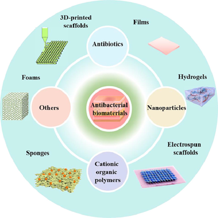
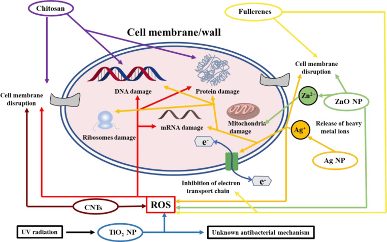
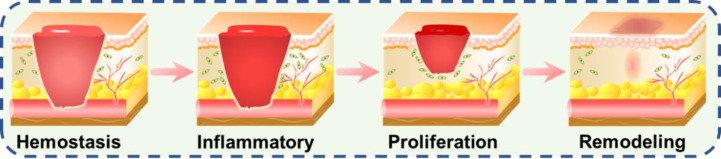
Bacterial infection and the ever-increasing bacterial resistance have imposed severe threat to human health. And bacterial contamination could significantly menace the wound healing process. Considering the sophisticated wound healing process, novel strategies for skin tissue engineering are focused on the integration of bioactive ingredients, antibacterial agents included, into biomaterials with different morphologies to improve cell behaviors and promote wound healing. However, a comprehensive review on anti-bacterial wound dressing to enhance wound healing has not been reported. In this review, various antibacterial biomaterials as wound dressings will be discussed. Different kinds of antibacterial agents, including antibiotics, nanoparticles (metal and metallic oxides, light-induced antibacterial agents), cationic organic agents, and others, and their recent advances are summarized. Biomaterial selection and fabrication of biomaterials with different structures and forms, including films, hydrogel, electrospun nanofibers, sponge, foam and three-dimension (3D) printed scaffold for skin regeneration, are elaborated discussed. Current challenges and the future perspectives are presented in this multidisciplinary field. We envision that this review will provide a general insight to the elegant design and further refinement of wound dressing.

Keywords: Antibacterial activity; Biomaterials; Skin tissue engineering; Wound dressing; Wound healing.


Conflict of interest statement

The authors declare no conflicts of interest.

Figures

Image, graphical abstract
Graphical abstract
Fig 1
Fig. 1
Antibacterial mechanism of chitosan, metal-based nanoparticles and carbon-based composites.
Fig 2
Fig. 2
Schematic illustrations of wound healing phases consist of hemostasis, inflammatory, proliferation and wound remodeling with scar tissue formation. Reproduced from with permission from American Chemical Society.
Fig 3
Fig. 3
(A) The shape memory behavior displays in practical applications of PCL-PEG-AT12. (B) Illustration of wound recovery by applying PCL-PEG-AT12 film. (C) Release behavior of vancomycin from the copolymers at 37 °C in pH 7.4 PBS. PCL-PEG-ATx: PCL-PEG-AT with different weight ratio of AT. Reproduced from with permission from Elsevier.
Fig 4
Fig. 4
The design and application of curcumin-loaded PLGA microspheres incorporated CS/aloe films. (A) The illustration for the preparation of the films. (B) The release behavior of curcumin. (C) Images of full-thickness skin wound treated with different samples during the healing process. Film-1: CS/aloe films. Film-2: CS/aloe-PLGA microspheres films. Film-3: curcumin-loaded Film-2. Reproduced from with permission from Elsevier.
Fig 5
Fig. 5
The topical thermal ablation of the TRIM films. (A) TRIM films with flat surface normally while reproducing corrugated microtopography during heating (via infrared-light irradiation). The corrugated surface would induce thermally activated disruption of the biofilm and facilitate the ablation of planktonic bacteria, while mitigating the thermal harm to host epithelium. (B) Fluorescence images show that S. aureus (green) attached within feature gaps when dispersed on films with different features. On surfaces with 5 µm and larger features, S. aureus formed aggregates. Inset: PDMS film with varied features. Scale bar: 50 µm. (C) Antibacterial efficiency of TRIM films or flat film. Reproduced from with permission from WILEY-VCH. (For interpretation of the references to colour in this figure legend, the reader is referred to the web version of this article.)
Fig 6
Fig. 6
(A) Inhibitor zones from sterilizing effectiveness test against E. coli and S. aureus. (B) Histomorphological analysis for regeneration of wounds treated with different samples. Reproduced from with permission from WILEY-VCH.
Fig 7
Fig. 7
The design and working principles of the smart flexible electronics-integrated double-layer wound dressing. (A) The smart dressing consists of a PDMS-encapsulated flexible electronic layer and an UV-responsive antibacterial hydrogel. (B) Schematic illustration of the integrated dressing for infected-wound monitoring and on-demand treatment. Reproduced from with permission from WILEY-VCH.
Fig 8
Fig. 8
The wound healing evaluation of PDA@Ag NPs/CPHs. (A) Photos of the diabetic feet treated with different samples (The control group is treated by pure carboxymethyl cellulose hydrogel). (B) Quantification of wound residual area. (C) Quantification of inflammatory cells. *P < 0.05, **P < 0.01, and ***P < 0.001. Reproduced from with permission from WILEY-VCH.
Fig 9
Fig. 9
Beneficial effects and application of the physical double-network hydrogel. (A) Shape-adaptive performance of PEGSD2/GTU5.0. Scale bar: 1 cm. (B) Photographs of hydrogel macroscopic self-healing performance when assisted with NIR irradiation (1.4 W cm−2). Scale bar: 1 cm. (C) The original state and withstanding joint bending of the soft and flexible hydrogel PEGSD2/GTU5.0. Scale bar: 1 cm. (D) Adhesive strength of the hydrogels on pig skin. (E) Representative photographs of rat full-thickness skin incision wounds at day 0, 7 and 14 post-surgery, scale bar: 5 mm. (F) Relative tensile strength of healed skins after treated for 14 d *P < 0.05 and ***P < 0.001. Reproduced from with permission from WILEY-VCH.
Fig 10
Fig. 10
(A) In vitro release kinetics of curcumin from the hydrogels in PBS at pH values of 7.4, 6.8 and 6.0. (B) Adhesive strength of different hydrogels. (C) Scheme of the self-healing process. (D) Schematic diagram of model drug (curcumin) released from hydrogel when it was applied on the joints. Reproduced from with permission from Elsevier.
Fig 11
Fig. 11
The electrospinning of SF–Mel/PCL nanofibers as wound dressing for skin repair and regeneration. Reproduced from with permission from The Royal Society of Chemistry.
Fig 12
Fig. 12
Pictures of the regenerated wound tissue on 7th, and 14th d by immunofluorescence staining labeling with (A) TNF-α (green) and (B) VEGF (green), yellow arrows show the expression of TNF-α and red arrows present VEGF. Reproduced from Ref. 217 with permission from Elsevier. (C) Schematic illustration of dressing with bifunctions of tumor therapy and skin tissue regeneration. Reproduced from with permission from American Chemical Society. (For interpretation of the references to colour in this figure legend, the reader is referred to the web version of this article.)
Fig 13
Fig. 13
Schematic illustration of preparation and biomedical application of the chitosan dressing containing ZnO/N-halamine hybride nanoparticles. Reproduced from with permission from American Chemical Society.
Fig 14
Fig. 14
Beneficial effects of asymmetric CS sponge. (A) Water contact angles of hydrophobic surface (a1) and hydrophilic surface (a2) of CS sponge, respectively. (B) The water vapor transmission rate of different materials (including sponges with 0.5%, 1%, 2% and 3% CS (w/v)). *P< 0.05. (C) Bacteria infiltration activity of CS (a), modified TMC NPs/CS (b) and modified CS (c) sponges against E. coli ((c1) and (c2)) and S. aureus ((c3) and (c4)). Reproduced from with permission from Elsevier. Elsevier.
Fig 15
Fig. 15
(A) SEM images of the surface and cross-section of PU foams with Ag NPs and human epidermal growth factors loaded. Reproduced from with permission from The Royal Society of Chemistry. (B) Schematic illustration of the synthesis of PU foams with cationic imidazolium diol. (C) Photographs of three cationic PU foams and the control one. (D) Computer simulation of the endotoxin adsorption mechanism displaying that the snapshots for the interaction process of the P. aeruginosa outer membrane (LPS molecules and lipid bilayer) with foam 3 at different simulation times (0, 240, and 600 ns). Reproduced from with permission from American Chemical Society.
Fig 16
Fig. 16
(A) Schematic representation of the 3D bioprinting, consolidation, and maturation steps of the bilayer skin structure (DE: dermal–epidermal junction; SE: surface–epidermal junction). Reproduced from with permission from WILEY-VCH. (B) Scheme of skin-equivalent integrated with perfusable vascular channels and culture device. Reproduced from with permission from Elsevier. (C) The schematic presentation for the preparation of Ag NPs-cross-linked hydrogel dressing. Reproduced from Ref. with permission from American Chemical Society.

References

    1. Norouzi M., Boroujeni S.M., Omidvarkordshouli N., Soleimani M. Advances in skin regeneration: application of electrospun scaffolds. Adv Healthcare Mater. 2015;4(8):1114–1133. - PubMed
    1. Yu R., Zhang H., Guo B. Conductive biomaterials as bioactive wound dressing for wound healing and skin tissue engineering. Nano-Micro Lett. 2022;14(1):1. - PMC - PubMed
    1. Guo B., Dong R., Bang Y., Li M. Haemostatic materials for wound healing applications. Nat Rev Chem. 2021;5(11):773–791. - PubMed
    1. Modaresifar K., Azizian S., Ganjian M., Fratila-Apachitei L.E., Zadpoor A.A. Bactericidal effects of nanopatterns: a systematic review. Acta Biomater. 2019;83:29–36. - PubMed
    1. Nussbaum S.R., Carter M.J., Fife C.E., DaVanzo J., Haught R., Nusgart M., et al. An economic evaluation of the impact, cost, and medicare policy implications of chronic nonhealing wounds. Value in Health. 2018;21(1):27–32. - PubMed

LinkOut - more resources