. 2022 Jun 24:6:23982128221106315.
doi: 10.1177/23982128221106315. eCollection 2022 Jan-Dec.

Septotemporal variation in modulation of synaptic transmission, paired-pulse ratio and frequency facilitation/depression by adenosine and GABAB receptors in the rat hippocampus

Affiliations

Septotemporal variation in modulation of synaptic transmission, paired-pulse ratio and frequency facilitation/depression by adenosine and GABAB receptors in the rat hippocampus

Maria A Samara et al. Brain Neurosci Adv. .

Abstract

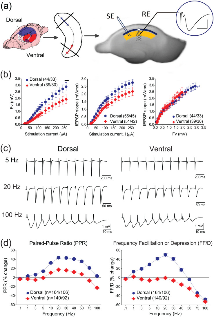
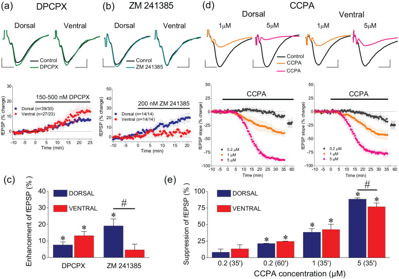
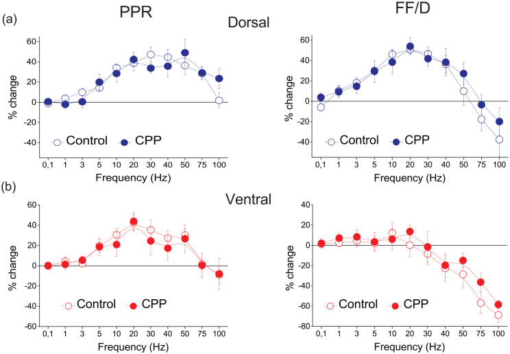
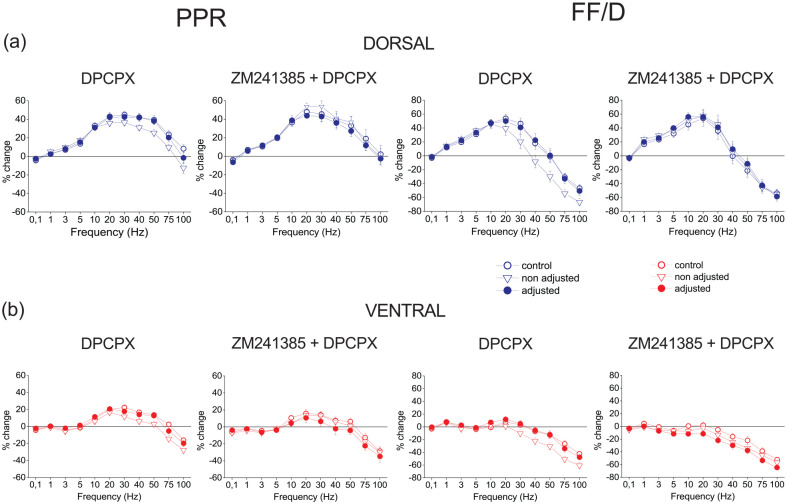
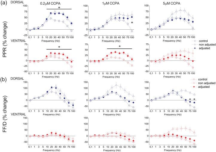
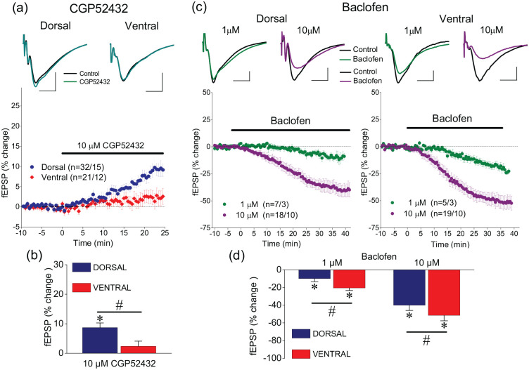
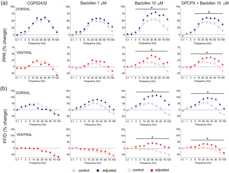
Short-term synaptic plasticity represents a fundamental mechanism in neural information processing and is regulated by neuromodulators. Here, using field recordings from the CA1 region of adult rat hippocampal slices, we show that excitatory synaptic transmission is suppressed by strong but not moderate activation of adenosine A1 receptors by 2-Chloro-N6-cyclopentyladenosine (CCPA) more in the dorsal than the ventral hippocampus; in contrast, both mild and strong activation of GABAB receptors by baclofen (1 μM, 10 μM) suppress synaptic transmission more in the ventral than the dorsal hippocampus. Using a 10-pulse stimulation train of variable frequency, we found that CCPA modulates short-term synaptic plasticity independently of the suppression of synaptic transmission in both segments of the hippocampus and at stimulation frequencies greater than 10 Hz. However, specifically regarding the paired-pulse ratio (PPR) and frequency facilitation/depression (FF/D) we found significant drug action before but not after adjusting conditioning responses to control levels. Activation of GABABRs by baclofen suppressed synaptic transmission more in the ventral than the dorsal hippocampus. Furthermore, relatively high (10 μM) but not low (1 μM) baclofen concentration enhanced both PPR and FF in both hippocampal segments at stimulation frequencies greater than 1 Hz, independently of the suppression of synaptic transmission by baclofen. These results show that A1Rs and GABABRs control synaptic transmission more effectively in the dorsal and the ventral hippocampus, respectively, and suggest that these receptors modulate PPR and FF/D at different frequency bands of afferent input, in both segments of the hippocampus.

Keywords: GABAb receptors; Hippocampus; adenosine receptors; dorsoventral; in vitro; longitudinal axis; neuromodulation; rat; septotemporal; short-term synaptic plasticity.


Conflict of interest statement

Declaration of conflicting interests: The author(s) declared no potential conflicts of interest with respect to the research, authorship, and/or publication of this article.

Figures

Figure 1.
Figure 1.
(a) Methods used to prepare dorsal and ventral hippocampal slices. Schematic drawing of the hippocampus in the rat brain and the portions of the dorsal and ventral hippocampus used to prepare slices (lines with arrowheads) transversely to the long axis of the structure are shown in the left and middle panels, respectively. In the right panel is shown a photograph of a ventral hippocampal slice illustrating the method used to stimulate Schaffer collaterals and record fEPSP (trace inside circle) in the stratum radiatum (yellow region), below stratum pyramidale (dark blue band) where pyramidal cell bodies are located. The extension of colored regions delineates the CA1 hippocampal subfield. SE, stimulation electrode; RE, recording electrode. Calibration bars: 1 mV, 5 ms. (b) Baseline measures in dorsal and ventral hippocampal slices. Input-output curves constructed by plotting fiber volley (Fv) and fEPSP as a function of stimulation current intensity (left and middle graph, respectively), and fEPSP as a function of Fv (right graph). Fv was significantly larger in dorsal than in ventral slices only at high stimulation current intensities (horizontal line in left graph; independent t-test, p < 0.05). (c) Examples of responses evoked by the stimulation frequency protocol, applied in dorsal and ventral hippocampal slices. Stimulation frequency consisted of a train of 10 pulses delivered at varying frequency. These examples illustrate synaptic responses (fEPSPs) elicited by stimulation trains delivered at three different frequencies: 5 Hz, 20 Hz, and 100 Hz. These two slices (dorsal and ventral) were obtained from the same right hippocampus of a rat. (d) Collective results, obtained under basal conditions from dorsal and ventral hippocampal slices, regarding the second and steady-state responses evoked by a stimulation train plotted as a function of stimulation frequency; the percent changes induced in the second and steady-state responses represent two forms of short-term synaptic plasticity: the paired-pulse ratio (PPR) and the frequency facilitation or depression (FF/D), respectively. The results presented in these diagrams correspond to the results for the 2nd and the average of 8th–10th responses, shown in Supplementary Figure 1 (which presents the percent changes of fEPSPs as a function of stimulation pulses). Data were obtained from 164 dorsal slices prepared from 106 rats and 140 ventral slices obtained from 92 rats. PPR ratio was significantly higher in the dorsal versus ventral hippocampus for all stimulation frequencies greater than 0.1 Hz (independent t-test, p < 0.001). Furthermore, the dorsal hippocampus showed frequency facilitation for stimulation frequencies 1–40 Hz and frequency depression at higher frequencies, while the ventral hippocampus consistently showed frequency depression; significant dorsoventral differences in FF/D were found for stimulation frequencies 1–50 Hz (independent t-test, p < 0.001). Results for additional statistical tests are given in the main text.
Figure 2.
Figure 2.
The control of synaptic transmission by adenosinergic neuromodulation differs between the dorsal and the ventral hippocampus. (a) Example fEPSP traces before and during application of the specific antagonist of A1Rs DPCPX, 150–500 nM (upper panel) and the time course of DPCPX action on fEPSP (lower panel) in dorsal and ventral hippocampal slices. (b) Example fEPSP traces before and during application of the specific antagonist of A2ARs ZM241385, 200 nM (upper panel) and the time course of ZM241385 action on fEPSP (lower panel) in dorsal and ventral hippocampal slices. (c) Blockade of A1Rs by DPCPX significantly enhances fEPSP in the dorsal and the ventral hippocampus, similarly, while blockade of A2ARs by ZM241385 significantly increased fEPSP only in the dorsal hippocampus. (d) Example fEPSP traces before and during application of 1 μM or 5 μM CCPA (upper traces) and the time course of drug action; CCPA was used at the concentrations of 0.2 μM, 1 μM and 5 μM. Calibration bars in panels (a), (b) and (d): 0.5 mV, 5 ms. Note that 0.2 μM CCPA was applied for longer time (i.e. 60 min, last 5 min shown in the two graphs) than higher drug concentrations, to reach steady state. (e) Exogenous application of CCPA produced a concentration-dependent suppression of fEPSP in both segments of the hippocampus; however, at the highest drug concentration used (5 μM), the suppression of fEPSP was significantly stronger in the dorsal than ventral hippocampus. Asterisks in (c) and (e) denote statistically significant drug effects (paired t-test, at p < 0.05), and hash symbol is denoting significant differences of drug effects between the dorsal and ventral hippocampus (independent t-test, at p < 0.05).
Figure 3.
Figure 3.
NMDA receptors are not involved in either PPR or FF/D, in the dorsal (a) or the ventral hippocampus (b). Results for PPR and FF/D are shown under blockade of NMDA receptors by 10 μM CPP (dorsal hippocampus, n = 5/3; ventral hippocampus, n = 5/3). Blockade of NMDA receptors produced no significant change in either PPR or FF/D in either the dorsal (paired t-test, p > 0.05) or the ventral hippocampus (paired t-test, p > 0.05).
Figure 4.
Figure 4.
Neither A1Rs nor A2ARs tonically modulate PPR or FF/D in either the dorsal (a) or the ventral hippocampus (b). Results on PPR and FF/D are shown under blockade of A1Rs by 150–500 nM DPCPX (dorsal hippocampus, n = 18/16; ventral hippocampus, n = 17/16) or under blockade of both A1Rs and A2ARs (by 200 nM ZM241385) (dorsal hippocampus, n = 15/15; ventral hippocampus, n = 15/15). Data under drug conditions were obtained before (open triangles) and after (filled circles) adjusting fEPSP to control levels.
Figure 5.
Figure 5.
CCPA modulates PPR and FF/D in the dorsal and the ventral hippocampus. The effects of CCPA on PPR and FF/D are shown in panels (a) and (b), respectively. Data under drug conditions were obtained before (open triangles) and after (filled circles) adjusting conditioning fEPSP to control levels (after their reduction by CCPA). CCPA was applied at the concentration of 0.2 μM (dorsal hippocampus, n = 11/5 and ventral hippocampus, n = 9/5), at the concentration of 1 μM (dorsal hippocampus, n = 24/18 and ventral hippocampus, n = 26/15) and at the concentration of 5 μM (dorsal hippocampus, n = 14/6 and ventral hippocampus, n = 10/6). Asterisks indicate statistically significant differences between control and drug conditions after adjusting conditioning fEPSP (paired t-test, at p < 0.05). Note that significant drug effects occur at stimulation frequencies greater than 10 Hz. The results of the statistical comparison between control and “non-adjusted” condition are described in the main text.
Figure 6.
Figure 6.
Tonic GABABR activation controls synaptic transmission only in the dorsal hippocampus while the effectiveness of exogenous GABABR activation is higher in the ventral than the dorsal hippocampus. (a–b) Blockade of GABABRs by 10 μM CGP52432 increases fEPSP in the dorsal hippocampus only. (c–d) Activation of GABABRs by 1 and 10 μM baclofen suppresses fEPSP more in the ventral than the dorsal hippocampus. Calibration bars in panels (a) and (c): 0.5 mV, 5 ms. Asterisks are denoting statistically significant drug effects (paired t-test, at p < 0.05), and hash symbols are denoting significant differences of drug effects between the dorsal and ventral hippocampus (independent t-test, at p < 0.05).
Figure 7.
Figure 7.
GABABRs modulate PPR or FF/D in the dorsal and the ventral hippocampus. Results on PPR and FF/D to stimulation train are shown in panels (a) and (b), respectively. Graphs arranged in columns show results obtained under blockade of GABABRs (10 μM CGP52432; dorsal hippocampus, n = 10/4 and ventral hippocampus, n = 6/3), activation of GABABRs by 1 μM baclofen (Baclofen 1 μM; dorsal hippocampus, n = 7/3 and ventral hippocampus, n = 9/3), 10 μM baclofen (Baclofen 10 μM; dorsal hippocampus, n = 11/6 and ventral hippocampus, n = 13/9), and application of 10 μM baclofen in the presence of 150 nM DPCPX (DPCPX + Baclofen 10 μM; dorsal hippocampus, n = 19/12 and ventral hippocampus, n = 20/12). Data under drug conditions were obtained after adjusting conditioning responses to control levels (after their reduction by baclofen). These results were similar to those obtained without adjusting conditioning responses (see Supplementary Figures 8 and 9). Asterisks denote statistically significant difference between control and drug conditions (paired t-test, p < 0.05).

Similar articles

Cited by

References

    1. Abbott LF, Regehr WG. (2004) Synaptic computation. Nature 431(7010): 796–803. - PubMed
    1. Abbott LF, Varela JA, Sen K, et al.. (1997) Synaptic depression and cortical gain control. Science 275(5297): 220–224. - PubMed
    1. Babiec WE, Jami SA, Guglietta R, et al.. (2017) Differential regulation of NMDA receptor-mediated transmission by SK channels underlies dorsal-ventral differences in dynamics of Schaffer collateral synaptic function. Journal of Neuroscience 37(7): 1950–1964. - PMC - PubMed
    1. Bannerman DM, Deacon RM, Offen S, et al.. (2002) Double dissociation of function within the hippocampus: Spatial memory and hyponeophagia. Behavioral Neuroscience 116(5): 884–901. - PubMed
    1. Bannerman DM, Sprengel R, Sanderson DJ, et al.. (2014) Hippocampal synaptic plasticity, spatial memory and anxiety. Nature Reviews. Neuroscience 15(3): 181–192. - PubMed

LinkOut - more resources