Review
. 2022 Feb 3;4(1):e210157.
doi: 10.1148/ryct.210157. eCollection 2022 Feb.

Major Aortopulmonary Collateral Arteries

Affiliations
Review

Major Aortopulmonary Collateral Arteries

Ajay Alex et al. Radiol Cardiothorac Imaging. .

Abstract

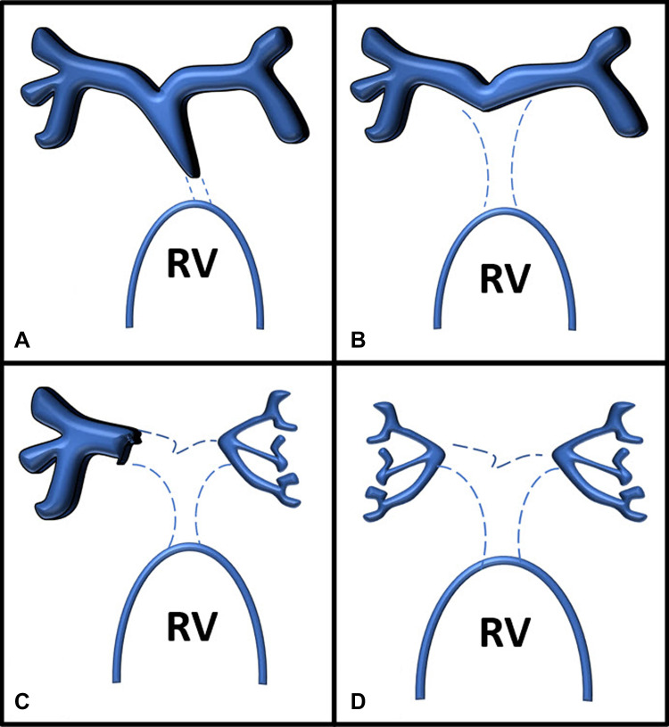
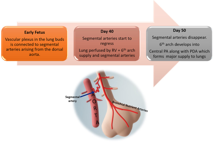
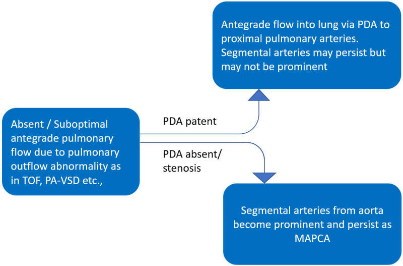
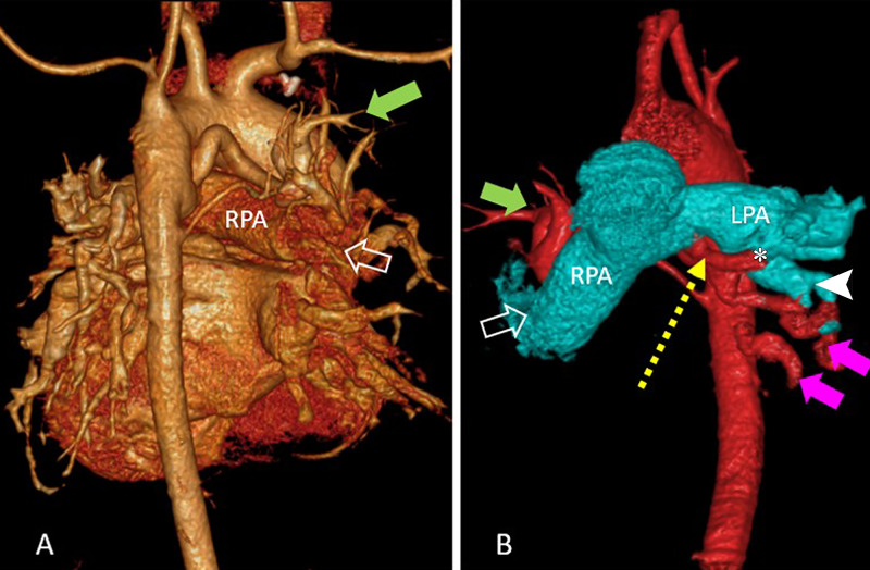
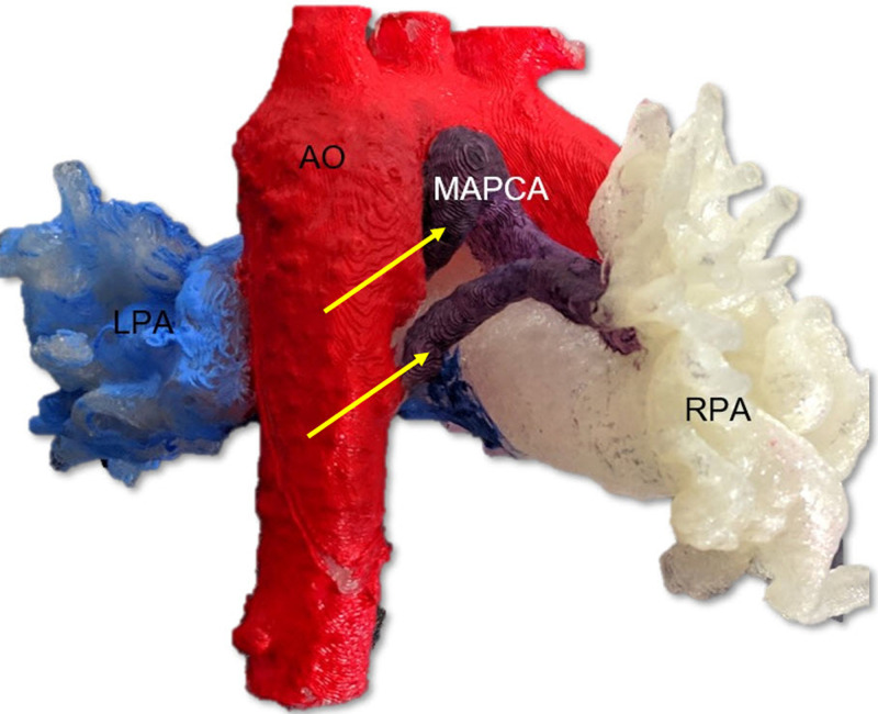
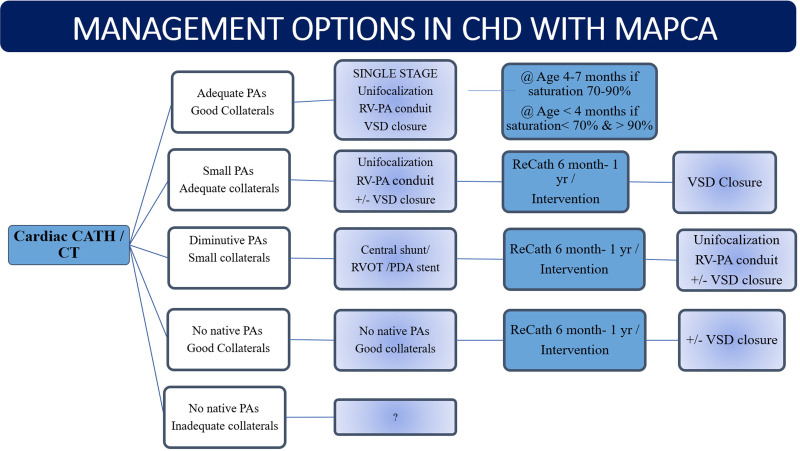
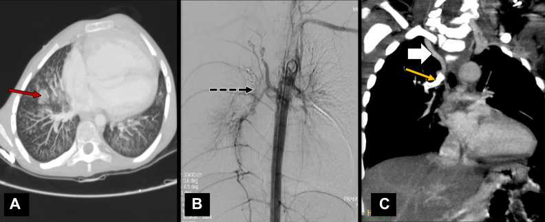
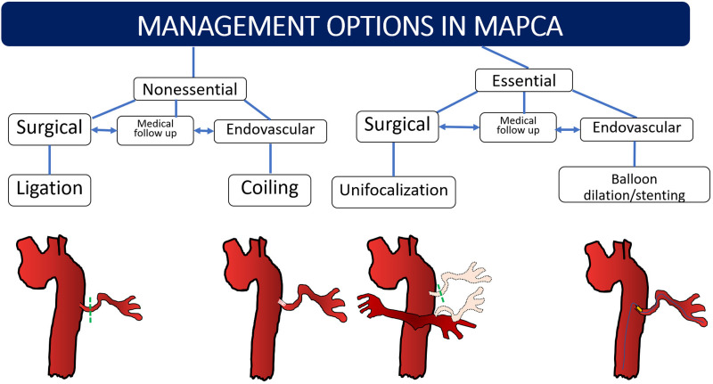
Major aortopulmonary collateral arteries (MAPCAs) are congenital vessels that arise from the aorta or its first-order branches and are distally connected to the pulmonary arterial vasculature, thereby providing pulmonary blood flow. MAPCAs are commonly associated with several congenital heart diseases that have compromised pulmonary circulation due to severe stenosis involving pulmonary valves or arteries or due to pulmonary atresia. Embryologically, MAPCAs are presumed to be persistent segmental arteries. MAPCAs can be imaged with CT and MRI, and such imaging findings are important for surgeons and interventionists. The management options for MAPCAs include unifocalization, surgical ligation, and endovascular interventions, such as coil embolization. This review highlights the role of reporting certain critical features of MAPCAs at CT and MRI, which will help to facilitate management decisions for systemic-to-pulmonary collateral vessels observed in patients with congenital heart disease. Keywords: Pediatrics, CT Angiography, Image Postprocessing, Interventional-Vascular, MR Angiography, Embolization, Stents, Cardiac, Vascular, Aorta © RSNA, 2022.

Keywords: Aorta; CT Angiography; Cardiac; Embolization; Image Postprocessing; Interventional-Vascular; MR Angiography; Pediatrics; Stents; Vascular.


Conflict of interest statement

Disclosures of Conflicts of Interest: A. Alex No relevant relationships. A. Ayyappan No relevant relationships. J.V. No relevant relationships. H.K. No relevant relationships. D.S. No relevant relationships. S.M. No relevant relationships.

Figures

Somerville classification of pulmonary arterial anatomy in pulmonary
atresia with ventricular septal defect. (A) Type 1, atresia of pulmonary valve.
(B) Type 2, atresia of pulmonary valve and main pulmonary artery. (C) Type 3,
atresia of pulmonary valve, main pulmonary artery, and one main branch. (D) Type
4, atresia of the pulmonary valve, main pulmonary artery, and both main
pulmonary artery branches. RV = right ventricle.
Figure 1:
Somerville classification of pulmonary arterial anatomy in pulmonary atresia with ventricular septal defect. (A) Type 1, atresia of pulmonary valve. (B) Type 2, atresia of pulmonary valve and main pulmonary artery. (C) Type 3, atresia of pulmonary valve, main pulmonary artery, and one main branch. (D) Type 4, atresia of the pulmonary valve, main pulmonary artery, and both main pulmonary artery branches. RV = right ventricle.
Flowchart depicts the normal pulmonary vasculature development with
regression of the segmental arteries as the pulmonary artery develops from the
sixth branchial arch. PA = pulmonary artery, PDA = patent ductus arteriosus, RV
= right ventricle.
Figure 2:
Flowchart depicts the normal pulmonary vasculature development with regression of the segmental arteries as the pulmonary artery develops from the sixth branchial arch. PA = pulmonary artery, PDA = patent ductus arteriosus, RV = right ventricle.
Flowchart shows the development of collateral vessels with regard to the
formation of ductus arteriosus. MAPCAs = major aortopulmonary collateral
arteries, PA-VSD = pulmonary atresia with ventricular septal defect, PDA =
patent ductus arteriosus, TOF = tetralogy of Fallot.
Figure 3:
Flowchart shows the development of collateral vessels with regard to the formation of ductus arteriosus. MAPCAs = major aortopulmonary collateral arteries, PA-VSD = pulmonary atresia with ventricular septal defect, PDA = patent ductus arteriosus, TOF = tetralogy of Fallot.
(A) Volume-rendered reconstruction image in posterior projection shows
branching of native pulmonary artery and major aortopulmonary collateral
arteries (MAPCAs) in a 9-year-old girl with tetralogy of Fallot and shows
predominant supply to right upper lobe from native pulmonary artery (open arrow)
along with a MAPCA (green solid arrow). (B) Volume-rendered reconstruction image
in anterior projection shows aorta and MAPCA colored in red and pulmonary
branches in cyan color with predominant supply of left lower lobe by MAPCA
(magenta solid arrows) than by native left pulmonary artery (white arrow head).
A communicating collateral (yellow dotted arrow) was observed joining the native
left pulmonary artery at hilum from its inferior surface (*). Supply to
right upper lobe from native pulmonary artery (open arrow) and MAPCA (green
solid arrow) are also labeled.
Figure 4:
(A) Volume-rendered reconstruction image in posterior projection shows branching of native pulmonary artery and major aortopulmonary collateral arteries (MAPCAs) in a 9-year-old girl with tetralogy of Fallot and shows predominant supply to right upper lobe from native pulmonary artery (open arrow) along with a MAPCA (green solid arrow). (B) Volume-rendered reconstruction image in anterior projection shows aorta and MAPCA colored in red and pulmonary branches in cyan color with predominant supply of left lower lobe by MAPCA (magenta solid arrows) than by native left pulmonary artery (white arrow head). A communicating collateral (yellow dotted arrow) was observed joining the native left pulmonary artery at hilum from its inferior surface (*). Supply to right upper lobe from native pulmonary artery (open arrow) and MAPCA (green solid arrow) are also labeled.
Diagram representing the common sites of major aortopulmonary
collateral arteries origin and classification. D4–D6 = dorsal
vertebrae 4 to 6, IMA = internal mammary artery.
Figure 5:
Diagram representing the common sites of major aortopulmonary collateral arteries origin and classification. D4–D6 = dorsal vertebrae 4 to 6, IMA = internal mammary artery.
(A) Volume-rendered CT image in a 10-year-old boy with
atrioventricular canal defect and pulmonary stenosis shows the prominent
collateral vessels from the left subclavian artery and left internal mammary
artery (white dotted arrows). (B) Coronal maximum intensity projection CT
image in a 7-year-old boy with double-outlet right ventricle and pulmonary
stenosis shows dilated left coronary artery (yellow arrows) supplying the
left lung. (C) Digital subtraction angiographic image in same patient as (B)
of the celiac artery ostium shows hypertrophied inferior phrenic branches
supplying bilateral lung parenchyma (black open arrows).
Figure 6:
(A) Volume-rendered CT image in a 10-year-old boy with atrioventricular canal defect and pulmonary stenosis shows the prominent collateral vessels from the left subclavian artery and left internal mammary artery (white dotted arrows). (B) Coronal maximum intensity projection CT image in a 7-year-old boy with double-outlet right ventricle and pulmonary stenosis shows dilated left coronary artery (yellow arrows) supplying the left lung. (C) Digital subtraction angiographic image in same patient as (B) of the celiac artery ostium shows hypertrophied inferior phrenic branches supplying bilateral lung parenchyma (black open arrows).
(A) Chest radiograph in frontal projection of a 4-year-old girl with long
segment pulmonary atresia and ventricular septal defect shows markedly increased
vascularity involving the right upper zone (white arrow) in comparison with rest
of the lung showing normal or mildly reduced vascularity. (B) Correlation with
coronal CT image shows dilated major aortopulmonary collateral arteries (white
arrow) independently supplying the right upper lobe segments. Child also had a
right-sided aortic arch.
Figure 7:
(A) Chest radiograph in frontal projection of a 4-year-old girl with long segment pulmonary atresia and ventricular septal defect shows markedly increased vascularity involving the right upper zone (white arrow) in comparison with rest of the lung showing normal or mildly reduced vascularity. (B) Correlation with coronal CT image shows dilated major aortopulmonary collateral arteries (white arrow) independently supplying the right upper lobe segments. Child also had a right-sided aortic arch.
A three-dimensional printed model in right-posterior projection from a
patient with pulmonary atresia with ventricular septal defect with major
aorto-pulmonary collateral arteries (MAPCA; yellow arrows) arising from the
aorta (Ao; colored in red), shown in relation to the right pulmonary arteries
(RPA; white color) and left pulmonary arteries (LPA; light blue color), aided in
surgical planning prior to unifocalization.
Figure 8:
A three-dimensional printed model in right-posterior projection from a patient with pulmonary atresia with ventricular septal defect with major aorto-pulmonary collateral arteries (MAPCA; yellow arrows) arising from the aorta (Ao; colored in red), shown in relation to the right pulmonary arteries (RPA; white color) and left pulmonary arteries (LPA; light blue color), aided in surgical planning prior to unifocalization.
(A) Coronal maximum intensity projection of multiphase contrast-enhanced
MR angiographic image in a 6-year-old girl with total anomalous pulmonary venous
return (course marked with red ★s), ventricular septal defect, and
hypoplastic left pulmonary artery (yellow curved arrow). Few major
aortopulmonary collateral arteries (dotted yellow arrows) arising from the
descending thoracic aorta were observed supplying right lower lobe and left
upper and lower lobes. The left-sided pulmonary veins were not adequately
opacified in this phase but were of normal size in subsequent phases, suggesting
slow flow to left lung. The right pulmonary artery was of normal caliber (white
arrow). (B) Axial maximum intensity projection of non–contrast-enhanced
balanced steady-state free precession angiographic image in a 2-year-old boy
with tetralogy of Fallot shows hypoplastic central pulmonary arteries (red open
arrows) and multiple major aortopulmonary collateral arteries (yellow dotted
arrows) arising from descending thoracic aorta (DTA) directly supplying lung and
communicating with native pulmonary artery. AA = ascending aorta.
Figure 9:
(A) Coronal maximum intensity projection of multiphase contrast-enhanced MR angiographic image in a 6-year-old girl with total anomalous pulmonary venous return (course marked with red ★s), ventricular septal defect, and hypoplastic left pulmonary artery (yellow curved arrow). Few major aortopulmonary collateral arteries (dotted yellow arrows) arising from the descending thoracic aorta were observed supplying right lower lobe and left upper and lower lobes. The left-sided pulmonary veins were not adequately opacified in this phase but were of normal size in subsequent phases, suggesting slow flow to left lung. The right pulmonary artery was of normal caliber (white arrow). (B) Axial maximum intensity projection of non–contrast-enhanced balanced steady-state free precession angiographic image in a 2-year-old boy with tetralogy of Fallot shows hypoplastic central pulmonary arteries (red open arrows) and multiple major aortopulmonary collateral arteries (yellow dotted arrows) arising from descending thoracic aorta (DTA) directly supplying lung and communicating with native pulmonary artery. AA = ascending aorta.
Summary of the key points that should be included with regard to
collateral arteries in the radiology report. MAPCA = major aortopulmonary
collateral artery, PA = pulmonary artery.
Figure 10:
Summary of the key points that should be included with regard to collateral arteries in the radiology report. MAPCA = major aortopulmonary collateral artery, PA = pulmonary artery.
(A) Sagittal oblique cardiac CT section in a 1-year-old boy shows
patent ductus arteriosus (rounded arrow) draining into left pulmonary artery
in a case of pulmonary atresia and ventricular septal defect. (B) Coronal
oblique cardiac CT image shows right pulmonary artery (black arrow) arising
from aorta in a 4-month-old girl, suggestive of hemitruncus. There was
coexisting main pulmonary artery (*) supply to left lung. (C)
Volume-rendered cardiac CT image shows isolated pulmonary supply from aorta
(white tailed arrow) to lung parenchyma in a 26-year-old man who presented
with hemoptysis and no structural heart disease, suggestive of type B
malinosculation. (D) Coronal projection of CT angiographic image in
volume-rendered reconstruction (excluding anterior ascending aorta for
visibility) in a 7-year-old boy with double-outlet right ventricle after
staged hemi-Fontan surgery shows small vessels in mediastinum with tortuous
courses (blue dotted arrows) having communication with superior caval vein
or its tributaries and veins draining into azygous vein, suggestive of
venovenous collaterals.
Figure 11:
(A) Sagittal oblique cardiac CT section in a 1-year-old boy shows patent ductus arteriosus (rounded arrow) draining into left pulmonary artery in a case of pulmonary atresia and ventricular septal defect. (B) Coronal oblique cardiac CT image shows right pulmonary artery (black arrow) arising from aorta in a 4-month-old girl, suggestive of hemitruncus. There was coexisting main pulmonary artery (*) supply to left lung. (C) Volume-rendered cardiac CT image shows isolated pulmonary supply from aorta (white tailed arrow) to lung parenchyma in a 26-year-old man who presented with hemoptysis and no structural heart disease, suggestive of type B malinosculation. (D) Coronal projection of CT angiographic image in volume-rendered reconstruction (excluding anterior ascending aorta for visibility) in a 7-year-old boy with double-outlet right ventricle after staged hemi-Fontan surgery shows small vessels in mediastinum with tortuous courses (blue dotted arrows) having communication with superior caval vein or its tributaries and veins draining into azygous vein, suggestive of venovenous collaterals.
Algorithm for surgical management of congenital heart disease (CHD) in
presence of major aortopulmonary collateral arteries (MAPCAs). A Nakata index of
less than 100 mm2/m2 or McGoon ratio of less than 1.2 is considered for central
shunt creation or right ventricle outflow tract or patent ductus arteriosus
(PDA) stent placement. A reduced Qp/Qs of less than 1 is an indication for
staging ventricular septal defect (VSD) closure. CATH = catheter, PA = pulmonary
artery, Qp/Qs = differential flow through right and left pulmonary arteries, RV
= right ventricle, RVOT = RV outflow tract.
Figure 12:
Algorithm for surgical management of congenital heart disease (CHD) in presence of major aortopulmonary collateral arteries (MAPCAs). A Nakata index of less than 100 mm2/m2 or McGoon ratio of less than 1.2 is considered for central shunt creation or right ventricle outflow tract or patent ductus arteriosus (PDA) stent placement. A reduced Qp/Qs of less than 1 is an indication for staging ventricular septal defect (VSD) closure. CATH = catheter, PA = pulmonary artery, Qp/Qs = differential flow through right and left pulmonary arteries, RV = right ventricle, RVOT = RV outflow tract.
(A) CT and (B) digital subtraction angiographic images in a 14-year-old
boy with tetralogy of Fallot and associated pulmonary infundibular stenosis. The
child presented with hemoptysis with lung parenchyma showing areas of pulmonary
hemorrhage (red arrow in A) in right lung. (B) Descending thoracic aortic
angiographic image shows the major aortopulmonary collateral arteries (MAPCAs)
arising at D6 level that were embolized using vascular coils (black dotted
arrow). (C) Coronal oblique projection of cardiac CT angiographic image in an
8-year-old boy with pulmonary atresia with ventricular septal defect shows a
large collateral vessel from right subclavian artery (white solid arrow)
supplying right upper lobe. The descending segment of this collateral vessel was
narrowed and had a stent placed (yellow arrow) to maintain saturation, as
central pulmonary arteries were poorly developed.
Figure 13:
(A) CT and (B) digital subtraction angiographic images in a 14-year-old boy with tetralogy of Fallot and associated pulmonary infundibular stenosis. The child presented with hemoptysis with lung parenchyma showing areas of pulmonary hemorrhage (red arrow in A) in right lung. (B) Descending thoracic aortic angiographic image shows the major aortopulmonary collateral arteries (MAPCAs) arising at D6 level that were embolized using vascular coils (black dotted arrow). (C) Coronal oblique projection of cardiac CT angiographic image in an 8-year-old boy with pulmonary atresia with ventricular septal defect shows a large collateral vessel from right subclavian artery (white solid arrow) supplying right upper lobe. The descending segment of this collateral vessel was narrowed and had a stent placed (yellow arrow) to maintain saturation, as central pulmonary arteries were poorly developed.
Summary of management options in major aortopulmonary collateral arteries
(MAPCAs) in general. Essential MAPCAs require either unifocalization or
endovascular stent placement or angioplasty. Nonessential and dilated MAPCAs are
coiled or ligated. Nonessential stenosed MAPCAs may be kept under medical
follow-up before or after surgery and addressed later. If all essential MAPCAs
cannot be unifocalized in a single stage, the remaining MAPCAs are followed-up
until the next stage of repair.
Figure 14:
Summary of management options in major aortopulmonary collateral arteries (MAPCAs) in general. Essential MAPCAs require either unifocalization or endovascular stent placement or angioplasty. Nonessential and dilated MAPCAs are coiled or ligated. Nonessential stenosed MAPCAs may be kept under medical follow-up before or after surgery and addressed later. If all essential MAPCAs cannot be unifocalized in a single stage, the remaining MAPCAs are followed-up until the next stage of repair.

References

    1. Wernovsky G , Anderson RH , Kumar K , et al. , eds. Anderson’s Pediatric Cardiology . 4th ed. Philadelphia, Pa: : Elsevier; , 2019. ; 660 – 663 .
    1. Marino B , Calabró R , Gagliardi MG , Bevilacqua M , Ballerini L , Marcelletti C . Patterns of pulmonary arterial anatomy and blood supply in complex congenital heart disease with pulmonary atresia . J Thorac Cardiovasc Surg 1987. ; 94 ( 4 ): 518 – 520 . - PubMed
    1. Ryan JR , Moe TG , Richardson R , Frakes DH , Nigro JJ , Pophal S . A novel approach to neonatal management of tetralogy of Fallot, with pulmonary atresia, and multiple aortopulmonary collaterals . JACC Cardiovasc Imaging 2015. ; 8 ( 1 ): 103 – 104 . - PubMed
    1. Seale AN , Ho SY , Shinebourne EA , Carvalho JS . Prenatal identification of the pulmonary arterial supply in tetralogy of Fallot with pulmonary atresia . Cardiol Young 2009. ; 19 ( 2 ): 185 – 191 . - PubMed
    1. Pelizzo G , Calcaterra V , Mannarino S , Moramarco LP , Leati G , Quaretti P . Aortopulmonary collateral artery in prenatal exposure to carbamazepine - endovascular therapy and technical considerations: a case report . J Med Case Rep 2015. ; 9 183 . - PMC - PubMed

LinkOut - more resources