Case Reports
. 2022 Feb 17;4(1):e210249.
doi: 10.1148/ryct.210249. eCollection 2022 Feb.

Left Circumflex Coronary Artery-to-Coronary Sinus Fistula with Coronary Sinus Ostial Atresia and a Persistent Left Superior Vena Cava in an Adult Patient

Affiliations
Case Reports

Left Circumflex Coronary Artery-to-Coronary Sinus Fistula with Coronary Sinus Ostial Atresia and a Persistent Left Superior Vena Cava in an Adult Patient

Vitor F Martins et al. Radiol Cardiothorac Imaging. .

Abstract

Understanding of coronary sinus (CS) anatomy and abnormalities is of critical importance due to their use in interventional procedures. Herein, the authors report a rare case of an asymptomatic 72-year-old man with a left circumflex coronary artery-to-CS fistula, together with CS ostial atresia and persistent left superior vena cava. These findings are described using both cardiac CT angiography and MRI with four-dimensional flow for anatomic and functional assessment. Keywords: Cardiac, Coronary Sinus, Aneurysms, Fistula, CT Angiography, MR Imaging Supplemental material is available for this article. © RSNA, 2022.

Keywords: Aneurysms; CT Angiography; Cardiac; Coronary Sinus; Fistula; MR Imaging.


Conflict of interest statement

Disclosures of Conflicts of Interest: V.F.M. No relevant relationships. A.H. Research grants from GE Healthcare and Bayer; patent, royalties, and licenses from Stanford University for “Comprehensive Cardiovascular Analysis with Volumetric Phase-Contrast MRI”; founder shares in Arterys. S.K. Deputy editor for Radiology: Cardiothoracic Imaging. S.S.B. No relevant relationships.

Figures

Axial cardiac CT angiographic images demonstrate the presence of a dilated
left circumflex coronary artery with fistula to a dilated coronary sinus (CS)
with ostial atresia and aneurysm. (A) Dilated and tortuous left circumflex
coronary artery (red arrows). (B) Dilated, opacified CS adjacent to the right
atrium (RA) without communication, demonstrating an ostial atresia (yellow
arrow). A large, partially thrombosed aneurysm is visualized with a patent
lumen. Red arrow indicates left circumflex coronary artery. (C) Drainage of the
CS (blue arrows) into lumen of a large, partially calcified, partially
thrombosed aneurysm. See Movie 1 for axial, sagittal, and coronal cuts, and see
Movie 2 for three-dimensional reconstruction. aA = ascending aorta, dA =
descending aorta, L = lumen of aneurysm, LA = left atrium, LV = left ventricle,
PA = pulmonary artery, rPA = right PA, RV = right ventricle, T = thrombus of
aneurysm.
Figure 1:
Axial cardiac CT angiographic images demonstrate the presence of a dilated left circumflex coronary artery with fistula to a dilated coronary sinus (CS) with ostial atresia and aneurysm. (A) Dilated and tortuous left circumflex coronary artery (red arrows). (B) Dilated, opacified CS adjacent to the right atrium (RA) without communication, demonstrating an ostial atresia (yellow arrow). A large, partially thrombosed aneurysm is visualized with a patent lumen. Red arrow indicates left circumflex coronary artery. (C) Drainage of the CS (blue arrows) into lumen of a large, partially calcified, partially thrombosed aneurysm. See Movie 1 for axial, sagittal, and coronal cuts, and see Movie 2 for three-dimensional reconstruction. aA = ascending aorta, dA = descending aorta, L = lumen of aneurysm, LA = left atrium, LV = left ventricle, PA = pulmonary artery, rPA = right PA, RV = right ventricle, T = thrombus of aneurysm.
Curved multiplanar reconstruction of cardiac CT angiographic image
demonstrates a partially thrombosed aneurysm (AN) draining superiorly through a
persistent left superior vena cava (red arrowhead), connecting to a left
brachiocephalic vein (LBCV).
Figure 2:
Curved multiplanar reconstruction of cardiac CT angiographic image demonstrates a partially thrombosed aneurysm (AN) draining superiorly through a persistent left superior vena cava (red arrowhead), connecting to a left brachiocephalic vein (LBCV).
Image from axial cardiac four-dimensional flow MRI demonstrates no flow
(ie, ostial atresia; yellow arrow) between the coronary sinus (CS) and the right
atrium (RA). The flow through cardiac structures can also be appreciated with
minimal flow within the lumen of the aneurysm (L), circular or turbulent flow
within the CS, and linear high flow within the left ventricle (LV) toward the LV
outflow tract. See Movie 3 for full loop. dA = descending aorta, RV = right
ventricle, T = thrombus of aneurysm.
Figure 3:
Image from axial cardiac four-dimensional flow MRI demonstrates no flow (ie, ostial atresia; yellow arrow) between the coronary sinus (CS) and the right atrium (RA). The flow through cardiac structures can also be appreciated with minimal flow within the lumen of the aneurysm (L), circular or turbulent flow within the CS, and linear high flow within the left ventricle (LV) toward the LV outflow tract. See Movie 3 for full loop. dA = descending aorta, RV = right ventricle, T = thrombus of aneurysm.
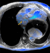
(A, B) Oblique reconstructions of cardiac four-dimensional flow MRI
demonstrate retrograde (cephalad) blood flow through the persistent left
superior vena cava (red arrowhead) into the left brachiocephalic vein (LBCV).
Red arrows indicate left circumflex artery. See Movie 4 (for A) and Movie 5 (for
B) for full loop. aA = ascending aorta, LV = left ventricle, PA = pulmonary
artery, RSVC = right superior vena cava, RV = right ventricle.
Figure 4:
(A, B) Oblique reconstructions of cardiac four-dimensional flow MRI demonstrate retrograde (cephalad) blood flow through the persistent left superior vena cava (red arrowhead) into the left brachiocephalic vein (LBCV). Red arrows indicate left circumflex artery. See Movie 4 (for A) and Movie 5 (for B) for full loop. aA = ascending aorta, LV = left ventricle, PA = pulmonary artery, RSVC = right superior vena cava, RV = right ventricle.

References

    1. Shah SS , Teague SD , Lu JC , Dorfman AL , Kazerooni EA , Agarwal PP . Imaging of the coronary sinus: normal anatomy and congenital abnormalities . RadioGraphics 2012. ; 32 ( 4 ): 991 – 1008 . - PubMed
    1. Mantini E , Grondin CM , Lillehei CW , Edwards JE . Congenital anomalies involving the coronary sinus . Circulation 1966. ; 33 ( 2 ): 317 – 327 . - PubMed
    1. Chen YA , Nguyen ET , Dennie C , et al. . Computed tomography and magnetic resonance imaging of the coronary sinus: anatomic variants and congenital anomalies . Insights Imaging 2014. ; 5 ( 5 ): 547 – 557 . - PMC - PubMed
    1. Xie M , Li L , Cheng TO , et al. . Coronary artery fistula: comparison of diagnostic accuracy by echocardiography versus coronary arteriography and surgery in 63 patients studied between 2002 and 2012 in a single medical center in China . Int J Cardiol 2014. ; 176 ( 2 ): 470 – 477 . - PubMed
    1. Santoscoy R , Walters HL 3rd , Ross RD , Lyons JM , Hakimi M . Coronary sinus ostial atresia with persistent left superior vena cava . Ann Thorac Surg 1996. ; 61 ( 3 ): 879 – 882 . - PubMed

Publication types