

Retinal Aging in 3× Tg-AD Mice Model of Alzheimer's Disease
- PMID: 35783138
- PMCID: PMC9244797
- DOI: 10.3389/fnagi.2022.832195
Retinal Aging in 3× Tg-AD Mice Model of Alzheimer's Disease
Abstract
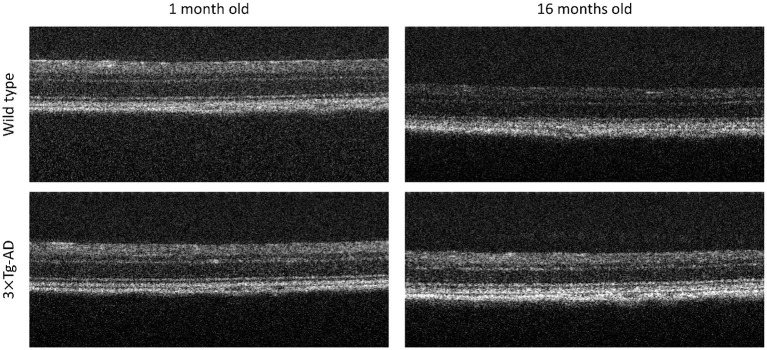
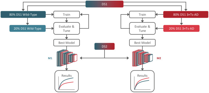
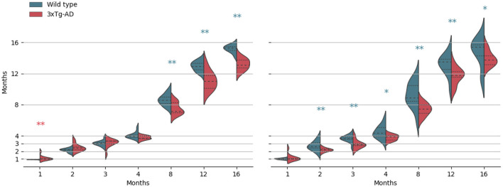
The retina, as part of the central nervous system (CNS), can be the perfect target for in vivo, in situ, and noninvasive neuropathology diagnosis and assessment of therapeutic efficacy. It has long been established that several age-related brain changes are more pronounced in Alzheimer's disease (AD). Nevertheless, in the retina such link is still under-explored. This study investigates the differences in the aging of the CNS through the retina of 3× Tg-AD and wild-type mice. A dedicated optical coherence tomograph imaged mice's retinas for 16 months. Two neural networks were developed to model independently each group's ages and were then applied to an independent set containing images from both groups. Our analysis shows a mean absolute error of 0.875±1.1 × 10⁻² and 1.112±1.4 × 10⁻² months, depending on training group. Our deep learning approach appears to be a reliable retinal OCT aging marker. We show that retina aging is distinct in the two classes: the presence of the three mutated human genes in the mouse genome has an impact on the aging of the retina. For mice over 4 months-old, transgenic mice consistently present a negative retina age-gap when compared to wild-type mice, regardless of training set. This appears to contradict AD observations in the brain. However, the 'black-box" nature of deep-learning implies that one cannot infer reasoning. We can only speculate that some healthy age-dependent neural adaptations may be altered in transgenic animals.
Keywords: Alzheimer's disease; age-gap; aging; animal model; artificial intelligence; deep learning; optical coherence tomography; retina.
Copyright © 2022 Guimarães, Serranho, Martins, Moreira, Ambrósio, Castelo-Branco and Bernardes.
Conflict of interest statement
The authors declare that the research was conducted in the absence of any commercial or financial relationships that could be construed as a potential conflict of interest.
Figures
References
-
- Burgaletto C., Platania C. B. M., Di Benedetto G., Munafó A., Giurdanella G., Federico C., et al. . (2021). Targeting the mirna-155/tnfsf10 network restrains inflammatory response in the retina in a mouse model of Alzheimer's disease. Cell Death Dis. 12, 1–15. 10.1038/s41419-021-04165-x - DOI - PMC - PubMed
-
- Chiquita S., Campos E. J., Castelhano J., Ribeiro M., Sereno J., Moreira P. I., et al. . (2019). Retinal thinning of inner sub-layers is associated with cortical atrophy in a mouse model of Alzheimer's disease: a longitudinal multimodal in vivo study. Alzheimer's Res. Ther. 11, 1–16. 10.1186/s13195-019-0542-8 - DOI - PMC - PubMed
-
- Deng J., Dong W., Socher R., Li L. -J., Li K., Fei-Fei L. (2009). Imagenet: a large-scale hierarchical image database, in 2009 IEEE Conference on Computer Vision and Pattern Recognition (Miami, FL: IEEE; ), 248–255. 10.1109/CVPR.2009.5206848 - DOI
LinkOut - more resources
Full Text Sources
Miscellaneous